
Article Figures & Data
Figures
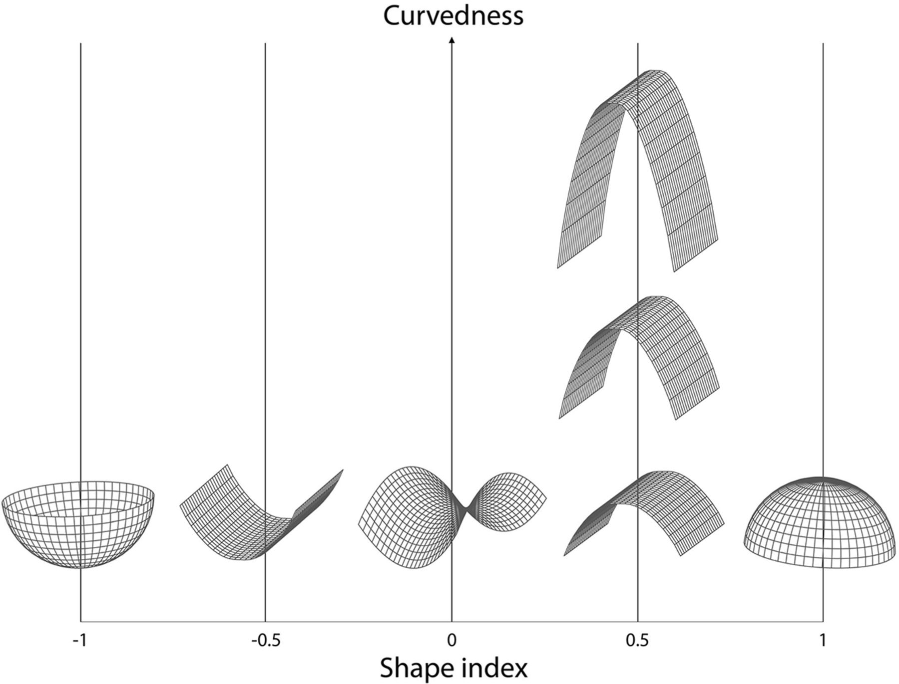
- FIG 1.
Shape index and curvedness. Shape index and curvedness values vary in 3D shapes. The shape index is a descriptor of the local shape of the surface of an object and is scale-invariant. Shape index values range from –1 (concave “cup”) through 0 (saddle point) to 1 (convex “dome”). The curvedness is a positive value to the local curvature of the surface, which usually lies between 0 and 1 and is dependent on the local scale of the object. These values are rotation- and translation-invariant.
- FIG 2.
Nongrowing UIA with statistically significant change in morphology. An example of a ROI around a UIA taken from baseline and follow-up TOF-MRAs made on a Philips 1.5T scanner (Intera/Achieva). The measured UIA shows statistically significant changes in morphology but was considered to be nongrowing (<1-mm change in length or width). The bulge (arrow) that becomes visible on follow-up results in more saddle points on the surface of the UIA, whereby the shape index decreases. The bulge also increases the curvature of the surface of the UIA, resulting in a slightly increased curvedness value.


Tables
Characteristic No. of patients 113 No. of aneurysms 127 (102 patient with 1 UIA, 8 with 2 and 3 with 3 UIAs) Sex (% women) 81 women, 32 men (72% women) Age at baseline (mean) (yr) 55 (range, 27 –77 ) UIA size at baseline (mean) (mm) 3.8 (SD, 1.9) Time between baseline and follow-up scan (median) (yr) 4.1 (range, 0.9–13.1 ) Location of aneurysm Anterior cerebral or communicating artery 25 (20%) ICA or posterior communicating artery 33 (26%) MCA 53 (42%) Posterior circulation 16 (13%) - Table 2:
Change in UIA morphology measurements in relation to continuous UIA 2D and 3D growtha
Change in Median (IQR) Correlation Coefficient (P Value) 2D Growth, Length 2D Growth, Width 3D Growth, Volume Volume (mm3) 1.60 (−3.70−10.80) 0.29 (<.01)b 0.28 (<.01)b − Area (mm2) 2.30 (−6.10−11.40) 0.25 (<.01)b 0.34 (<.01)b 0.90 (<.01)b Compactness 1 0.50 (−1.70−2.60)c 0.10 (.28) −0.01 (.89) 0.15 (.09) Compactness 2 0.01 (−0.04−0.07) 0.09 (.30) −0.02 (.86) 0.15 (.10) Elongation 0.01 (−0.03−0.04) 0.01 (.89) 0.02 (.79) 0.09 (.33) Flatness 0.00 (−0.03−0.04) 0.00 (.97) 0.05 (.58) 0.19 (.03)b Sphericity 0.01 (−0.02−0.04) 0.10 (.27) −0.01 (.94) 0.15 (.09) Shape index 0.00 (−0.03−0.01) −0.09 (.32) −0.17 (.06) −0.33 (<.01)b Curvedness −0.01 (−0.18−.07) −0.12 (.16) −0.15 (.09) −0.33 (<.01)b Change in Growing Nongrowing P Value Volume (mm3) 21.92 (4.8−33.23)b 1.42 (−4.26−9.76)b .01b Area (mm2) 28.09 (−4.23−35.37)b 2.07 (−6.47−9.89)b <.01b Compactness 1 0.40 (−1.35−3.85)b 0.00 (−1.65−2.33)c .21 Compactness 2 0.01 (−0.03−0.11) 0.01 (−0.04−0.06) .22 Elongation −0.02 (−0.05−0.02) 0.02 (−0.03−0.04) .42 Flatness 0.01 (−0.04−0.04) 0.00 (−0.03−0.04) .37 Sphericity 0.01 (−0.02−0.05) 0.01 (−0.02−0.03) .20 Shape index 0.00 (−0.13−0.00) 0.00 (−0.03−0.01) .06 Curvedness −0.14 (−0.37−0.01)b −0.01 (−0.16−0.09)b .03b ↵a Comparing change in 3D quantified morphology of stable and growing UIAs. Values are written as median (IQR). Growth was defined as an increase of at least 1 mm in either width or length of the UIA. P values refer to the relation between parameters of the growing and stable UIAs using a t test or Mann-Whitney U test.
↵b P values are statistically significant.
↵c x 103.


Jump to section
Related Articles
Cited By...
- Intra-Aneurysmal High-Resolution 4D MR Flow Imaging for Hemodynamic Imaging Markers in Intracranial Aneurysm Instability
- Deep learning-based cerebral aneurysm segmentation and morphological analysis with three-dimensional rotational angiography
- Deep learning-based cerebral aneurysm segmentation and morphological analysis with three-dimensional rotational angiography