
Article Figures & Data
Figures
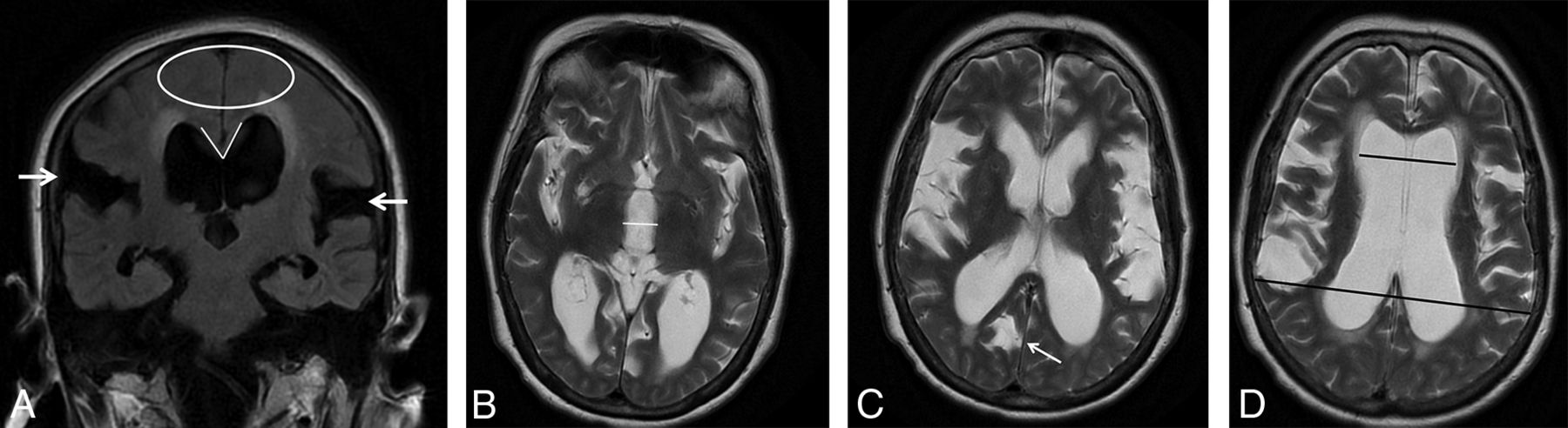
- FIG 1.
MR imaging of a 79-year-old female shunt-responder with an NPH Radscale score of 11. A, Coronal FLAIR image shows marked bilateral Sylvian fissure dilation (white arrows), apical narrowing of sulci (white elipse), a narrow CA (65°), and marked periventricular hyperintensities. B–D, Axial T2 images at different levels show measurement of third ventricle diameter (white line), sulcal dilation (white arrow), and measurements used for the Evans ratio calculation (black lines).
- FIG 2.
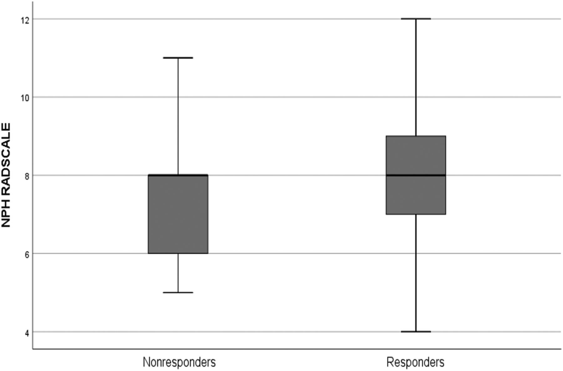
A boxplot of NPH Radscale values (y-axis) for responders and nonresponders, respectively. The thick black line is the median; and box upper and lower margins are 75th and 25th percentiles, respectively. Whiskers represent upper and lower ranges.


Tables
- Table 1:
Distribution of different imaging parameters for the entire population for responders and nonresponders
Score Total Responders Nonresponders P Value Patients (No.) 119 94 25 EIa 0.39 (0.046) 0.39 (0.046) 0.38 (0.046) .28c CAa 73.0 (17.4) 71.9 (16.7) 77.2 (19.7) .23c Third ventricle diametera 14.2 (3.14) 14.3 (3.00) 14.1 (3.72) .75c Mean temporal horn diametera 8.14 (2.40) 8.38 (2.40) 7.26 (2.22) .03c NPH Radscalea 8.17 (1.56) 8.35 (1.53) 7.48 (1.53) .02c DESHb Present 60 (50.4%) 49 (52.1%) 11 (44%) .47d Not present 59 (49.6%) 45 (47.9%) 14 (56%) Focal bulging Present 14 (11.8%) 12 (12.8%) 2 (8.0%) .73e Not present 105 (88.2%) 82 (87.2%) 23 (92.0%) Focally enlarged sulcib Present 22 (18.5%) 17 (18.1%) 5 (20.0%) .78e Not present 97 (81.5%) 77 (81.9%) 20 (80.0%) Sylvian fissure dilationb Present 101 (84.9%) 83 (88.3%) 18 (72.0% .05e Not present 18 (15.1%) 11 (11.7%) 7 (28%) Narrow sulcib 2 47 (39.5%) 29 (30.9%) 4 (16.0%) .19f 1 39 (32.8%) 30 (31.9%) 9 (36.0%) 0 33 (27.7%) 35 (37.2%) 12 (48.0%) PVWMLb 2 55 (46.2%) 47 (50.0%) 8 (32.0%) .16f 1 47 (39.5%) 34 (36.2%) 13 (52.0%) 0 17 (14.3%) 13 (13.8%) 4 (16.0%) MR Imaging (n = 74) CT (n = 19) ICC LB UB ICC LB UB EI 0.914 0.863 0.946 0.974 0.932 0.990 CA 0.934 0.895 0.958 0.856 0.626 0.945 Mean temporal horn diameter 0.943 0.909 0.964 0.917 0.784 0.968 Third ventricle diameter 0.918 0.869 0.948 0.966 0.911 0.987 NPH Radscale 0.858 0.774 0.910 0.819 0.531 0.930 Note:—LB indicates lower bound; UB, upper bound; ICC, intraclass correlation coefficient.
MR Imaging (n = 74) CT (n = 19) κ UB LB κ UB LB DESH 0.569 0.402 0.736 0.607 0.247 0.967 Focal bulging 0.354 0.090 0.617 –0.145 –0.282 –0.006 Focally enlarged sulci 0.617 0.393 0.841 0.650 0.292 1.008 Sylvian fissure dilation 0.617 0.393 0.841 0.477 0.025 0.979 Note:—κ Indicates Kappa statistics; LB, lower bound; UB, upper bound; DESH, disproportionately enlarged subarchmoid space hydroceplaus.
- Table 4:
Spearman rank statistics for non-binary categorical evaluations for MR imaging and CT
MR Imaging (n = 74) CT (n = 19) Spearman Rank UB LB Spearman Rank UB LB Narrow sulci 0.626 0.464 0.747 0.787 0.518 0.914 PVWML 0.691 0.550 0.794 0.826 0.596 0.930 Note:—LB indicates lower bound; UB, upper bound; pvWML, periventricular white matter hyperintensities.


Jump to section
Related Articles
Cited By...
- No citing articles found.