Advertisement

Main menu

  • Home
  • Content
    • Current Issue
    • Accepted Manuscripts
    • Article Preview
    • Past Issue Archive
    • Video Articles
    • AJNR Case Collection
    • Case of the Week Archive
    • Case of the Month Archive
    • Classic Case Archive
  • Special Collections
    • AJNR Awards
    • Low-Field MRI
    • Alzheimer Disease
    • ASNR Foundation Special Collection
    • Photon-Counting CT
    • View All
  • Multimedia
    • AJNR Podcasts
    • AJNR SCANtastic
    • Trainee Corner
    • MRI Safety Corner
    • Imaging Protocols
  • For Authors
    • Submit a Manuscript
    • Submit a Video Article
    • Submit an eLetter to the Editor/Response
    • Manuscript Submission Guidelines
    • Statistical Tips
    • Fast Publishing of Accepted Manuscripts
    • Graphical Abstract Preparation
    • Imaging Protocol Submission
    • Author Policies
  • About Us
    • About AJNR
    • Editorial Board
    • Editorial Board Alumni
  • More
    • Become a Reviewer/Academy of Reviewers
    • Subscribers
    • Permissions
    • Alerts
    • Feedback
    • Advertisers
    • ASNR Home

User menu

  • Alerts
  • Log in

Search

  • Advanced search
American Journal of Neuroradiology

American Journal of Neuroradiology

ASHNR American Society of Functional Neuroradiology ASHNR American Society of Pediatric Neuroradiology ASSR
  • Alerts
  • Log in

Advanced Search

  • Home
  • Content
    • Current Issue
    • Accepted Manuscripts
    • Article Preview
    • Past Issue Archive
    • Video Articles
    • AJNR Case Collection
    • Case of the Week Archive
    • Case of the Month Archive
    • Classic Case Archive
  • Special Collections
    • AJNR Awards
    • Low-Field MRI
    • Alzheimer Disease
    • ASNR Foundation Special Collection
    • Photon-Counting CT
    • View All
  • Multimedia
    • AJNR Podcasts
    • AJNR SCANtastic
    • Trainee Corner
    • MRI Safety Corner
    • Imaging Protocols
  • For Authors
    • Submit a Manuscript
    • Submit a Video Article
    • Submit an eLetter to the Editor/Response
    • Manuscript Submission Guidelines
    • Statistical Tips
    • Fast Publishing of Accepted Manuscripts
    • Graphical Abstract Preparation
    • Imaging Protocol Submission
    • Author Policies
  • About Us
    • About AJNR
    • Editorial Board
    • Editorial Board Alumni
  • More
    • Become a Reviewer/Academy of Reviewers
    • Subscribers
    • Permissions
    • Alerts
    • Feedback
    • Advertisers
    • ASNR Home
  • Follow AJNR on Twitter
  • Visit AJNR on Facebook
  • Follow AJNR on Instagram
  • Join AJNR on LinkedIn
  • RSS Feeds

AJNR Awards, New Junior Editors, and more. Read the latest AJNR updates

Research ArticleAdult Brain

Intracranial Vessel Segmentation in 3D High-Resolution T1 Black-Blood MRI

S. Elsheikh, H. Urbach and M. Reisert
American Journal of Neuroradiology December 2022, 43 (12) 1719-1721; DOI: https://doi.org/10.3174/ajnr.A7700
S. Elsheikh
aFrom the Departments of Neuroradiology (S.E., H.U.)
  • Find this author on Google Scholar
  • Find this author on PubMed
  • Search for this author on this site
  • ORCID record for S. Elsheikh
H. Urbach
aFrom the Departments of Neuroradiology (S.E., H.U.)
  • Find this author on Google Scholar
  • Find this author on PubMed
  • Search for this author on this site
  • ORCID record for H. Urbach
M. Reisert
bMedical Physics, Functional Neurosurgery, and Stereotaxy (M.R.), Faculty of Medicine, Medical Center–University of Freiburg, Freiburg, Germany
  • Find this author on Google Scholar
  • Find this author on PubMed
  • Search for this author on this site
  • ORCID record for M. Reisert
  • Article
  • Figures & Data
  • Supplemental
  • Info & Metrics
  • Responses
  • References
  • PDF
Loading

Abstract

SUMMARY: We demonstrate the feasibility of intracranial vascular segmentation based on the hypointense signal in non-contrast-enhanced black-blood MR imaging using convolutional neural networks. We selected 37 cases. Qualitatively, we observed no degradation due to stent artifacts, a comparable recognition of an aneurysm recurrence with TOF-MRA, and consistent success in the differentiation of intracranial arteries and veins. False-positive and false-negative results were observed. Quantitatively, our model achieved a promising Dice similarity coefficient of 0.72.

ABBREVIATIONS:

BBMRI
black-blood compressed-sensing MRI
CNN
convolutional neural networks
DSC
Dice similarity coefficient

Automated segmentation techniques of the cerebral vasculature are an area of interest, with numerous previous publications.1 Currently, convolutional neural networks (CNN) are the criterion standard for medical image segmentation.2 Publications applying CNN segmentation of the cerebral vessels were largely based on TOF-MRA.3

Recently, a black-blood compressed-sensing MRI (BBMRI) sequence was introduced. It allows isotropic voxels of 0.5 mm3, suppression of signal within the vessels, and covering a large volume. These advantages are useful in diverse clinical applications.4 We aimed to test the feasibility of intracranial vascular segmentation based on the hypointense signal in BBMRI using CNN.

Technical Report

After obtaining institutional review board approval, we retrospectively searched our PACS for examinations including BBMRI. We selected 37 (training, 26; testing, 11 randomly assigned [repeat examinations of the same patients were manually assigned to the training data set]) cases. The imaging indication was post-coiling examination in 34 cases, vasculitis in 2 cases, and dissection in 1 case.

All images were scanned on a 3T MR imaging (Magnetom Prisma; Siemens).4 The image matrix was 384 × 384 × 256; voxel size, 0.55× 0.55× 0.6 mm3.

We limited the volume of interest to cover the proximal course of the intracranial vessels and to extend beyond the circle of Willis (Online Supplemental Data). A binary mask was manually created in the Montreal Neurological Institute space (Online Supplemental Data) and then transformed to the individual patient space; this was used to define the cropped volume. Ground truth annotation was performed by a neuroradiologist (S.E.) with 15 years of experience in neurovascular imaging.

We used a hierarchic, multiscale, 3D CNN motivated by Yu et al.5 Four scales of nested patches with a matrix size of 323 voxels were used. Scale sizes ranged from 105.6 × 105.6 × 76.8 mm3 to 17.6 × 17.6 × 19.2 mm3. The 2 intermediate scales were exponentially interpolated. The scales were randomly selected but with a 50% probability that the scale center lay within the target label. In each scale, a UNET-type architecture similar to that in Ronneberger et al2 was used. The feature dimensions were 8, 16, 16, 32, 64. Max pooling in the encoding layers and transposed convolutions in the decoding layers were used. The input to the network was the BBMRI contrast normalized by the global signal mean. The output channels of each scale were forwarded as the input to the next scale (Online Supplemental Data). We trained the network using the Adam optimizer,6 with a rate of 0.001 and binary cross-entropy. The network application used a random patching scheme, in which in each layer, only the highest probability (50%) daughter patches were further processed (https://bitbucket.org/reisert/patchwork/).

For evaluation of our test results, we measured the overlap and spatial distance metrics7,8 using the Deepmind library (https://github.com/deepmind/surface-distance).

Illustrative Findings in Testing Data Set

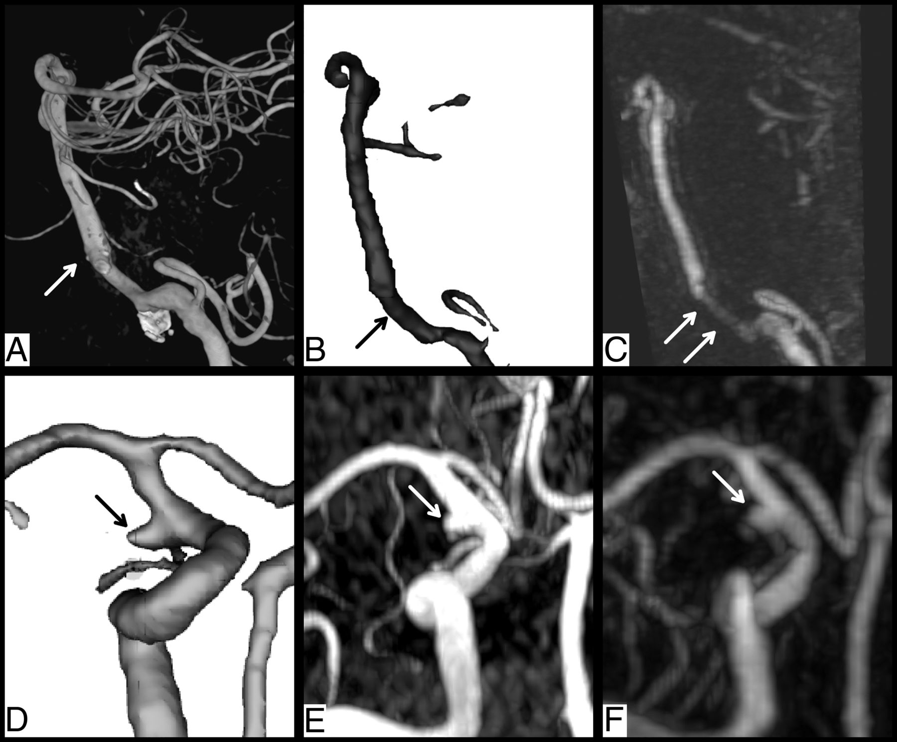
Segmentation of the vessel lumen following stent-assisted coiling of a ruptured dissecting aneurysm of the right vertebral artery was possible. The corresponding contrast-enhanced MRA showed signal degradation in the corresponding segment (Fig 1A–C). In another example, a small recurrence following coiling of a ruptured right posterior communicating artery aneurysm was successfully segmented. The segmentation was comparable with that in the TOF-MRA and the contrast-enhanced MRA (Fig 1D–F; overview, Fig 2). We observed a consistent true-positive and true-negative segmentation of intracranial arterial and venous structures in the volume of interest (Online Supplemental Data).

FIG 1.
  • Download figure
  • Open in new tab
  • Download powerpoint
FIG 1.

Sample images of advantageous findings. Identification of the vessel lumen following stent placement during treatment of a ruptured, dissecting aneurysm of the right vertebral artery in a test subject. A, 3D-rendering of rotational DSA in a lateral oblique projection following stent-assisted coiling (arrow, distal stent markers). B, Volume-rendering of the BBMRI model prediction (arrow, vessel lumen within the stent). C, Contrast-enhanced MRA shows partial signal degradation within the stent (arrows). D–F, Correct identification of a small recurrence (arrows) 6 months following coiling of a ruptured right posterior communicating artery aneurysm in a test subject. Volume-rendering in a coronal, oblique view of the BBMRI model prediction (D), of the TOF-MRA (E), and of the contrast medium–enhanced MRA (F).

FIG 2.
  • Download figure
  • Open in new tab
  • Download powerpoint
FIG 2.

Anterior-posterior 3D-rendering of the intracranial vessel tree. A, BBMRI model prediction. B, TOF-MRA. C, Contrast-enhanced MRA.

False-negative results were encountered in small-diameter vessels and in the distal vertebral arteries. They were rarely encountered in larger-diameter vessels. False-positive results were seen in areas showing low signal intensity in close proximity to the vessels, eg, metal artifacts following aneurysm clipping and in nearby bony structures (Online Supplemental Data).

Our model achieved Dice similarity coefficients (DSCs) of 0.77 and 0.72 in the training and testing data sets, respectively. The average evaluation metrics and corresponding plot are available in the Online Supplemental Data.

DISCUSSION

In this preliminary work, we demonstrate the feasibility of automated segmentation of the cerebral vasculature based on the negative contrast of the vessels in the non-contrast-enhanced BBMRI sequence. To our knowledge, this has not been previously attempted.

Qualitatively, vascular evaluation in the BBMRI is feasible. Differentiation between arterial and venous structures as well as recognition of morphologic changes (eg, aneurysm recurrence) were possible. Signal degradation following intracranial stent placement was not encountered (Fig 1 and Online Supplemental Data). The segmentation of smaller vessels and the distal vertebral arteries as well as the differentiation between vessels and nearby structures of low signal intensity (bony structures and aneurysm clips) were less accurate (Online Supplemental Data).

Quantitatively, our model achieved promising DSCs of 0.77 and of 0.72 in the training and testing data sets, respectively, indicating negligible overfitting. Our results are comparable with those of other TOF-based published works using CNN or thresholding techniques (DSC, 0.73–0.78). A more complex segmentation pipeline achieved a better DSC, reaching 0.93.3,9,10

A morphologic, flow-independent visualization of the cerebral vessels in the BBMRI has potential advantages. It avoids the flow-related artifacts and the stent-related signal degradation in TOF-MRA.11,12 The large FOV and the high resolution4 could allow segmentation of a large volume of interest. Furthermore, no application of contrast medium is required. Future prospects include further optimization of the machine learning parameters using a larger and more diverse data set, expanding the volume of interest, and testing performance in various intracranial pathologies.

CONCLUSIONS

CNN segmentation of the arteries of the circle of Willis and its branches in non-contrast-enhanced BBMRI with accuracy comparable with that of TOF-based segmentation techniques is feasible and promising.

Footnotes

  • Disclosure forms provided by the authors are available with the full text and PDF of this article at www.ajnr.org.

References

  1. 1.↵
    1. Lesage D,
    2. Angelini ED,
    3. Bloch I, et al
    . A review of 3D vessel lumen segmentation techniques: models, features and extraction schemes. Med Image Anal 2009;13:819–45 doi:10.1016/j.media.2009.07.011 pmid:19818675
    CrossRefPubMed
  2. 2.↵
    1. Navab N,
    2. Hornegger J,
    3. Wells WM
    1. Ronneberger O,
    2. Fischer P,
    3. Brox T
    . U-net: Convolutional networks for biomedical image segmentation. In: Navab N, Hornegger J, Wells WM, et al., eds. Medical Image Computing and Computer-Assisted Intervention–MICCAI 2015. Springer; 2015:234–41
  3. 3.↵
    1. Cardoso MJ,
    2. Arbel T,
    3. Lee SL
    , et al., eds. Intravascular Imaging and Computer Assisted Stenting, and Large-Scale Annotation of Biomedical Data and Expert Label Synthesis. Springer; 2017 doi:10.1007/978-3-319-67534-3
    CrossRef
  4. 4.↵
    1. Guggenberger K,
    2. Krafft AJ,
    3. Ludwig U, et al
    . High-resolution compressed-sensing T1 black-blood MRI. Clin Neuroradiol 2021;31:207–16 doi:10.1007/s00062-019-00867-0 pmid:31853612
    CrossRefPubMed
  5. 5.↵
    1. Xu Y,
    2. Gong M,
    3. Fu H, et al
    1. Xu Y,
    2. Gong M,
    3. Fu H, et al
    . Multi-scale masked 3-D U-Net for brain tumor segmentation. In: Xu Y, Gong M, Fu H, et al. Brainlesion: Glioma, Multiple Sclerosis, Stroke and Traumatic Brain Injuries. Springer; 2019:222–33 doi:10.1007/978-3-030-11726-9_20
    CrossRef
  6. 6.↵
    1. Kingma DP,
    2. Ba J
    . Adam: A Method for Stochastic Optimization. In: Proceedings of the 3rd International Conference for Learning Representations, San Diego, California. May 7–9, 2015
  7. 7.↵
    1. Taha AA,
    2. Hanbury A
    . Metrics for evaluating 3D medical image segmentation: analysis, selection, and tool. BMC Med Imaging 2015;15:29 doi:10.1186/s12880-015-0068-x
    CrossRefPubMed
  8. 8.↵
    1. Yeghiazaryan V,
    2. Voiculescu I
    . Family of boundary overlap metrics for the evaluation of medical image segmentation. J Med Imaging (Bellingham) 2018;5:015006 doi:10.1117/1.JMI.5.1.015006 pmid:29487883
    CrossRefPubMed
  9. 9.↵
    1. Chen L,
    2. Xie Y,
    3. Sun J, et al
    . 3D intracranial artery segmentation using a convolutional autoencoder. In: Proceedings of the 2017 IEEE International Conference on Bioinformatics and Biomedicine (BIBM), Kansas City, Kansas. November 13–16, 2017 doi:10.1109/BIBM.2017.8217741
    CrossRef
  10. 10.↵
    1. Zhang B,
    2. Liu S,
    3. Zhou S, et al
    . Cerebrovascular segmentation from TOF-MRA using model- and data-driven method via sparse labels. Neurocomputing 2020;380:162–79 doi:10.1016/j.neucom.2019.10.092
    CrossRef
  11. 11.↵
    1. Özsarlak Ö,
    2. Van Goethem JW,
    3. Maes M, et al
    . MR angiography of the intracranial vessels: technical aspects and clinical applications. Neuroradiology 2004;46:955–72 doi:10.1007/s00234-004-1297-9 pmid:15580489
    CrossRefPubMed
  12. 12.↵
    1. Marciano D,
    2. Soize S,
    3. Metaxas G, et al
    . Follow-up of intracranial aneurysms treated with stent-assisted coiling: comparison of contrast-enhanced MRA, time-of-flight MRA, and digital subtraction angiography. J Neuroradiol 2017;44:44–51 doi:10.1016/j.neurad.2016.10.004 pmid:27836654
    CrossRefPubMed
  • Received January 21, 2022.
  • Accepted after revision October 5, 2022.
  • © 2022 by American Journal of Neuroradiology
PreviousNext
Back to top

In this issue

American Journal of Neuroradiology: 43 (12)
American Journal of Neuroradiology
Vol. 43, Issue 12
1 Dec 2022
  • Table of Contents
  • Index by author
  • Complete Issue (PDF)
Advertisement
Print
Download PDF
Email Article

Thank you for your interest in spreading the word on American Journal of Neuroradiology.

NOTE: We only request your email address so that the person you are recommending the page to knows that you wanted them to see it, and that it is not junk mail. We do not capture any email address.

Enter multiple addresses on separate lines or separate them with commas.
Intracranial Vessel Segmentation in 3D High-Resolution T1 Black-Blood MRI
(Your Name) has sent you a message from American Journal of Neuroradiology
(Your Name) thought you would like to see the American Journal of Neuroradiology web site.
CAPTCHA
This question is for testing whether or not you are a human visitor and to prevent automated spam submissions.
Cite this article
S. Elsheikh, H. Urbach, M. Reisert
Intracranial Vessel Segmentation in 3D High-Resolution T1 Black-Blood MRI
American Journal of Neuroradiology Dec 2022, 43 (12) 1719-1721; DOI: 10.3174/ajnr.A7700

Citation Manager Formats

  • BibTeX
  • Bookends
  • EasyBib
  • EndNote (tagged)
  • EndNote 8 (xml)
  • Medlars
  • Mendeley
  • Papers
  • RefWorks Tagged
  • Ref Manager
  • RIS
  • Zotero
0 Responses
Respond to this article
Share
Bookmark this article
3D Vessel Segmentation in High-Res T1 MRI
S. Elsheikh, H. Urbach, M. Reisert
American Journal of Neuroradiology Dec 2022, 43 (12) 1719-1721; DOI: 10.3174/ajnr.A7700
del.icio.us logo Twitter logo Facebook logo Mendeley logo
  • Tweet Widget
  • Facebook Like
  • Google Plus One
Purchase

Jump to section

  • Article
    • Abstract
    • ABBREVIATIONS:
    • DISCUSSION
    • CONCLUSIONS
    • Footnotes
    • References
  • Figures & Data
  • Supplemental
  • Info & Metrics
  • Responses
  • References
  • PDF

Related Articles

  • PubMed
  • Google Scholar

Cited By...

  • No citing articles found.
  • Crossref (2)
  • Google Scholar

This article has been cited by the following articles in journals that are participating in Crossref Cited-by Linking.

  • A 2.5D Self-Training Strategy for Carotid Artery Segmentation in T1-Weighted Brain Magnetic Resonance Images
    Adriel Silva de Araújo, Márcio Sarroglia Pinho, Ana Maria Marques da Silva, Luis Felipe Fiorentini, Jefferson Becker
    Journal of Imaging 2024 10 7
  • Accuracy of automated segmentation and volumetry of acute intracerebral hemorrhage following minimally invasive surgery using a patch-based convolutional neural network in a small dataset
    Samer Elsheikh, Ahmed Elbaz, Alexander Rau, Theo Demerath, Christian Fung, Elias Kellner, Horst Urbach, Marco Reisert
    Neuroradiology 2024 66 4

More in this TOC Section

  • Diagnostic Neuroradiology of Monoclonal Antibodies
  • Clinical Outcomes After Chiari I Decompression
  • Segmentation of Brain Metastases with BLAST
Show more ADULT BRAIN

Similar Articles

Advertisement

Indexed Content

  • Current Issue
  • Accepted Manuscripts
  • Article Preview
  • Past Issues
  • Editorials
  • Editor's Choice
  • Fellows' Journal Club
  • Letters to the Editor
  • Video Articles

Cases

  • Case Collection
  • Archive - Case of the Week
  • Archive - Case of the Month
  • Archive - Classic Case

More from AJNR

  • Trainee Corner
  • Imaging Protocols
  • MRI Safety Corner
  • Book Reviews

Multimedia

  • AJNR Podcasts
  • AJNR Scantastics

Resources

  • Turnaround Time
  • Submit a Manuscript
  • Submit a Video Article
  • Submit an eLetter to the Editor/Response
  • Manuscript Submission Guidelines
  • Statistical Tips
  • Fast Publishing of Accepted Manuscripts
  • Graphical Abstract Preparation
  • Imaging Protocol Submission
  • Evidence-Based Medicine Level Guide
  • Publishing Checklists
  • Author Policies
  • Become a Reviewer/Academy of Reviewers
  • News and Updates

About Us

  • About AJNR
  • Editorial Board
  • Editorial Board Alumni
  • Alerts
  • Permissions
  • Not an AJNR Subscriber? Join Now
  • Advertise with Us
  • Librarian Resources
  • Feedback
  • Terms and Conditions
  • AJNR Editorial Board Alumni

American Society of Neuroradiology

  • Not an ASNR Member? Join Now

© 2025 by the American Society of Neuroradiology All rights, including for text and data mining, AI training, and similar technologies, are reserved.
Print ISSN: 0195-6108 Online ISSN: 1936-959X

Powered by HighWire