
Article Figures & Data
Figures
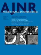
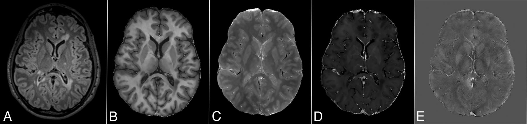
- FIG 1.
An example of quantitative MR imaging maps. Along with findings of a conventional FLAIR sequence (A) are examples of R1 (B), PD (C), R2* (D), and QSM (E) maps from a 22 -year-old man with MS.
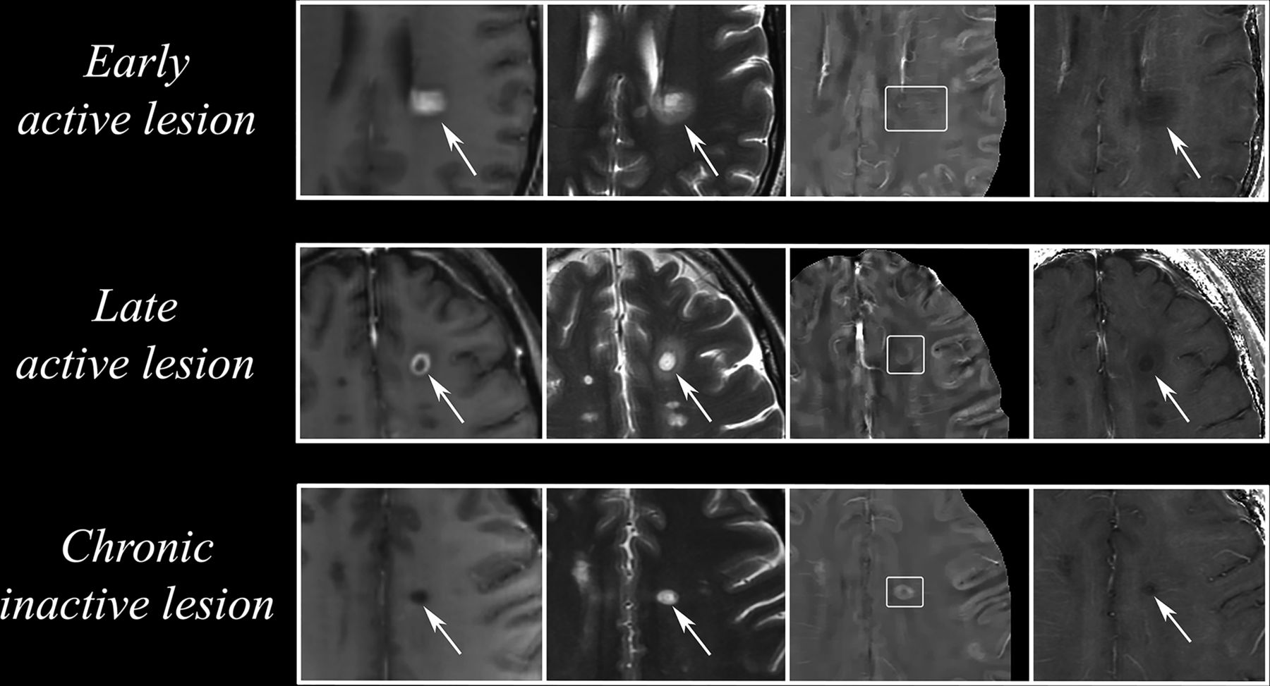
- FIG 2.
Conventional and quantitative MR imaging findings of WM lesions at different stages. In the upper row, conventional findings (postgadolinium T1-weighted and precontrast T2-weighted, first and second images from left to right respectively) of a typical pattern of nodular enhancement in an early active lesion (arrows) showing isointense signal in QSM (third image, white box) and mild hypointensity in R2* map (fourth image). In late active lesions (middle row, arrows), a peripheral pattern of enhancement is present, coupled with an area of increased signal at QSM and a slightly more pronounced hypointensity on R2* maps compared with the previous stage. As lesion staging further increases, the lesion eventually enters its chronic inactive stage (lower row, arrows), characterized by absent gadolinium enhancement, a QSM hyperintensity, and a hypointense R2* signal. Modified with permission from Zhang et al.45
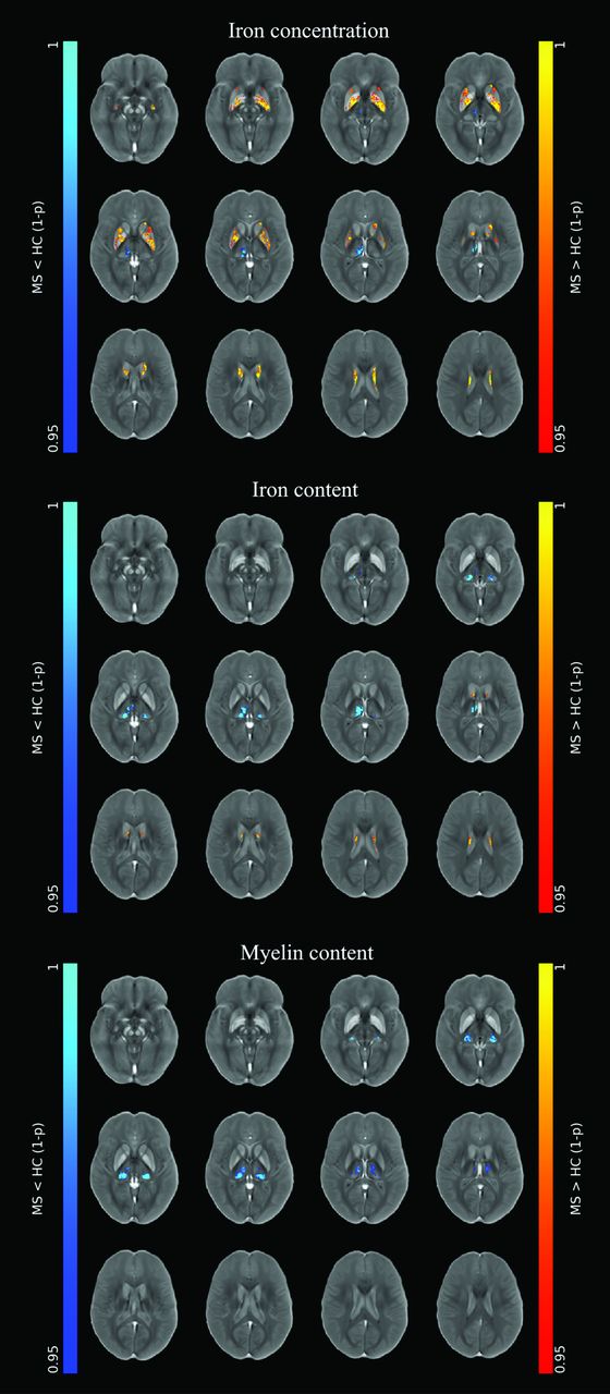
- FIG 3.
Pattern of iron concentration, iron content, and myelin content changes in deep gray matter nuclei in MS. Results of voxelwise analyses comparing patients with MS with healthy controls, showing the presence of an increased iron concentration at the level of the basal ganglia (red-yellow), coupled with a decrease in iron and myelin content mainly affecting the thalami and, in particular, the pulvinar nuclei (blue-light blue). Modified with permission from Pontillo et al.59 HC indicates healthy control; 1-ρ, 1 minus P value.


Tables
Site Pathologic Processes and qMRI Correlates WM lesions Early active Decreased R1, R2, and R2* values, along with increased PD, reflecting initial myelin degradation and edema43,45 Late active Decreased R1 and R2 values, coupled with increased PD, with myelin debris removal that determines further R2* decrease and QSM increase43,45,51 Chronic active Further R1 and R2 decrease, with PD increase, due to demyelination progression;43 increased R2* and QSM at the periphery of the lesions due to iron-laden microglia and macrophages45,46 Chronic inactive Compared with chronic active, R2* decreases with high QSM values; across time, susceptibility values gradually become similar to those in NAWM46 NAWM Decreased R1 and R2 values, with increased PD, compared with the WM of healthy controls,50,51 reflecting edema and myelin loss secondary to inflammatory infiltration. During active phases of the disease, iron is released from oligodendrocytes and begins to accumulate in newly forming lesions, causing an R2* decrease50 and no relevant modification of QSM signal52 Site Pathologic Processes and qMRI Correlates Deep gray matter Basal ganglia Increased R2*58,64-66,70,71 and QSM values61,64,66,70,72 indicating increased iron concentrations, with atrophy that might play a role in causing these changes57,68 Thalamus The more recent findings suggest the presence of reduced iron content and concentration, along with demyelination, as shown by decreased R1, R2*, and QSM values56-61 Cortical gray matter Cortical lesions Reduced R2* signal due to demyelination and iron depletion;74,75 cortical lesions are more heterogeneous on QSM, with decreased-to-increased values, depending on the level of inflammatory activity78 Normal-appearing cortex Demyelination and iron depletion lead to reduced R1 and R2* values,50 with a gradient indicating the WM interface83


Jump to section
Related Articles
Cited By...
- No citing articles found.