Advertisement

Main menu

  • Home
  • Content
    • Current Issue
    • Accepted Manuscripts
    • Article Preview
    • Past Issue Archive
    • Video Articles
    • AJNR Case Collection
    • Case of the Week Archive
    • Case of the Month Archive
    • Classic Case Archive
  • Special Collections
    • AJNR Awards
    • Low-Field MRI
    • Alzheimer Disease
    • ASNR Foundation Special Collection
    • Photon-Counting CT
    • View All
  • Multimedia
    • AJNR Podcasts
    • AJNR SCANtastic
    • Trainee Corner
    • MRI Safety Corner
    • Imaging Protocols
  • For Authors
    • Submit a Manuscript
    • Submit a Video Article
    • Submit an eLetter to the Editor/Response
    • Manuscript Submission Guidelines
    • Statistical Tips
    • Fast Publishing of Accepted Manuscripts
    • Graphical Abstract Preparation
    • Imaging Protocol Submission
    • Author Policies
  • About Us
    • About AJNR
    • Editorial Board
    • Editorial Board Alumni
  • More
    • Become a Reviewer/Academy of Reviewers
    • Subscribers
    • Permissions
    • Alerts
    • Feedback
    • Advertisers
    • ASNR Home

User menu

  • Alerts
  • Log in

Search

  • Advanced search
American Journal of Neuroradiology

American Journal of Neuroradiology

ASHNR American Society of Functional Neuroradiology ASHNR American Society of Pediatric Neuroradiology ASSR
  • Alerts
  • Log in

Advanced Search

  • Home
  • Content
    • Current Issue
    • Accepted Manuscripts
    • Article Preview
    • Past Issue Archive
    • Video Articles
    • AJNR Case Collection
    • Case of the Week Archive
    • Case of the Month Archive
    • Classic Case Archive
  • Special Collections
    • AJNR Awards
    • Low-Field MRI
    • Alzheimer Disease
    • ASNR Foundation Special Collection
    • Photon-Counting CT
    • View All
  • Multimedia
    • AJNR Podcasts
    • AJNR SCANtastic
    • Trainee Corner
    • MRI Safety Corner
    • Imaging Protocols
  • For Authors
    • Submit a Manuscript
    • Submit a Video Article
    • Submit an eLetter to the Editor/Response
    • Manuscript Submission Guidelines
    • Statistical Tips
    • Fast Publishing of Accepted Manuscripts
    • Graphical Abstract Preparation
    • Imaging Protocol Submission
    • Author Policies
  • About Us
    • About AJNR
    • Editorial Board
    • Editorial Board Alumni
  • More
    • Become a Reviewer/Academy of Reviewers
    • Subscribers
    • Permissions
    • Alerts
    • Feedback
    • Advertisers
    • ASNR Home
  • Follow AJNR on Twitter
  • Visit AJNR on Facebook
  • Follow AJNR on Instagram
  • Join AJNR on LinkedIn
  • RSS Feeds

AJNR Awards, New Junior Editors, and more. Read the latest AJNR updates

Research ArticleAdult Brain
Open Access

MRI-Visible Perivascular Spaces in the Centrum Semiovale Are Associated with Brain Amyloid Deposition in Patients with Alzheimer Disease–Related Cognitive Impairment

H.J. Kim, H. Cho, M. Park, J.W. Kim, S.J. Ahn, C.H. Lyoo, S.H. Suh and Y.H. Ryu
American Journal of Neuroradiology July 2021, 42 (7) 1231-1238; DOI: https://doi.org/10.3174/ajnr.A7155
H.J. Kim
aFrom the Department of Nuclear Medicine (H.J.K., Y.H.R.)
dDepartment of Nuclear Medicine (H.J.K.), Yongin Severance Hospital, Yonsei University College of Medicine, Yongin-si, South Korea
  • Find this author on Google Scholar
  • Find this author on PubMed
  • Search for this author on this site
  • ORCID record for H.J. Kim
H. Cho
bNeurology (H.C., C.H.L.)
  • Find this author on Google Scholar
  • Find this author on PubMed
  • Search for this author on this site
  • ORCID record for H. Cho
M. Park
cRadiology (M.P., J.W.K., S.J.A., S.H.S.), Gangnam Severance Hospital, Yonsei University College of Medicine, Seoul, South Korea
  • Find this author on Google Scholar
  • Find this author on PubMed
  • Search for this author on this site
  • ORCID record for M. Park
J.W. Kim
cRadiology (M.P., J.W.K., S.J.A., S.H.S.), Gangnam Severance Hospital, Yonsei University College of Medicine, Seoul, South Korea
  • Find this author on Google Scholar
  • Find this author on PubMed
  • Search for this author on this site
  • ORCID record for J.W. Kim
S.J. Ahn
cRadiology (M.P., J.W.K., S.J.A., S.H.S.), Gangnam Severance Hospital, Yonsei University College of Medicine, Seoul, South Korea
  • Find this author on Google Scholar
  • Find this author on PubMed
  • Search for this author on this site
  • ORCID record for S.J. Ahn
C.H. Lyoo
bNeurology (H.C., C.H.L.)
  • Find this author on Google Scholar
  • Find this author on PubMed
  • Search for this author on this site
  • ORCID record for C.H. Lyoo
S.H. Suh
cRadiology (M.P., J.W.K., S.J.A., S.H.S.), Gangnam Severance Hospital, Yonsei University College of Medicine, Seoul, South Korea
  • Find this author on Google Scholar
  • Find this author on PubMed
  • Search for this author on this site
  • ORCID record for S.H. Suh
Y.H. Ryu
aFrom the Department of Nuclear Medicine (H.J.K., Y.H.R.)
  • Find this author on Google Scholar
  • Find this author on PubMed
  • Search for this author on this site
  • ORCID record for Y.H. Ryu
  • Article
  • Figures & Data
  • Supplemental
  • Info & Metrics
  • Responses
  • References
  • PDF
Loading

Article Figures & Data

Figures

  • Tables
  • FIG 1.
    • Download figure
    • Open in new tab
    • Download powerpoint
    FIG 1.

    Patient-inclusion flowchart.

  • FIG 2.
    • Download figure
    • Open in new tab
    • Download powerpoint
    FIG 2.

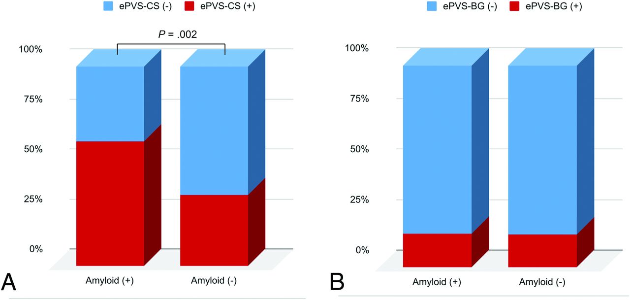
    Comparisons of the presence of MR imaging–visible PVS-CS (A) and MR imaging–visible PVS in the basal ganglia (B) based on the β-amyloid status. The enlarged perivascular spaces in the centrum semiovale (ePVS-CS) were significantly higher in the patient group positive for β-amyloid than in the patient group negative for it, whereas the high degree of enlarged perivascular spaces in the basal ganglia (ePVS-BG) did not differ between the groups positive and negative for β-amyloid.

  • FIG 3.
    • Download figure
    • Open in new tab
    • Download powerpoint
    FIG 3.

    Examples of perivascular space patterns with the corresponding [18F] FBB PET image. The axial T2-weighted MR imaging shows a high degree of MR imaging–visible PVS-CS (A), and the corresponding [18F] FBB PET (B) shows pronounced β-amyloid deposition. Axial T2-weighted MR imaging shows a low degree of MR imaging–visible PVS-CS (C) and the [18F] FBB PET scan (D) shows low β-amyloid deposition.

Tables

  • Figures
    • View popup
    Table 1:

    Baseline characteristics of the groups positive and negative for brain β-amyloid

    Amyloid-NegativeAmyloid-PositiveP Value
    (No.) (%)67 (46.5%)77 (53.5%)
    Age (mean) (SD) (yr)71.3 (10.6)75.4 (7.6).010
    Female sex (No.) (%)44 (65.7%)44 (57.1%).297
    Hypertension (No.) (%)25 (37.3%)36 (46.8%).254
    Diabetes mellitus (No.) (%)10 (14.9%)16 (20.8%).364
    Hyperlipidemia (No.) (%)9 (13.4%)11 (14.3%).883
    Previous stroke (No.) (%)7 (10.4%)4 (5.2%).238
    APOE ε4 presence (No.) (%)13 (19.4%)33 (42.9%).001
    High degree of MR imaging–visible PVS-CS (No.) (%)24 (35.8%)48 (62.3%).002
    High degree of MR imaging–visible PVS-BG (No.) (%)11 (16.4%)13 (16.9%).297
    AD (No.) (%)19 (28.4%)47 (61.0%)<.001
    MMSE (median) (IQR)26 (23–28)24 (20–26)<.001
    CDR (median) (IQR)0.5 (0.5–0.5)0.5 (0.5–1.0).019
    CDR-SB (median) (IQR)1.5 (0.5–3.0)3.0 (1.5–4.5)<.001
    Lacunes (median) (IQR)0 (0–0)0 (0–0).778
    cSS present (No.) (%)1 (1.5%)6 (7.8%).081
    Lobar CMB (median) (IQR)0 (0–0)0 (0–1).117
    Deep CMB (median) (IQR)0 (0–0)0 (0–0).160
    WMH presence (No.) (%)27 (40.3%)47 (61.0%).013
    • Note:—IQR indicates interquartile range; PVS-BG, perivascular space in the basal ganglia; CMB, cerebral microbleed; CDR, Clinical Dementia Rating Scale; CDR-SB, Clinical Dementia Rating Scale–Sum of Boxes; cSS, cortical superficial siderosis.

    • View popup
    Table 2:

    Logistic regression analysis for the predictors of β-amyloid positivity

    UnivariableMultivariable
    OR (95% CI)P ValueOR (95% CI)P Value
    Age (yr)1.051 (1.012–1.092).0101.050 (1.004–1.098).034
    Sex
     FemaleReference group
     Male1.435 (0.729–2.823).2961.561 (0.699–3.489).278
    Hypertension (present)1.475 (0.757–2.875).254
    Diabetes (present)1.495 (0.627–3.564).364
    Hyperlipidemia (present)1.074 (0.416–2.774).883
    Previous stroke (present)0.470 (0.131–1.681).245
    APOE ε4 allele (present)3.526 (1.630–7.627).0014.583 (1.945–10.796)<.001
    High degree of MR imaging–visible PVS-CS (score, ≥3)2.966 (1.503–5.851).0022.307 (1.036–5.136).041
    High degree of MR imaging–visible PVS-BG (score, ≥3)1.034 (0.429–2.492).940
    WMH (present)2.321 (1.188–4.533).014
    Lacunes (for 1 number higher)0.937 (0.726–1.209).618
    cSS (present)5.577 (0.654–47.565).116
    Lobar CMB (present)1.100 (0.934–1.295).253
    Deep CMB (present)0.688 (0.428–1.108).124
    • Note:—CMB indicates cerebral microbleed; cSS, cortical superficial siderosis; MRI-visible PVS-BG, enlarged perivascular space in the basal ganglia; MRI-visible PVS-CS, enlarged perivascular space in the semi ovale.

PreviousNext
Back to top

In this issue

American Journal of Neuroradiology: 42 (7)
American Journal of Neuroradiology
Vol. 42, Issue 7
1 Jul 2021
  • Table of Contents
  • Index by author
  • Complete Issue (PDF)
Advertisement
Print
Download PDF
Email Article

Thank you for your interest in spreading the word on American Journal of Neuroradiology.

NOTE: We only request your email address so that the person you are recommending the page to knows that you wanted them to see it, and that it is not junk mail. We do not capture any email address.

Enter multiple addresses on separate lines or separate them with commas.
MRI-Visible Perivascular Spaces in the Centrum Semiovale Are Associated with Brain Amyloid Deposition in Patients with Alzheimer Disease–Related Cognitive Impairment
(Your Name) has sent you a message from American Journal of Neuroradiology
(Your Name) thought you would like to see the American Journal of Neuroradiology web site.
CAPTCHA
This question is for testing whether or not you are a human visitor and to prevent automated spam submissions.
Cite this article
H.J. Kim, H. Cho, M. Park, J.W. Kim, S.J. Ahn, C.H. Lyoo, S.H. Suh, Y.H. Ryu
MRI-Visible Perivascular Spaces in the Centrum Semiovale Are Associated with Brain Amyloid Deposition in Patients with Alzheimer Disease–Related Cognitive Impairment
American Journal of Neuroradiology Jul 2021, 42 (7) 1231-1238; DOI: 10.3174/ajnr.A7155

Citation Manager Formats

  • BibTeX
  • Bookends
  • EasyBib
  • EndNote (tagged)
  • EndNote 8 (xml)
  • Medlars
  • Mendeley
  • Papers
  • RefWorks Tagged
  • Ref Manager
  • RIS
  • Zotero
0 Responses
Respond to this article
Share
Bookmark this article
MRI-Visible Perivascular Spaces in the Centrum Semiovale Are Associated with Brain Amyloid Deposition in Patients with Alzheimer Disease–Related Cognitive Impairment
H.J. Kim, H. Cho, M. Park, J.W. Kim, S.J. Ahn, C.H. Lyoo, S.H. Suh, Y.H. Ryu
American Journal of Neuroradiology Jul 2021, 42 (7) 1231-1238; DOI: 10.3174/ajnr.A7155
del.icio.us logo Twitter logo Facebook logo Mendeley logo
  • Tweet Widget
  • Facebook Like
  • Google Plus One
Purchase

Jump to section

  • Article
    • Abstract
    • ABBREVIATIONS:
    • MATERIALS AND METHODS
    • RESULTS
    • DISCUSSION
    • CONCLUSIONS
    • Footnotes
    • References
  • Figures & Data
  • Supplemental
  • Info & Metrics
  • Responses
  • References
  • PDF

Related Articles

  • PubMed
  • Google Scholar

Cited By...

  • The association between enlarged perivascular spaces and muscle sympathetic nerve activity in normotensive and hypertensive humans
  • Perivascular spaces in Alzheimers disease are associated with inflammatory, stress-related, and hypertension biomarkers
  • Crossref
  • Google Scholar

This article has not yet been cited by articles in journals that are participating in Crossref Cited-by Linking.

More in this TOC Section

  • Diagnostic Neuroradiology of Monoclonal Antibodies
  • Clinical Outcomes After Chiari I Decompression
  • Segmentation of Brain Metastases with BLAST
Show more Adult Brain

Similar Articles

Advertisement

Indexed Content

  • Current Issue
  • Accepted Manuscripts
  • Article Preview
  • Past Issues
  • Editorials
  • Editor's Choice
  • Fellows' Journal Club
  • Letters to the Editor
  • Video Articles

Cases

  • Case Collection
  • Archive - Case of the Week
  • Archive - Case of the Month
  • Archive - Classic Case

More from AJNR

  • Trainee Corner
  • Imaging Protocols
  • MRI Safety Corner
  • Book Reviews

Multimedia

  • AJNR Podcasts
  • AJNR Scantastics

Resources

  • Turnaround Time
  • Submit a Manuscript
  • Submit a Video Article
  • Submit an eLetter to the Editor/Response
  • Manuscript Submission Guidelines
  • Statistical Tips
  • Fast Publishing of Accepted Manuscripts
  • Graphical Abstract Preparation
  • Imaging Protocol Submission
  • Evidence-Based Medicine Level Guide
  • Publishing Checklists
  • Author Policies
  • Become a Reviewer/Academy of Reviewers
  • News and Updates

About Us

  • About AJNR
  • Editorial Board
  • Editorial Board Alumni
  • Alerts
  • Permissions
  • Not an AJNR Subscriber? Join Now
  • Advertise with Us
  • Librarian Resources
  • Feedback
  • Terms and Conditions
  • AJNR Editorial Board Alumni

American Society of Neuroradiology

  • Not an ASNR Member? Join Now

© 2025 by the American Society of Neuroradiology All rights, including for text and data mining, AI training, and similar technologies, are reserved.
Print ISSN: 0195-6108 Online ISSN: 1936-959X

Powered by HighWire