
Article Figures & Data
Figures
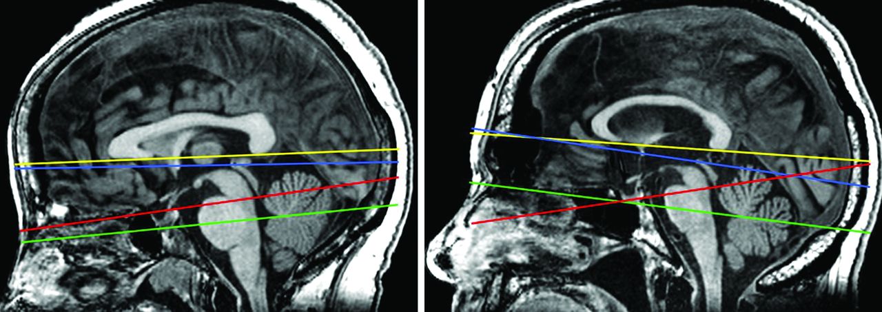
- FIG 1.
Definition of standard transverse imaging planes: bicallosal (yellow), bicommissural (blue), Hy-Fa (green), and perpendicular to the posterior brain stem margin (red), shown on midsagittal MR images of 2 different individuals. Note the differences in their mutual alignment between the 2 subjects.
- FIG 2.
Graphic representation of biomarker measurements. A, Maximum frontal horn diameter (FHD) and inner skull diameter (ISD). B, Frontal horn vertical diameter (FHVD) measured to the midpoint of the foramen of Monro. C, Maximum supratentorial intracranial diameter (MSID) measured perpendicular to the bicommissural line (dotted line). D, Maximum vertical widths of the supraventricular brain (SVW), cella media (CMW), and temporal horn (THW), respectively; callosal angle (CA); and DESH (curved dashed lines, rated as severe, grade 2 in this case) measured on the coronal section passing through the posterior commissure. E, Simplified callosal angle (simpCA) measured at the corpus callosum midpoint on the coronal section paralleling the PBSM (F, dotted line). Note that for each biomarker, 4 measurement values have been obtained from 4 different sections (as defined in Fig 1). To keep the illustration simple, we show measurements on sections aligned parallel and perpendicular to the bicommissural line, except for the simpCA (E and F). For details, see the Materials and Methods section.
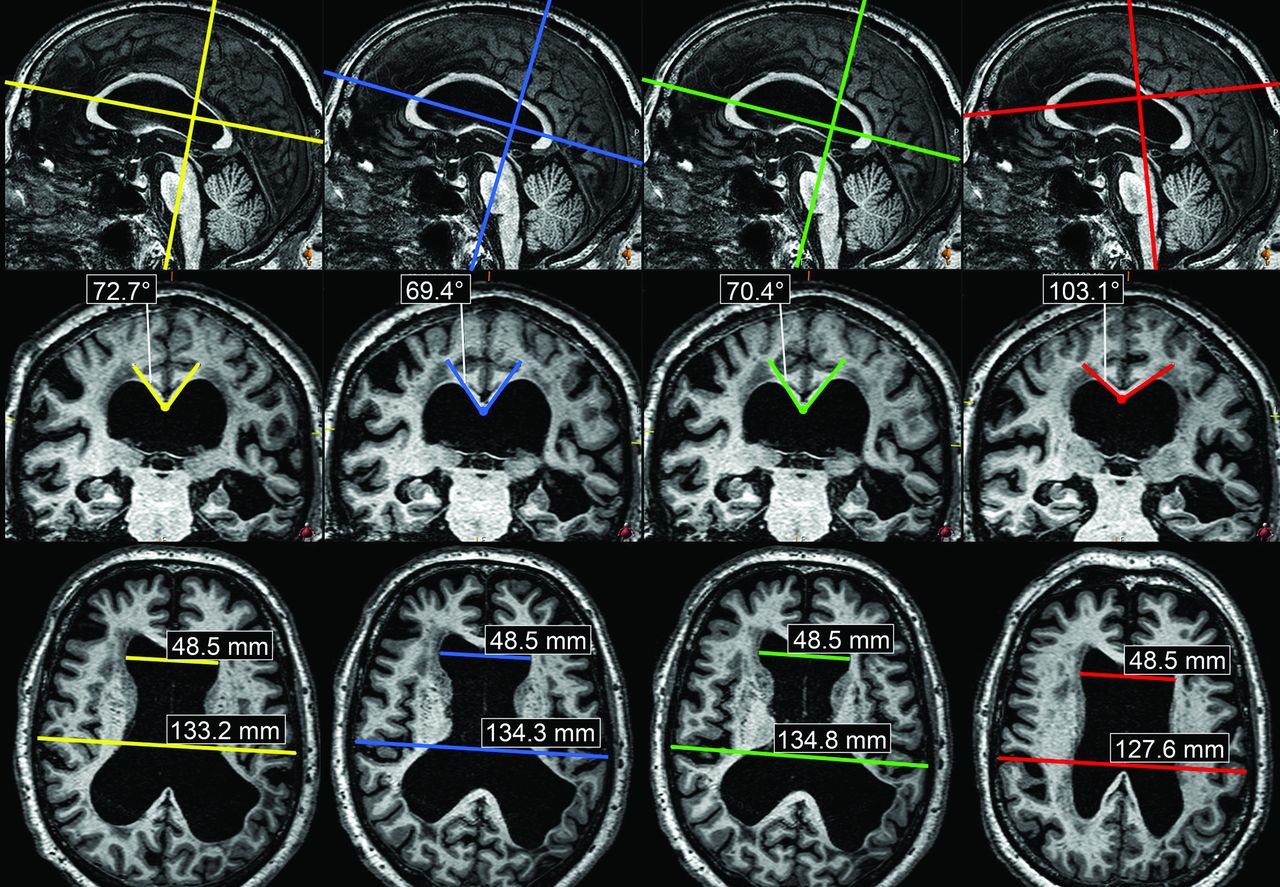
- FIG 3.
Variability of the CA (middle row) and EI (lower row) as measured on bicallosal (yellow), bicommissural (blue), Hy-Fa (green), and PBSM (red) sections of a single subject with iNPH. The CA yields 73°, 69°, 70°, and 103° in the 4 respective planes (maximum percentage difference of 39.5%). The EI shows a less variable range of values: 0.364, 0.361, 0.359, and 0.380, respectively (maximum difference of 5.7%). Color lines on the sagittal sections (upper row) refer to the alignment of the respective coronal and transverse sections below. The disproportion between Sylvian and parasagittal convexity subarachnoid spaces (DESH) has been rated moderate in this case (grade 1).


Tables
Biomarker Mean Repeated Measures ANOVA (P Values) HC iNPH iNPH/HC Angulation Angulation iNPH/HC DESH 0.12 1.31 <.001 .463 .463 simpCA 129.8 98.2 <.001 .264 .950 FHD 39.1 51.2 <.001 .151 .757 ZEI 0.286 0.420 <.001 .075 .792 CMW 17.4 34.5 <.001 .056 .49 BVR 2.44 0.92 <.001 .005 .106 THW 7.6 12.5 <.001 .002 .026 CTR 2.33 2.86 <.001 .002 .235 EI 0.293 0.384 <.001 <.001 .971 FHVD 25.7 38.8 <.001 <.001 .059 CA 116.6 74.7 <.001 <.001 <.001 SVW 39.6 30.9 <.001 <.001 .288 Note:—HC indicates healthy control; iNPH, idiopathic normal pressure hydrocephalus.
↵a Please refer to the Results section for detailed explanation. Two-way repeated measures ANOVA: angulation is a within-subjects factor (Angulation), and disease status is a between-subjects factor (iNPH/HC). Angulation by disease status interaction term (Angulation iNPH/HC) is significant for THW and CA only, with larger effects observed in the iNPH group for both biomarkers. Data are sorted in descending angulation P value order (column 5: “Angulation”). Decreasing P values indicate increasing statistical differences between measurements obtained at different section planes.
- Table 2:
Pair-wise comparisons between different planes performed with Bonferroni correction for biomarkers showing significant dependence on angulation (P < .05) in the repeated measures ANOVA (as per Table 1)a
Biomarker Bi-Call, Bi-Comm Bi-Call, Hy-Fa Bi-Call, PBSM Bi-Comm, Hy-Fa Bi-Comm, PBSM Hy-Fa, PBSM DESH NS NS NS NS NS NS simpCA NS NS NS NS NS NS FHD NS NS NS NS NS NS ZEI NS NS NS NS NS NS CMW NS NS NS NS NS NS BVR NS .007 .009 NS NS <.001 –0.04 ± 0.01 0.07 ± 0.02 0.11 ± 0.02 THW NS NS NS .002 .014 NS 0.24 ± 0.06 0.32 ± 0.10 CTR NS NS NS NS .004 NS –0.12 ± 0.03 EI NS NS <.001 NS <.001 <.001 –0.005 ± 0.001 –0.005 ± 0.001 –0.005 ± 0.001 FHVD NS NS .014 NS <.001 <.001 –0.52 ± 0.17 –0.92 ± 0.20 –0.79 ± 0.17 CA .013 NS <.001 NS <.001 <.001 –1.94 ± 0.61 –7.06 ± 0.87 –9.01 ± 1.07 –8.24 ± 0.91 SVW .034 NS .005 NS <.001 .001 –0.45 ± 0.16 0.97 ± 0.28 1.42 ± 0.30 1.26 ± 0.32 Note:—Bi-Call indicates bicallosal; Bi-Comm, bicommissural; Hy-Fa, hypophysis-fastigium; PBSM, posterior brain stem margin; NS, not significant.
↵a P values (upper digit) and means ± standard errors of paired differences are calculated (n = 80) and presented for those paired tests that are statistically significant.


Jump to section
Related Articles
Cited By...
- No citing articles found.