
Article Figures & Data
Figures
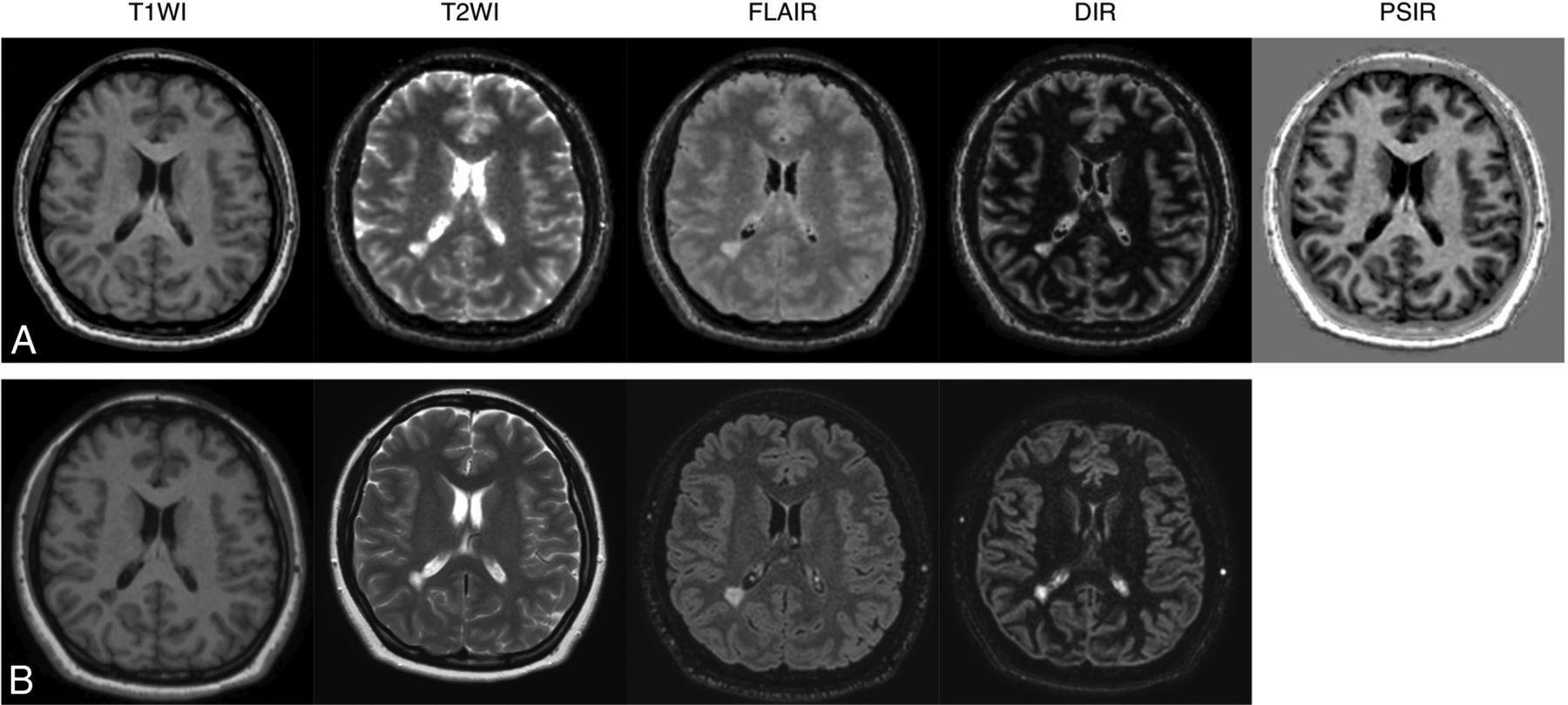
- FIG 1.
Representative examples of quantitative synthetic (A) and conventional (B) MR imaging in a 35-year-old woman with MS. The overall image quality of synthetic T1WI, T2WI, FLAIR, DIR, and PSIR were scored as 5, 3, 3, 5, and 5 by reader 1 and 5, 3, 4, 5, and 5 by reader 2. All of the conventional contrast-weighted images were scored as 5 by both readers.
- FIG 2.
Comparison of overall image quality for conventional and synthetic MR imaging in patients with MS. Each contrast-weighted image in 23 patients was rated on a 5-point Likert scale by 2 readers.
- FIG 3.
Comparison of structural delineations between conventional and synthetic MR imaging in patients with MS. Each target structure in 23 patients was rated for each contrast-weighted image on a 5-point Likert scale by 2 readers.
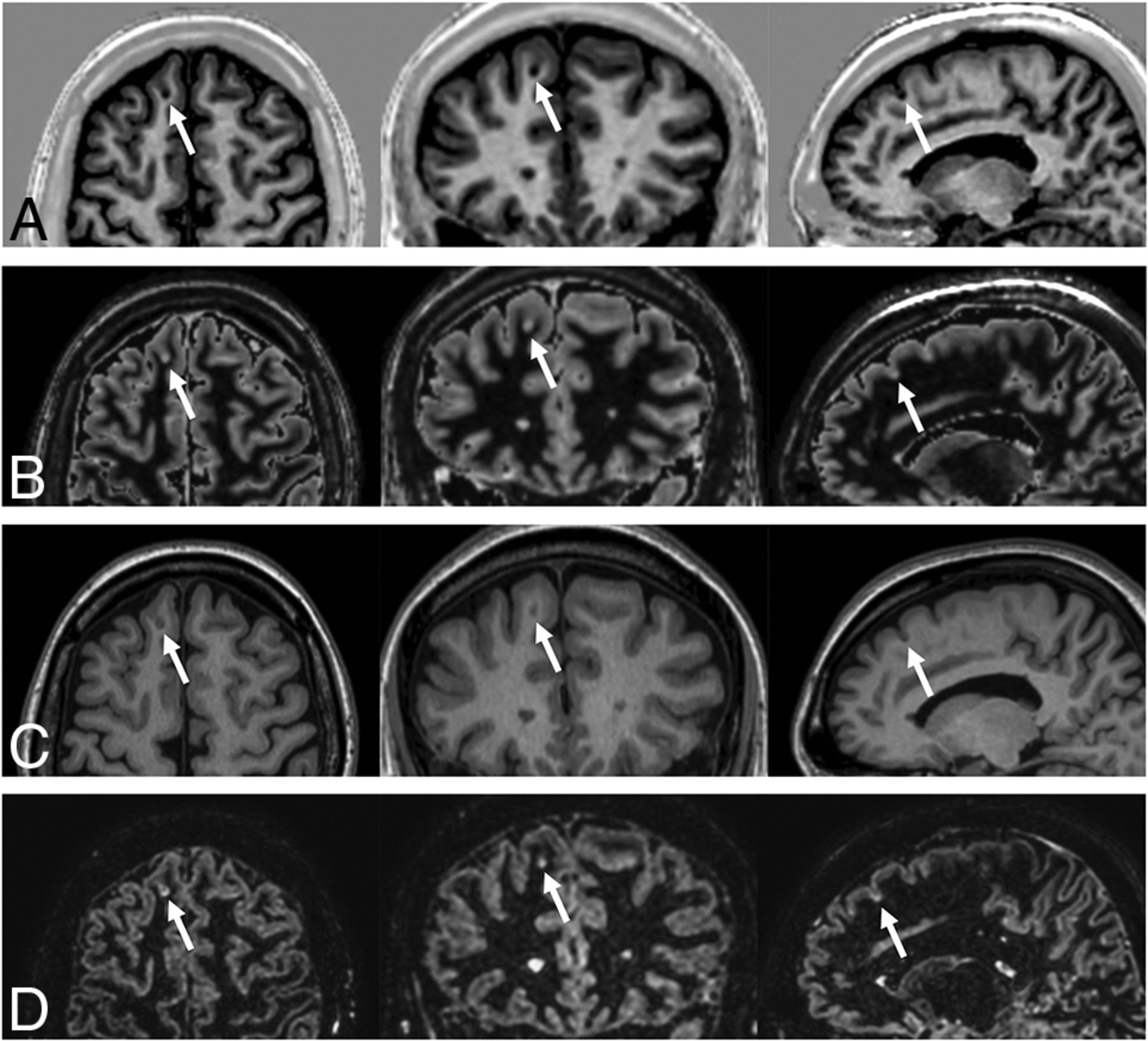
- FIG 4.
Example of an MS cortical lesion (arrows) in synthetic PSIR (A) and DIR (B) images. Conventional T1WI (C) and DIR (D) images are shown for reference. (Left) axial, (middle) coronal, and (right) sagittal views.
- FIG 5.
Representative examples of lesion segmentation in a 35-year-old woman with MS. Lesions were automatically segmented on synthetic and conventional FLAIR images. The segmented lesions are overlaid on the images used for segmentation. A, Lesions overlaid on 3D synthetic FLAIR images. B, Lesions overlaid on conventional 3D FLAIR images. Minimal differences are observed between segmented lesions of 3D synthetic and conventional FLAIR images.


Tables
Parameters 3D-QALAS 3D T1WI 3D FLAIR 3D DIR TSE T2WI Acquisition plane 3D axial 3D sagittal 3D sagittal 3D sagittal 2D axial Image matrix 192 × 192 256 × 256 208 × 208 176 × 174 368 × 230 FOV (mm) 256 256 256 250 230 Section thickness (mm) 1.3 1 1.2 1.5 5 Voxel size (mm) 1.3 × 1.3 × 1.3 1.0 × 1.0 × 1.0 1.2 × 1.2 × 1.2 1.4 × 1.4 × 1.5 0.6 × 1.0 TR (ms) 6.2 8.2 4800 5500 4082 TE (ms) 2.8 3.8 371 306 90 TI (ms) a – 1660 2510/480 – Flip angle (degree) 4 10 90 90 90 Bandwidth (Hz/pixel) 249 191 910 1076 167 Averages 1 1 1 2 3 Scanning time 9:06 6:20 5:22 5:03 1:42 Note: — – indicates no value.
↵a Inversion delay times, 100 ms, 1000 ms, 1900 ms, and 2800 ms; T2 prep echo time, 100 ms.
Characteristics Findings Participants (n) 23 Sex (male/female) 6/17 Age (years) 41.3 ± 9.8 (range, 19–59) Disease duration (years) 10.1 ± 5.2 Subtype (RR/SP/PP) 20/1/2 EDSS score (range) [0, 8.5] (median 1.5) Note—EDSS indicates Expanded Disability Status Scale; PP, primary-progressive; RR, relapsing-remitting; SP, secondary-progressive.
Data are shown as mean ± SD unless otherwise specified.


