Advertisement

Main menu

  • Home
  • Content
    • Current Issue
    • Accepted Manuscripts
    • Article Preview
    • Past Issue Archive
    • Video Articles
    • AJNR Case Collection
    • Case of the Week Archive
    • Case of the Month Archive
    • Classic Case Archive
  • Special Collections
    • AJNR Awards
    • Low-Field MRI
    • Alzheimer Disease
    • ASNR Foundation Special Collection
    • Photon-Counting CT
    • View All
  • Multimedia
    • AJNR Podcasts
    • AJNR SCANtastic
    • Trainee Corner
    • MRI Safety Corner
    • Imaging Protocols
  • For Authors
    • Submit a Manuscript
    • Submit a Video Article
    • Submit an eLetter to the Editor/Response
    • Manuscript Submission Guidelines
    • Statistical Tips
    • Fast Publishing of Accepted Manuscripts
    • Graphical Abstract Preparation
    • Imaging Protocol Submission
    • Author Policies
  • About Us
    • About AJNR
    • Editorial Board
    • Editorial Board Alumni
  • More
    • Become a Reviewer/Academy of Reviewers
    • Subscribers
    • Permissions
    • Alerts
    • Feedback
    • Advertisers
    • ASNR Home

User menu

  • Alerts
  • Log in

Search

  • Advanced search
American Journal of Neuroradiology

American Journal of Neuroradiology

ASHNR American Society of Functional Neuroradiology ASHNR American Society of Pediatric Neuroradiology ASSR
  • Alerts
  • Log in

Advanced Search

  • Home
  • Content
    • Current Issue
    • Accepted Manuscripts
    • Article Preview
    • Past Issue Archive
    • Video Articles
    • AJNR Case Collection
    • Case of the Week Archive
    • Case of the Month Archive
    • Classic Case Archive
  • Special Collections
    • AJNR Awards
    • Low-Field MRI
    • Alzheimer Disease
    • ASNR Foundation Special Collection
    • Photon-Counting CT
    • View All
  • Multimedia
    • AJNR Podcasts
    • AJNR SCANtastic
    • Trainee Corner
    • MRI Safety Corner
    • Imaging Protocols
  • For Authors
    • Submit a Manuscript
    • Submit a Video Article
    • Submit an eLetter to the Editor/Response
    • Manuscript Submission Guidelines
    • Statistical Tips
    • Fast Publishing of Accepted Manuscripts
    • Graphical Abstract Preparation
    • Imaging Protocol Submission
    • Author Policies
  • About Us
    • About AJNR
    • Editorial Board
    • Editorial Board Alumni
  • More
    • Become a Reviewer/Academy of Reviewers
    • Subscribers
    • Permissions
    • Alerts
    • Feedback
    • Advertisers
    • ASNR Home
  • Follow AJNR on Twitter
  • Visit AJNR on Facebook
  • Follow AJNR on Instagram
  • Join AJNR on LinkedIn
  • RSS Feeds

AJNR Awards, New Junior Editors, and more. Read the latest AJNR updates

Research ArticleAdult Brain
Open Access

Performance of Standardized Relative CBV for Quantifying Regional Histologic Tumor Burden in Recurrent High-Grade Glioma: Comparison against Normalized Relative CBV Using Image-Localized Stereotactic Biopsies

J.M. Hoxworth, J.M. Eschbacher, A.C. Gonzales, K.W. Singleton, G.D. Leon, K.A. Smith, A.M. Stokes, Y. Zhou, G.L. Mazza, A.B. Porter, M.M. Mrugala, R.S. Zimmerman, B.R. Bendok, D.P. Patra, C. Krishna, J.L. Boxerman, L.C. Baxter, K.R. Swanson, C.C. Quarles, K.M. Schmainda and L.S. Hu
American Journal of Neuroradiology March 2020, 41 (3) 408-415; DOI: https://doi.org/10.3174/ajnr.A6486
J.M. Hoxworth
aFrom the Departments of Radiology (J.M.H., Y.Z., L.S.H.)
  • Find this author on Google Scholar
  • Find this author on PubMed
  • Search for this author on this site
  • ORCID record for J.M. Hoxworth
J.M. Eschbacher
fDepartments of Pathology (J.M.E.)
  • Find this author on Google Scholar
  • Find this author on PubMed
  • Search for this author on this site
  • ORCID record for J.M. Eschbacher
A.C. Gonzales
bPsychiatry and Psychology (A.C.G.)
  • Find this author on Google Scholar
  • Find this author on PubMed
  • Search for this author on this site
  • ORCID record for A.C. Gonzales
K.W. Singleton
ePrecision Neurotherapeutics Lab (K.W.S., G.D.L., B.R.B., K.R.S.), Mayo Clinic in Arizona, Phoenix, Arizona
  • Find this author on Google Scholar
  • Find this author on PubMed
  • Search for this author on this site
  • ORCID record for K.W. Singleton
G.D. Leon
ePrecision Neurotherapeutics Lab (K.W.S., G.D.L., B.R.B., K.R.S.), Mayo Clinic in Arizona, Phoenix, Arizona
  • Find this author on Google Scholar
  • Find this author on PubMed
  • Search for this author on this site
  • ORCID record for G.D. Leon
K.A. Smith
iKeller Center for Imaging Innovation (A.M.S.), Barrow Neurological Institute, Phoenix, Arizona
  • Find this author on Google Scholar
  • Find this author on PubMed
  • Search for this author on this site
  • ORCID record for K.A. Smith
A.M. Stokes
iKeller Center for Imaging Innovation (A.M.S.), Barrow Neurological Institute, Phoenix, Arizona
  • Find this author on Google Scholar
  • Find this author on PubMed
  • Search for this author on this site
  • ORCID record for A.M. Stokes
Y. Zhou
aFrom the Departments of Radiology (J.M.H., Y.Z., L.S.H.)
  • Find this author on Google Scholar
  • Find this author on PubMed
  • Search for this author on this site
  • ORCID record for Y. Zhou
G.L. Mazza
jDepartment of Health Sciences Research (G.L.M.), Division of Biomedical Statistics and Informatics, Mayo Clinic Scottsdale, Scottsdale, Arizona
  • Find this author on Google Scholar
  • Find this author on PubMed
  • Search for this author on this site
  • ORCID record for G.L. Mazza
A.B. Porter
cNeuro-Oncology (A.B.P., M.M.M.)
  • Find this author on Google Scholar
  • Find this author on PubMed
  • Search for this author on this site
  • ORCID record for A.B. Porter
M.M. Mrugala
cNeuro-Oncology (A.B.P., M.M.M.)
  • Find this author on Google Scholar
  • Find this author on PubMed
  • Search for this author on this site
  • ORCID record for M.M. Mrugala
R.S. Zimmerman
dNeurosurgery (R.S.Z., C.K.)
  • Find this author on Google Scholar
  • Find this author on PubMed
  • Search for this author on this site
  • ORCID record for R.S. Zimmerman
B.R. Bendok
ePrecision Neurotherapeutics Lab (K.W.S., G.D.L., B.R.B., K.R.S.), Mayo Clinic in Arizona, Phoenix, Arizona
  • Find this author on Google Scholar
  • Find this author on PubMed
  • Search for this author on this site
  • ORCID record for B.R. Bendok
D.P. Patra
kDepartments of Neurosurgery (D.P.P.)
  • Find this author on Google Scholar
  • Find this author on PubMed
  • Search for this author on this site
  • ORCID record for D.P. Patra
C. Krishna
dNeurosurgery (R.S.Z., C.K.)
  • Find this author on Google Scholar
  • Find this author on PubMed
  • Search for this author on this site
  • ORCID record for C. Krishna
J.L. Boxerman
mDepartment of Diagnostic Imaging (J.L.B.), Rhode Island Hospital, Providence, Rhode Island
  • Find this author on Google Scholar
  • Find this author on PubMed
  • Search for this author on this site
  • ORCID record for J.L. Boxerman
L.C. Baxter
lNeuropsychology (L.C.B.), Mayo Clinic Hospital, Phoenix, Arizona
  • Find this author on Google Scholar
  • Find this author on PubMed
  • Search for this author on this site
  • ORCID record for L.C. Baxter
K.R. Swanson
ePrecision Neurotherapeutics Lab (K.W.S., G.D.L., B.R.B., K.R.S.), Mayo Clinic in Arizona, Phoenix, Arizona
  • Find this author on Google Scholar
  • Find this author on PubMed
  • Search for this author on this site
  • ORCID record for K.R. Swanson
C.C. Quarles
hNeurobiology (C.C.Q.)
  • Find this author on Google Scholar
  • Find this author on PubMed
  • Search for this author on this site
  • ORCID record for C.C. Quarles
K.M. Schmainda
nDepartment of Radiology (K.M.S.), Medical College of Wisconsin, Milwaukee, Wisconsin.
  • Find this author on Google Scholar
  • Find this author on PubMed
  • Search for this author on this site
  • ORCID record for K.M. Schmainda
L.S. Hu
aFrom the Departments of Radiology (J.M.H., Y.Z., L.S.H.)
  • Find this author on Google Scholar
  • Find this author on PubMed
  • Search for this author on this site
  • ORCID record for L.S. Hu
  • Article
  • Figures & Data
  • Supplemental
  • Info & Metrics
  • Responses
  • References
  • PDF
Loading

Article Figures & Data

Figures

  • Tables
  • Fig 1.
    • Download figure
    • Open in new tab
    • Download powerpoint
    Fig 1.

    FTB maps in 3 separate biopsy cases correlating with low, medium, and high histologic tumor content. The 3 rows correspond to 3 separate patients and 3 separate biopsy locations, as shown by the green square ROIs on the anatomic postcontrast images in the far-left column (1A, 1B, 1C). The middle and far right columns show the biopsy locations (green square ROIs) in relation to the normalized (Norm) FTB (1B, 2B, 3B) and standardized (Std) FTB (1C, 2C, 3C) maps. On the FTB maps, blue corresponds to predicted PTRE regions with low rCBV ≤1.0. The yellow (1.75 ≥ rCBV >1.0) and red (rCBV >1.75) correspond to predicted tumor regions. For this study, FTB was defined as the percentage of both yellow and red voxels relative to all voxels within an ROI.
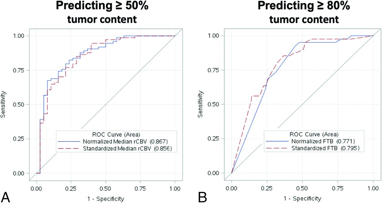
  • Fig 2.
    • Download figure
    • Open in new tab
    • Download powerpoint
    Fig 2.

    ROC analyses of the top-performing rCBV metrics for predicting  ≥ 50% histologic tumor content (A) and  ≥ 80% histologic tumor content (B). A, ROC curve for predicting ≥50% histologic tumor content using normalized (blue curve) and standardized (red dotted curve) median rCBV. B, ROC curve for predicting ≥80% histologic tumor content using normalized (blue curve) and standardized (red dotted curve) FTB. The standardized metrics show similar performance compared with corresponding normalized metrics.

Tables

  • Figures
    • View popup
    Table 1:

    Patient demographics and distribution of recurrent tumors by pathology and grade

    Total No.Age (yr)Men (Total)Women (Total)
    Patient demographics38Mean = 49.3, Range = 24–691919
    Pathology and grade
     Recurrent ODG (GIII)3Mean = 4621
     Recurrent astrocytoma (GIII)2Mean = 5211
     Recurrent GBM (GIV)33Mean = 501617
    • Note:—ODG indicates oligodendroglioma; GIII, WHO Grade III; GIV, WHO Grade IV.

    • View popup
    Table 2:

    Summary of predictive performance for normalized and standardized rCBV metrics

    rCBV MetricHistologic Tumor Content (0%–100%), Pearson Coefficient (P Value)Tumor Content ≥50%, ROC-AUC (Optimal Threshold)Tumor Content ≥80%, ROC-AUC (Optimal Threshold)
    FTB
     Normalizedr = 0.63 (<.001)0.80 (0.72)0.77 (0.84)
     Standardizedr = 0.66 (<.001)0.82 (0.56)0.80 (0.64)
    Mean rCBV
     Normalizedr = 0.45 (<.001)0.86 (1.558)0.75 (1.603)
     Standardizedr = 0.53 (<.001)0.85 (1.187)0.78 (1.187)
    Median rCBV
     Normalizedr = 0.48 (<.001)0.87 (1.68)0.77 (1.378)
     Standardizedr = 0.55 (<.001)0.86 (1.071)0.79 (1.10)
PreviousNext
Back to top

In this issue

American Journal of Neuroradiology: 41 (3)
American Journal of Neuroradiology
Vol. 41, Issue 3
1 Mar 2020
  • Table of Contents
  • Index by author
  • Complete Issue (PDF)
Advertisement
Print
Download PDF
Email Article

Thank you for your interest in spreading the word on American Journal of Neuroradiology.

NOTE: We only request your email address so that the person you are recommending the page to knows that you wanted them to see it, and that it is not junk mail. We do not capture any email address.

Enter multiple addresses on separate lines or separate them with commas.
Performance of Standardized Relative CBV for Quantifying Regional Histologic Tumor Burden in Recurrent High-Grade Glioma: Comparison against Normalized Relative CBV Using Image-Localized Stereotactic Biopsies
(Your Name) has sent you a message from American Journal of Neuroradiology
(Your Name) thought you would like to see the American Journal of Neuroradiology web site.
CAPTCHA
This question is for testing whether or not you are a human visitor and to prevent automated spam submissions.
Cite this article
J.M. Hoxworth, J.M. Eschbacher, A.C. Gonzales, K.W. Singleton, G.D. Leon, K.A. Smith, A.M. Stokes, Y. Zhou, G.L. Mazza, A.B. Porter, M.M. Mrugala, R.S. Zimmerman, B.R. Bendok, D.P. Patra, C. Krishna, J.L. Boxerman, L.C. Baxter, K.R. Swanson, C.C. Quarles, K.M. Schmainda, L.S. Hu
Performance of Standardized Relative CBV for Quantifying Regional Histologic Tumor Burden in Recurrent High-Grade Glioma: Comparison against Normalized Relative CBV Using Image-Localized Stereotactic Biopsies
American Journal of Neuroradiology Mar 2020, 41 (3) 408-415; DOI: 10.3174/ajnr.A6486

Citation Manager Formats

  • BibTeX
  • Bookends
  • EasyBib
  • EndNote (tagged)
  • EndNote 8 (xml)
  • Medlars
  • Mendeley
  • Papers
  • RefWorks Tagged
  • Ref Manager
  • RIS
  • Zotero
0 Responses
Respond to this article
Share
Bookmark this article
Performance of Standardized Relative CBV for Quantifying Regional Histologic Tumor Burden in Recurrent High-Grade Glioma: Comparison against Normalized Relative CBV Using Image-Localized Stereotactic Biopsies
J.M. Hoxworth, J.M. Eschbacher, A.C. Gonzales, K.W. Singleton, G.D. Leon, K.A. Smith, A.M. Stokes, Y. Zhou, G.L. Mazza, A.B. Porter, M.M. Mrugala, R.S. Zimmerman, B.R. Bendok, D.P. Patra, C. Krishna, J.L. Boxerman, L.C. Baxter, K.R. Swanson, C.C. Quarles, K.M. Schmainda, L.S. Hu
American Journal of Neuroradiology Mar 2020, 41 (3) 408-415; DOI: 10.3174/ajnr.A6486
del.icio.us logo Twitter logo Facebook logo Mendeley logo
  • Tweet Widget
  • Facebook Like
  • Google Plus One
Purchase

Jump to section

  • Article
    • Abstract
    • ABBREVIATIONS:
    • MATERIALS AND METHODS
    • RESULTS
    • DISCUSSION
    • CONCLUSIONS
    • Footnotes
    • References
  • Figures & Data
  • Supplemental
  • Info & Metrics
  • Responses
  • References
  • PDF

Related Articles

  • PubMed
  • Google Scholar

Cited By...

  • Multisite Benchmark Study for Standardized Relative CBV in Untreated Brain Metastases Using the DSC-MRI Consensus Acquisition Protocol
  • Identification of a Single-Dose, Low-Flip-Angle-Based CBV Threshold for Fractional Tumor Burden Mapping in Recurrent Glioblastoma
  • DSC Perfusion MRI-Derived Fractional Tumor Burden and Relative CBV Differentiate Tumor Progression and Radiation Necrosis in Brain Metastases Treated with Stereotactic Radiosurgery
  • Crossref (29)
  • Google Scholar

This article has been cited by the following articles in journals that are participating in Crossref Cited-by Linking.

  • High-Grade Glioma Treatment Response Monitoring Biomarkers: A Position Statement on the Evidence Supporting the Use of Advanced MRI Techniques in the Clinic, and the Latest Bench-to-Bedside Developments. Part 1: Perfusion and Diffusion Techniques
    Otto M. Henriksen, María del Mar Álvarez-Torres, Patricia Figueiredo, Gilbert Hangel, Vera C. Keil, Ruben E. Nechifor, Frank Riemer, Kathleen M. Schmainda, Esther A. H. Warnert, Evita C. Wiegers, Thomas C. Booth
    Frontiers in Oncology 2022 12
  • Opportunities for improving brain cancer treatment outcomes through imaging-based mathematical modeling of the delivery of radiotherapy and immunotherapy
    David A. Hormuth, Maguy Farhat, Chase Christenson, Brandon Curl, C. Chad Quarles, Caroline Chung, Thomas E. Yankeelov
    Advanced Drug Delivery Reviews 2022 187
  • Assessment and prediction of glioblastoma therapy response: challenges and opportunities
    Dan Qi, Jing Li, C Chad Quarles, Ekokobe Fonkem, Erxi Wu
    Brain 2023 146 4
  • Integrated molecular and multiparametric MRI mapping of high-grade glioma identifies regional biologic signatures
    Leland S. Hu, Fulvio D’Angelo, Taylor M. Weiskittel, Francesca P. Caruso, Shannon P. Fortin Ensign, Mylan R. Blomquist, Matthew J. Flick, Lujia Wang, Christopher P. Sereduk, Kevin Meng-Lin, Gustavo De Leon, Ashley Nespodzany, Javier C. Urcuyo, Ashlyn C Gonzales, Lee Curtin, Erika M. Lewis, Kyle W. Singleton, Timothy Dondlinger, Aliya Anil, Natenael B. Semmineh, Teresa Noviello, Reyna A. Patel, Panwen Wang, Junwen Wang, Jennifer M. Eschbacher, Andrea Hawkins-Daarud, Pamela R. Jackson, Itamar S. Grunfeld, Christian Elrod, Gina L. Mazza, Sam C. McGee, Lisa Paulson, Kamala Clark-Swanson, Yvette Lassiter-Morris, Kris A. Smith, Peter Nakaji, Bernard R. Bendok, Richard S. Zimmerman, Chandan Krishna, Devi P. Patra, Naresh P. Patel, Mark Lyons, Matthew Neal, Kliment Donev, Maciej M. Mrugala, Alyx B. Porter, Scott C. Beeman, Todd R. Jensen, Kathleen M. Schmainda, Yuxiang Zhou, Leslie C. Baxter, Christopher L. Plaisier, Jing Li, Hu Li, Anna Lasorella, C. Chad Quarles, Kristin R. Swanson, Michele Ceccarelli, Antonio Iavarone, Nhan L. Tran
    Nature Communications 2023 14 1
  • Challenges, limitations, and pitfalls of PET and advanced MRI in patients with brain tumors: A report of the PET/RANO group
    Norbert Galldiks, Timothy J Kaufmann, Philipp Vollmuth, Philipp Lohmann, Marion Smits, Michael C Veronesi, Karl-Josef Langen, Roberta Rudà, Nathalie L Albert, Elke Hattingen, Ian Law, Markus Hutterer, Riccardo Soffietti, Michael A Vogelbaum, Patrick Y Wen, Michael Weller, Joerg-Christian Tonn
    Neuro-Oncology 2024 26 7
  • Update on neuroimaging in brain tumours
    Marion Smits
    Current Opinion in Neurology 2021 34 4
  • Conventional and Advanced Imaging Techniques in Post-treatment Glioma Imaging
    Anna Y. Li, Michael Iv
    Frontiers in Radiology 2022 2
  • DSC Perfusion MRI–Derived Fractional Tumor Burden and Relative CBV Differentiate Tumor Progression and Radiation Necrosis in Brain Metastases Treated with Stereotactic Radiosurgery
    F. Kuo, N.N. Ng, S. Nagpal, E.L. Pollom, S. Soltys, M. Hayden-Gephart, G. Li, D.E. Born, Michael
    American Journal of Neuroradiology 2022 43 5
  • A multi-reader comparison of normal-appearing white matter normalization techniques for perfusion and diffusion MRI in brain tumors
    Nicholas S. Cho, Akifumi Hagiwara, Francesco Sanvito, Benjamin M. Ellingson
    Neuroradiology 2023 65 3
  • Magnetic Resonance Imaging Mapping of Brain Tumor Burden: Clinical Implications for Neurosurgical Management: Case Report
    Jennifer M Connelly, Melissa A Prah, Fernando Santos-Pinheiro, Wade Mueller, Elizabeth Cochran, Kathleen M Schmainda
    Neurosurgery Open 2021 2 4

More in this TOC Section

Adult Brain

  • Diagnostic Neuroradiology of Monoclonal Antibodies
  • NCCT vs. MRI for Brain Atrophy in Acute Stroke
  • Clinical Outcomes After Chiari I Decompression
Show more Adult Brain

Functional

  • Kurtosis and Epileptogenic Tubers: A Pilot Study
  • Glutaric Aciduria Type 1: DK vs. Conventional MRI
  • WM Neuroaxonal Loss in Type 1 Diabetes
Show more Functional

Similar Articles

Advertisement

Indexed Content

  • Current Issue
  • Accepted Manuscripts
  • Article Preview
  • Past Issues
  • Editorials
  • Editor's Choice
  • Fellows' Journal Club
  • Letters to the Editor
  • Video Articles

Cases

  • Case Collection
  • Archive - Case of the Week
  • Archive - Case of the Month
  • Archive - Classic Case

More from AJNR

  • Trainee Corner
  • Imaging Protocols
  • MRI Safety Corner

Multimedia

  • AJNR Podcasts
  • AJNR Scantastics

Resources

  • Turnaround Time
  • Submit a Manuscript
  • Submit a Video Article
  • Submit an eLetter to the Editor/Response
  • Manuscript Submission Guidelines
  • Statistical Tips
  • Fast Publishing of Accepted Manuscripts
  • Graphical Abstract Preparation
  • Imaging Protocol Submission
  • Evidence-Based Medicine Level Guide
  • Publishing Checklists
  • Author Policies
  • Become a Reviewer/Academy of Reviewers
  • News and Updates

About Us

  • About AJNR
  • Editorial Board
  • Editorial Board Alumni
  • Alerts
  • Permissions
  • Not an AJNR Subscriber? Join Now
  • Advertise with Us
  • Librarian Resources
  • Feedback
  • Terms and Conditions
  • AJNR Editorial Board Alumni

American Society of Neuroradiology

  • Not an ASNR Member? Join Now

© 2025 by the American Society of Neuroradiology All rights, including for text and data mining, AI training, and similar technologies, are reserved.
Print ISSN: 0195-6108 Online ISSN: 1936-959X

Powered by HighWire