Advertisement

Main menu

  • Home
  • Content
    • Current Issue
    • Accepted Manuscripts
    • Article Preview
    • Past Issue Archive
    • Video Articles
    • AJNR Case Collection
    • Case of the Week Archive
    • Case of the Month Archive
    • Classic Case Archive
  • Special Collections
    • AJNR Awards
    • Low-Field MRI
    • Alzheimer Disease
    • ASNR Foundation Special Collection
    • Photon-Counting CT
    • View All
  • Multimedia
    • AJNR Podcasts
    • AJNR SCANtastic
    • Trainee Corner
    • MRI Safety Corner
    • Imaging Protocols
  • For Authors
    • Submit a Manuscript
    • Submit a Video Article
    • Submit an eLetter to the Editor/Response
    • Manuscript Submission Guidelines
    • Statistical Tips
    • Fast Publishing of Accepted Manuscripts
    • Graphical Abstract Preparation
    • Imaging Protocol Submission
    • Author Policies
  • About Us
    • About AJNR
    • Editorial Board
    • Editorial Board Alumni
  • More
    • Become a Reviewer/Academy of Reviewers
    • Subscribers
    • Permissions
    • Alerts
    • Feedback
    • Advertisers
    • ASNR Home

User menu

  • Alerts
  • Log in

Search

  • Advanced search
American Journal of Neuroradiology

American Journal of Neuroradiology

ASHNR American Society of Functional Neuroradiology ASHNR American Society of Pediatric Neuroradiology ASSR
  • Alerts
  • Log in

Advanced Search

  • Home
  • Content
    • Current Issue
    • Accepted Manuscripts
    • Article Preview
    • Past Issue Archive
    • Video Articles
    • AJNR Case Collection
    • Case of the Week Archive
    • Case of the Month Archive
    • Classic Case Archive
  • Special Collections
    • AJNR Awards
    • Low-Field MRI
    • Alzheimer Disease
    • ASNR Foundation Special Collection
    • Photon-Counting CT
    • View All
  • Multimedia
    • AJNR Podcasts
    • AJNR SCANtastic
    • Trainee Corner
    • MRI Safety Corner
    • Imaging Protocols
  • For Authors
    • Submit a Manuscript
    • Submit a Video Article
    • Submit an eLetter to the Editor/Response
    • Manuscript Submission Guidelines
    • Statistical Tips
    • Fast Publishing of Accepted Manuscripts
    • Graphical Abstract Preparation
    • Imaging Protocol Submission
    • Author Policies
  • About Us
    • About AJNR
    • Editorial Board
    • Editorial Board Alumni
  • More
    • Become a Reviewer/Academy of Reviewers
    • Subscribers
    • Permissions
    • Alerts
    • Feedback
    • Advertisers
    • ASNR Home
  • Follow AJNR on Twitter
  • Visit AJNR on Facebook
  • Follow AJNR on Instagram
  • Join AJNR on LinkedIn
  • RSS Feeds

AJNR Awards, New Junior Editors, and more. Read the latest AJNR updates

Research ArticleAdult Brain
Open Access

COVID-19–Associated PRES–like Encephalopathy with Perivascular Gadolinium Enhancement

G. Conte, S. Avignone, M. Carbonara, M. Meneri, F. Ortolano, C. Cinnante and F. Triulzi
American Journal of Neuroradiology December 2020, 41 (12) 2206-2208; DOI: https://doi.org/10.3174/ajnr.A6762
G. Conte
aNeuroradiology Unit (G.C., S.A., C.C., F.T.)
  • Find this author on Google Scholar
  • Find this author on PubMed
  • Search for this author on this site
  • ORCID record for G. Conte
S. Avignone
aNeuroradiology Unit (G.C., S.A., C.C., F.T.)
  • Find this author on Google Scholar
  • Find this author on PubMed
  • Search for this author on this site
  • ORCID record for S. Avignone
M. Carbonara
bNeuroscience Intensive Care Unit (M.C., F.O.), Department of Anesthesia and Critical Care
  • Find this author on Google Scholar
  • Find this author on PubMed
  • Search for this author on this site
  • ORCID record for M. Carbonara
M. Meneri
cNeurology Unit (M.M.), Fondazione Institute for Research, Hospitalization and Health Care Ca' Granda Ospedale Maggiore Policlinico, Milan, Italy
  • Find this author on Google Scholar
  • Find this author on PubMed
  • Search for this author on this site
  • ORCID record for M. Meneri
F. Ortolano
bNeuroscience Intensive Care Unit (M.C., F.O.), Department of Anesthesia and Critical Care
  • Find this author on Google Scholar
  • Find this author on PubMed
  • Search for this author on this site
  • ORCID record for F. Ortolano
C. Cinnante
aNeuroradiology Unit (G.C., S.A., C.C., F.T.)
  • Find this author on Google Scholar
  • Find this author on PubMed
  • Search for this author on this site
  • ORCID record for C. Cinnante
F. Triulzi
aNeuroradiology Unit (G.C., S.A., C.C., F.T.)
  • Find this author on Google Scholar
  • Find this author on PubMed
  • Search for this author on this site
  • ORCID record for F. Triulzi
  • Article
  • Figures & Data
  • Supplemental
  • Info & Metrics
  • Responses
  • References
  • PDF
Loading

Abstract

SUMMARY: We describe the case of a 63-year-old woman who developed a coronavirus disease 2019–associated acute encephalopathy with perivascular gadolinium enhancement.

ABBREVIATIONS:

ICU
intensive care unit
PRES
posterior reversible encephalopathy syndrome
SARS-Cov-2
Severe Acute Respiratory Syndrome coronavirus 2

A 63-year-old woman was admitted to our hospital on day 7 (day 1 is the first day of symptoms) with fever, dry cough, fatigue, and subjective dyspnea. A nasopharyngeal swab for Severe Acute Respiratory Syndrome coronavirus 2 (SARS-Cov-2) was positive. Arterial blood gas analysis in room air showed mild hypocapnia (partial pressure of carbon dioxide, 31), causing mild respiratory alkalosis (pH = 7.48) and a low level of oxygen (partial pressure of carbon dioxide, 67). A chest x-ray was performed, showing bilateral ground-glass opacities. The patient had hypertension, but no other comorbidities were identified.

She was first admitted to the emergency ward and treated with a continuous positive airway pressure helmet; lopinavir/ritonavir was started as compassionate use approved by the ethics committee, as well as empirical piperacillin-tazobactam. Her respiratory parameters progressively worsened during the next days until she showed dyspnea, and her PaO2/FiO2 ratio was 110 with a continuous positive airway pressure helmet at 15 cm of water and an inspiratory fraction of oxygen of 70%. On day 11, she was intubated and transferred to the intensive care unit (ICU). During her ICU stay, she initially received mechanical ventilation at a medium level of positive end-expiratory pressure (up to 14) and inhaled nitric oxide and underwent two 20-hour prone-position cycles. She also developed a moderately acute kidney injury (measured creatinine clearance of 28 mL/minute), which never required replacement therapy, and high levels of hepatic enzymes and bilirubin, which caused us to stop lopinavir/ritonavir. An angiotensin-converting-enzyme inhibitor was introduced to reduce blood pressure in the ventilator weaning phase after 15 days on sedative drugs and opioids.

Regarding the treatment given in the ICU that was different from the usual care, she received anakinra (an interleukin 1 antagonist) due to a persistent high level of C-reactive Protein and ferritin and a high dose of subcutaneous heparin to target an activated partial thromboplastin time ratio of 2. On day 24, antibiotics were suspended until she developed ventilator-associated pneumonia by methicillin-sensitive Staphylococcus aureus, and cefazolin was given. Gradually, her clinical conditions improved, and she was weaned from the ventilator on day 28, after 24 hours of spontaneous breathing. On day 29, she was transferred back to medium intensity care in relatively good condition: afebrile, low-flow oxygen therapy via nasal cannula with normal peripheral capillary oxygen saturation and respiratory mechanics, normal blood pressure, and diuresis with acute kidney and hepatic injuries improving.

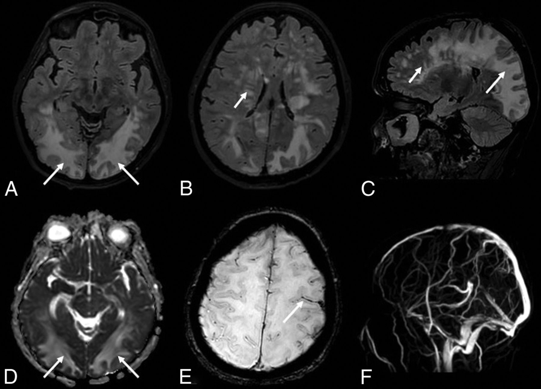
On day 30, she presented with left head and eye deviation and continuous tonic seizures on the left side of the body. After ineffective treatment with diazepam and lacosamide, the patient was sedated, intubated, and transferred again to the ICU. A phenytoin IV load was started, followed by enteral maintenance dosages, associated with diazepam infusion with clinical resolution. An electroencephalogram showed periodic lateralized epileptiform discharges in the right posterior regions, without clinical correlation. A lumbar puncture revealed only a slightly elevated protein level (70 mg/dL). A polymerase chain reaction test for virus detection in the CSF, including SARS-Cov-2, was negative. MR imaging showed cortical swelling and restricted diffusivity in the posterior regions of the right hemisphere, corresponding to the epileptic focus (On-line Figure). We observed multiple, partially confluent, tumefactive lesions of the sub- and supratentorial white matter hemispheres, with prevalence in the posterior hemisphere regions, which appeared as increased signal intensity on T2-weighted images, consistent with brain edema. The susceptibility-weighted images showed a subarachnoid blood effusion along the left precentral sulcus and excluded intraparenchymal hemorrhagic petecchiae. MR venography excluded intracranial thrombosis (Fig 1). The contrast-enhanced T1-weighted black-blood sequence showed a miliariform perivascular gadolinium enhancement in the supra- and subtentorial white matter. Neither wall enhancement of the large intracranial arterial vessels nor leptomeningeal enhancement was observed on postcontrast sequences (Fig 2).

FIG 1.
  • Download figure
  • Open in new tab
  • Download powerpoint
FIG 1.

The FLAIR sequence shows multiple lesions of the white matter of both hemispheres (A–C), more striking in the posterior regions with a tumefactive appearance (long arrows) and with a multifocal perivascular pattern in the deep white matter regions (short arrows). The lesions (arrows) show high values on the apparent diffusion coefficient map (D), suggesting increased water content in the parenchyma. The SWI sequence (E) shows a subarachnoid blood effusion along the left precentral sulcus (arrow). MR venography (F) excludes intracranial thrombosis.

FIG 2.
  • Download figure
  • Open in new tab
  • Download powerpoint
FIG 2.

The contrast-enhanced 3D conventional T1-weighted sequence (A–C) shows multiple spotlike gadolinium enhancement (arrows) in the posterior white matter and left hemisphere. The postcontrast 3D-T1-weighted black-blood sequence (D–F) shows a stratiform perivascular gadolinium enhancement in the white matter of both cerebral hemispheres. Neither wall enhancement of the large intracranial arterial vessels nor leptomeningeal enhancement are observed on postcontrast sequences.

The patient gradually improved. On day 41, she was extubated and was discharged to the ward 2 days later with residual mild left hemiparesis and left lateral hemianopsia. A control electroencephalogram showed only sporadic sharp waves and spikes in the right posterior regions. At follow-up, on MR imaging on day 49, the cerebellar lesions disappeared, cerebral lesions were markedly reduced, perivascular enhancement was not detected, and diffusion-weighted imaging findings were negative. On day 61, the patient was discharged from the hospital and referred to rehabilitation. At 180-day follow-up, the patient was at home, without motor deficits but with partial visual loss in the left eye (extended Glasgow Outcome Scale, 4; mRS score, 3).

We made a diagnosis of posterior reversible encephalopathy syndrome (PRES)-like encephalopathy with peculiar MR imaging findings. The miliariform perivascular postgadolinium enhancement has not previously been reported among typical and atypical MR imaging findings in PRES.1,2 The perivascular enhancement might be a sign of immune-mediated small-vessel damage, leading to altered integrity of the blood-brain barrier and brain edema, as supposed in PRES.3

Footnotes

  • Disclosures: Marco Carbonara—UNRELATED: Travel/Accommodations/Meeting Expenses Unrelated to Activities Listed: European Union, Comments: In the context of CENTER-TB1–related activities.

Indicates open access to non-subscribers at www.ajnr.org

References

  1. 1.↵
    1. Bartynski WS
    . Posterior reversible encephalopathy syndrome, Part 1: fundamental imaging and clinical features. AJNR Am J Neuroradiol 2008;29:1036–42 doi:10.3174/ajnr.A0928 pmid:18356474
    Abstract/FREE Full Text
  2. 2.↵
    1. Saad AF,
    2. Chaudhari R,
    3. Wintermark M
    . Imaging of atypical and complicated posterior reversible encephalopathy syndrome. Front Neurol 2019;10:964 doi:10.3389/fneur.2019.00964 pmid:31551919
    CrossRefPubMed
  3. 3.↵
    1. Fugate JE,
    2. Rabinstein AA
    . Posterior reversible encephalopathy syndrome: clinical and radiological manifestations, pathophysiology, and outstanding questions. Lancet Neurol 2015;14:914–25 doi:10.1016/S1474-4422(15)00111-8 pmid:26184985
    CrossRefPubMed
  • Received June 6, 2020.
  • Accepted after revision July 14, 2020.
  • © 2020 by American Journal of Neuroradiology
PreviousNext
Back to top

In this issue

American Journal of Neuroradiology: 41 (12)
American Journal of Neuroradiology
Vol. 41, Issue 12
1 Dec 2020
  • Table of Contents
  • Index by author
  • Complete Issue (PDF)
Advertisement
Print
Download PDF
Email Article

Thank you for your interest in spreading the word on American Journal of Neuroradiology.

NOTE: We only request your email address so that the person you are recommending the page to knows that you wanted them to see it, and that it is not junk mail. We do not capture any email address.

Enter multiple addresses on separate lines or separate them with commas.
COVID-19–Associated PRES–like Encephalopathy with Perivascular Gadolinium Enhancement
(Your Name) has sent you a message from American Journal of Neuroradiology
(Your Name) thought you would like to see the American Journal of Neuroradiology web site.
CAPTCHA
This question is for testing whether or not you are a human visitor and to prevent automated spam submissions.
Cite this article
G. Conte, S. Avignone, M. Carbonara, M. Meneri, F. Ortolano, C. Cinnante, F. Triulzi
COVID-19–Associated PRES–like Encephalopathy with Perivascular Gadolinium Enhancement
American Journal of Neuroradiology Dec 2020, 41 (12) 2206-2208; DOI: 10.3174/ajnr.A6762

Citation Manager Formats

  • BibTeX
  • Bookends
  • EasyBib
  • EndNote (tagged)
  • EndNote 8 (xml)
  • Medlars
  • Mendeley
  • Papers
  • RefWorks Tagged
  • Ref Manager
  • RIS
  • Zotero
0 Responses
Respond to this article
Share
Bookmark this article
COVID-19–Associated PRES–like Encephalopathy with Perivascular Gadolinium Enhancement
G. Conte, S. Avignone, M. Carbonara, M. Meneri, F. Ortolano, C. Cinnante, F. Triulzi
American Journal of Neuroradiology Dec 2020, 41 (12) 2206-2208; DOI: 10.3174/ajnr.A6762
del.icio.us logo Twitter logo Facebook logo Mendeley logo
  • Tweet Widget
  • Facebook Like
  • Google Plus One
Purchase

Jump to section

  • Article
    • Abstract
    • ABBREVIATIONS:
    • Footnotes
    • References
  • Figures & Data
  • Supplemental
  • Info & Metrics
  • Responses
  • References
  • PDF

Related Articles

  • PubMed
  • Google Scholar

Cited By...

  • No citing articles found.
  • Crossref (21)
  • Google Scholar

This article has been cited by the following articles in journals that are participating in Crossref Cited-by Linking.

  • The SARS-CoV-2 main protease Mpro causes microvascular brain pathology by cleaving NEMO in brain endothelial cells
    Jan Wenzel, Josephine Lampe, Helge Müller-Fielitz, Raphael Schuster, Marietta Zille, Kristin Müller, Markus Krohn, Jakob Körbelin, Linlin Zhang, Ümit Özorhan, Vanessa Neve, Julian U. G. Wagner, Denisa Bojkova, Mariana Shumliakivska, Yun Jiang, Anke Fähnrich, Fabian Ott, Valentin Sencio, Cyril Robil, Susanne Pfefferle, Florent Sauve, Caio Fernando Ferreira Coêlho, Jonas Franz, Frauke Spiecker, Beate Lembrich, Sonja Binder, Nina Feller, Peter König, Hauke Busch, Ludovic Collin, Roberto Villaseñor, Olaf Jöhren, Hermann C. Altmeppen, Manolis Pasparakis, Stefanie Dimmeler, Jindrich Cinatl, Klaus Püschel, Matija Zelic, Dimitry Ofengeim, Christine Stadelmann, François Trottein, Ruben Nogueiras, Rolf Hilgenfeld, Markus Glatzel, Vincent Prevot, Markus Schwaninger
    Nature Neuroscience 2021 24 11
  • Cerebrospinal fluid in COVID-19: A systematic review of the literature
    Ariane Lewis, Jennifer Frontera, Dimitris G. Placantonakis, Jennifer Lighter, Steven Galetta, Laura Balcer, Kara R. Melmed
    Journal of the Neurological Sciences 2021 421
  • Understanding on the possible routes for SARS CoV-2 invasion via ACE2 in the host linked with multiple organs damage
    Nikhil Kirtipal, Sanjay Kumar, Sumit Kumar Dubey, Vivek Dhar Dwivedi, K. Gireesh Babu, Petr Malý, Shiv Bharadwaj
    Infection, Genetics and Evolution 2022 99
  • Persistent visual dysfunction following posterior reversible encephalopathy syndrome due to COVID‐19: Case series and literature review
    Alison M. Hixon, Ashesh A. Thaker, Victoria S. Pelak
    European Journal of Neurology 2021 28 10
  • COVID-19-Associated Neurological Manifestations: An Emerging Electroencephalographic Literature
    Geoffroy Vellieux, Romain Sonneville, Sérafima Vledouts, Pierre Jaquet, Anny Rouvel-Tallec, Marie-Pia d’Ortho
    Frontiers in Physiology 2021 11
  • Brain MRI findings in COVID-19 patients with PRES: A systematic review
    Rubaya Yeahia, Javin Schefflein, Patrick Chiarolanzio, Anna Rozenstein, William Gomes, Sana Ali, Hasit Mehta, Fawaz Al-Mufti, Andrew McClelland, Edwin Gulko
    Clinical Imaging 2022 81
  • Cerebrospinal fluid findings in patients with seizure in the setting of COVID-19: A review of the literature
    Elizabeth Carroll, Kara R. Melmed, Jennifer Frontera, Dimitris G. Placantonakis, Steven Galetta, Laura Balcer, Ariane Lewis
    Seizure 2021 89
  • Posterior reversible encephalopathy syndrome and COVID-19: A series of 6 cases from Lombardy, Italy
    Antonio Colombo, Filippo Martinelli Boneschi, Sandro Beretta, Nereo Bresolin, Maurizio Versino, Lorenzo Lorusso, Diego Spagnoli, Giulia Nastasi, Davide Vallauri, Stefania Rota, Maria Repaci, Massimo Ferrarini, Mattia Pozzato, Lucia Princiotta Cariddi, Payam Tabaee Damavandi, Federico Carimati, Paola Banfi, Alessandro Clemenzi, Margherita Marelli, Andrea Giorgianni, Gabriele Vinacci, Marco Mauri, Paola Melzi, Maria Di Stefano, Antonio Tetto, Margherita Canesi, Andrea Salmaggi
    eNeurologicalSci 2021 22
  • Understanding the Role of Blood Vessels in the Neurologic Manifestations of Coronavirus Disease 2019 (COVID-19)
    Hannah A.B. Whitmore, Leo A. Kim
    The American Journal of Pathology 2021 191 11
  • COVID‐19 associated brain/spinal cord lesions and leptomeningeal enhancement: A meta‐analysis of the relationship to CSF SARS‐CoV‐2
    Ariane Lewis, Rajan Jain, Jennifer Frontera, Dimitris G. Placantonakis, Steven Galetta, Laura Balcer, Kara R. Melmed
    Journal of Neuroimaging 2021 31 5

More in this TOC Section

  • Diagnostic Neuroradiology of Monoclonal Antibodies
  • Cerebral ADC Changes in Fabry Disease
  • ML for Glioma Molecular Subtype Prediction
Show more ADULT BRAIN

Similar Articles

Advertisement

Indexed Content

  • Current Issue
  • Accepted Manuscripts
  • Article Preview
  • Past Issues
  • Editorials
  • Editor's Choice
  • Fellows' Journal Club
  • Letters to the Editor
  • Video Articles

Cases

  • Case Collection
  • Archive - Case of the Week
  • Archive - Case of the Month
  • Archive - Classic Case

More from AJNR

  • Trainee Corner
  • Imaging Protocols
  • MRI Safety Corner
  • Book Reviews

Multimedia

  • AJNR Podcasts
  • AJNR Scantastics

Resources

  • Turnaround Time
  • Submit a Manuscript
  • Submit a Video Article
  • Submit an eLetter to the Editor/Response
  • Manuscript Submission Guidelines
  • Statistical Tips
  • Fast Publishing of Accepted Manuscripts
  • Graphical Abstract Preparation
  • Imaging Protocol Submission
  • Evidence-Based Medicine Level Guide
  • Publishing Checklists
  • Author Policies
  • Become a Reviewer/Academy of Reviewers
  • News and Updates

About Us

  • About AJNR
  • Editorial Board
  • Editorial Board Alumni
  • Alerts
  • Permissions
  • Not an AJNR Subscriber? Join Now
  • Advertise with Us
  • Librarian Resources
  • Feedback
  • Terms and Conditions
  • AJNR Editorial Board Alumni

American Society of Neuroradiology

  • Not an ASNR Member? Join Now

© 2025 by the American Society of Neuroradiology All rights, including for text and data mining, AI training, and similar technologies, are reserved.
Print ISSN: 0195-6108 Online ISSN: 1936-959X

Powered by HighWire