
Article Figures & Data
Figures
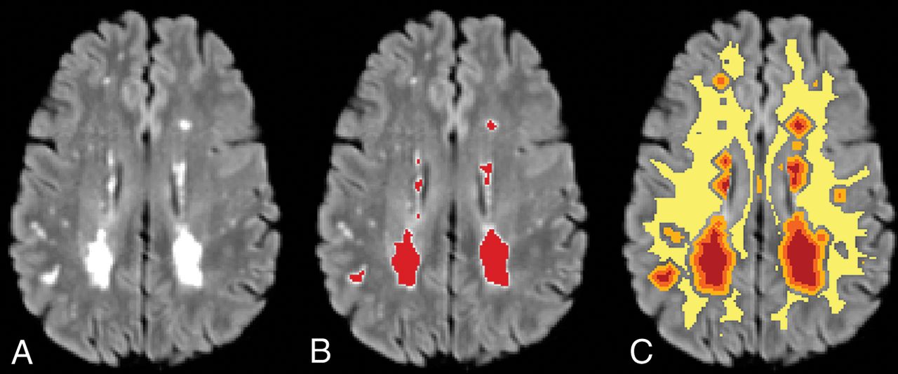
- Fig 1.
Sample FLAIR image showing WML (A); WML segmentation in red (B); and WML (red), 0–2 mm (orange) and 2–4 mm (light orange) penumbra, and dNAWM (yellow) (C). dNAWM is eroded from GM to eliminate partial volume effects among these tissue types.
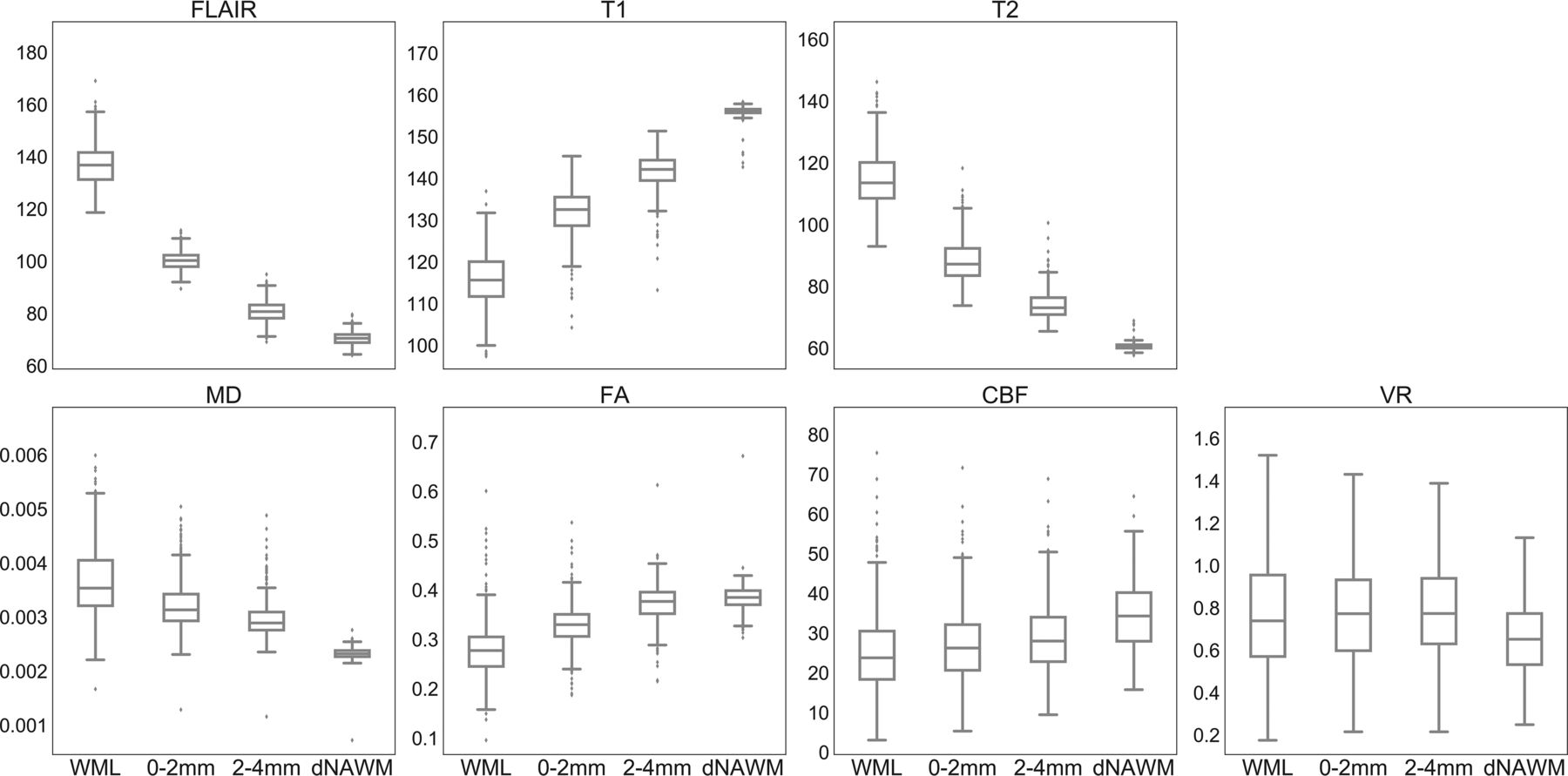
- Fig 2.
Boxplots of mean intensity values for each region. CBF is measured in cubic millimeters/100 g/min, and VR shows the mean percentage signal change. For vascular reactivity, outliers are excluded (comprising ∼10% of the sample) to better demonstrate differences among means. A graph with outliers is available (On-line Fig 2). For each parameter, mean values are significantly different among all pair-wise comparisons between ROIs with corrected P < .001, except for VR in which comparison of WML and dNAWM shows corrected P = .001, and there is no significant difference among WMLs and the 2 penumbra regions (P > .4).
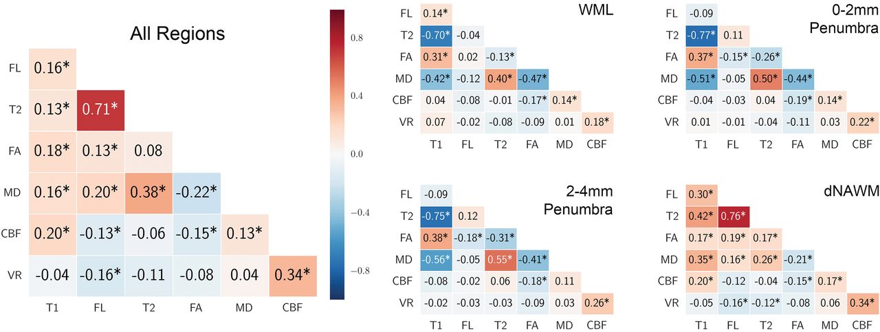
- Fig 3.
Correlations among the 7 MR imaging variables (Pearson r). Correlation is shown for all voxels in all regions combined and separately for each region. Positive correlations are shaded red, and negative correlations are shaded blue. The asterisk indicates Bonferroni-Holm corrected P < .05.


Tables
- Table 1:
Demographic and risk factor data of the subjects from CARDIA with MRI included in this study
Total KPDR UMN P Value No. 463 213 250 NA Mean age (yr) 50.5 ± 3.4 50.5 ± 3.4 50.5 ± 3.3 .98 Race Black 36% 38% 34% .48 White 64% 62% 66% Sex Male 47% 47% 46% .98 Female 53% 53% 54% Mean BMI 28.3 ± 5.3 27.6 ± 5.4 28.9 ± 5.2 .009 Mean systolic/diastolic blood pressure (mm Hg) 118 ± 15/74 ± 12 118 ± 14/74 ± 11 119 ± 16/73 ± 12 .58/.66 Diabetic (%) 7.6% 6.9% 8.2% .61 Smoking history .005 Never 60% 67% 53% Former 25% 22% 28% Current 15% 11% 19% Sedentary behavior (>75th percentile) 19% 18% 20% .55 Hypercholesterolemia 69% 71% 68% .38 Mean risk factor score (range, 0–6) 2.1 ± 1.3 1.9 ± 1.2 2.2 ± 1.3 .06 Note:—KPDR indicates Kaiser-Permanente Division of Research; UMN, University of Minnesota; NA, not applicable.
WML Penumbra (0–2 mm) Penumbra (2–4 mm) dNAWM Regression Coefficient Mean volume (cm3) 0.738 (0.842) 2.78 (2.26) 5.81 (4.21) 219 (3.68) NA [0.661–0.815] [2.57–2.99] [5.43–6.19] [186–253] FLAIR intensity 136 (10.1) 98.9 (3.50) 79.4 (3.71) 70.8 (2.58) −0.854 [135–137] [98.6–99.2] [79.1–79.7] [70.6–71.0] T1 intensity 116 (6.43) 132 (5.80) 142 (4.50) 156 (1.31) 0.824 [115–117] [132–133] [142–142] [156–156] T2 intensity 115 (8.62) 88.3 (6.82) 74.1 (4.75) 60.6 (1.02) −0.734 [114–115] [87.7–88.9] [73.7–74.5] [60.5–60.7] FA 0.279 (0.0554) 0.328 (0.0411) 0.374 (0.0380) 0.384 (0.0254) 0.630 [0.273–0.284] [0.324–0.331] [0.371–0.377] [0.382–0.386] MD (10−3) 3.68 (0.662) 3.23 (0.445) 2.97 (0.344) 2.33 (0.112) −0.578 [3.62–3.73] [3.19–3.27] [2.93–3.00] [2.32–2.34] CBF (mL/100 g/min) 25.2 (10.3) 26.9 (9.37) 28.8 (9.06) 34.4 (8.56) 0.306 [24.3–26.1] [26.1–27.7] [28.0–29.6] [33.6–35.2] VR 0.776 (0.493) 0.779 (0.379) 0.790 (0.328) 0.681 (0.377) 0.192 (Mean % change) [0.731–0.821] [0.745–0.813] [0.760–0.820] [0.647–0.715] Note:—NA indicates not applicable.
↵a SD is in parentheses, and 95% confidence intervals for intensity parameters are in brackets. Kruskal-Wallis tests were significant for all parameters, and pair-wise comparisons had P < .001 except that the WML and penumbral ROIs did not show significant differences in VR. The slope of linear regression across the 4 regions for intensity variables using a median value is shown in the last column; regression analyses were all statistically significant with P < .001.


