
Article Figures & Data
Figures
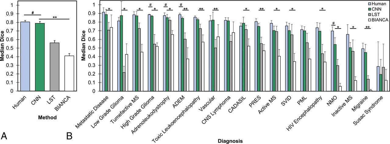
- Fig 1.
Performance of the CNN compared with human manual segmentation and other automated FLAIR segmentation methods. A, Median Dice scores across all validation cases. The asterisks denotes P < .05 for paired 2-tailed t tests compared with the CNN. The hashtag denotes P < .05 for human performance compared with the CNN. B, Median Dice scores across validation cases separated by underlying diagnosis. The asterisk denotes P < .05 (FDR-corrected for multiple comparisons) for the CNN compared with 1 method, and double asterisks denote P < .05 (FDR-corrected for multiple comparisons) for CNN compared with both methods using paired 2-tailed t tests. The hashtag separately denotes P < .05 (FDR-corrected for multiple comparisons) for human performance compared with the CNN. Error bars represent 1 standard error of the mean) across cases. ADEM indicates acute disseminated encephalomyelitis; PRES, posterior reversible encephalopathy syndrome; PML, progressive multifocal leukoencephalopathy; NMO, neuromyelitis optica.
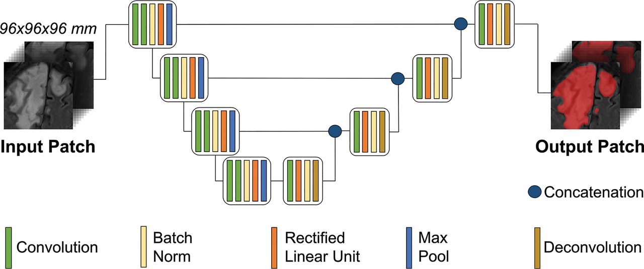
- Fig 2.
Schematic of the CNN U-net architecture. The architecture uses a 3D region-based approach for training and validation. The sample MR FLAIR images are from a patient with progressive multifocal leukoencephalopathy. Max indicates maximum.
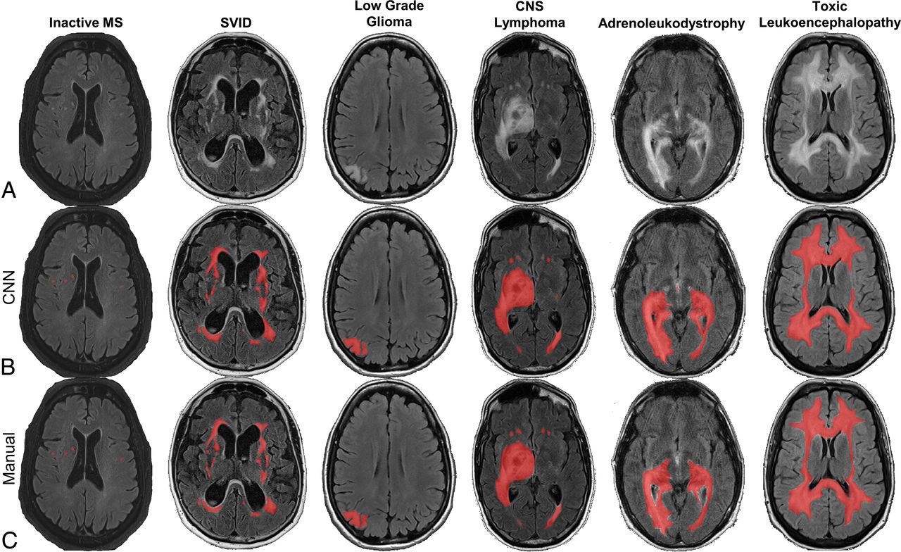
- Fig 3.
Representative slices from validation samples of FLAIR MR brain images (A) with CNN-based (B) and manual lesion segmentations (C), with predicted or ground truth lesion segmentations overlaid in red. The CNN performs well on a variety of different neurologic disorders, here shown in cases of multiple sclerosis, SVID, low grade-glioma, primary CNS lymphoma, adrenoleukodystrophy, and toxic leukoencephalopathy.
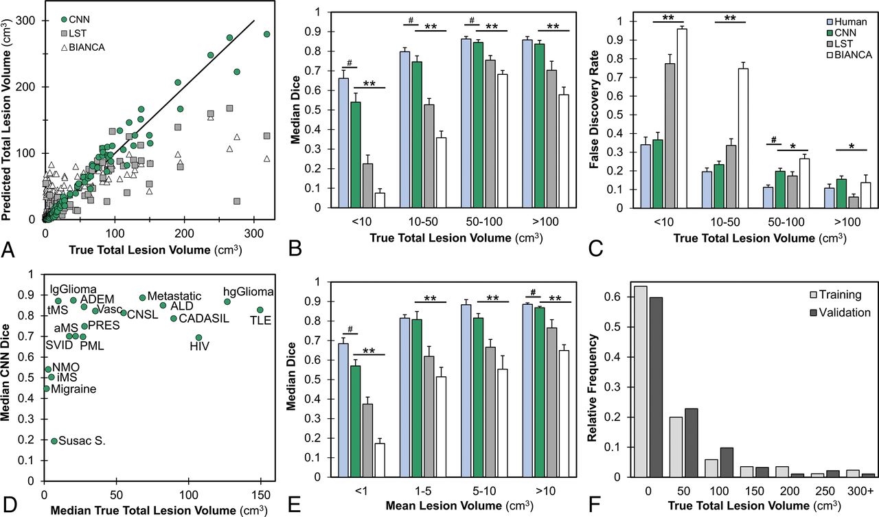
- Fig 4.
Performance of segmentation methods according to lesion characteristics. A, Scatterplot of predicted-versus-true total lesion volume with CNN (green circle) (Spearman correlation ρ = 0.985, best fit line slope β = 0.958), LST (gray square) ρ = 0.862, β = 0.490), and BIANCA (white triangle) (ρ = 0.655, β = 0.277) with the y = x line. Note clustering of CNN points along the y = x line, representing low deviation of CNN-based volume estimates from manual lesion volumes. B, Median Dice scores of cases stratified by total lesion volume. C, False discovery rate stratified by total lesion volume. D, Scatterplot of median CNN Dice score versus median true total lesion volume per diagnostic group. E, Median Dice scores of cases grouped by mean individual lesion volume. F, Histogram of lesion volumes in training and validation datasets. Error bars in all panels represent ±1 standard error of the mean across cases. The asterisk denotes P < .01 for the CNN compared with 1 method, and double asterisks denote P < .01 for CNN compared with both methods using 1-way group ANOVA and paired 2-tailed t tests. The hashtag separately denotes P < .05 for human performance compared with the CNN. ADEM indicates acute disseminated encephalomyelitis; ALD, adrenoleukodystrophy; TLE, toxic leukoencephalopathy; aMS, active MS: tMS, tumefactive MS; PRES, posterior reversible encephalopathy syndrome; iMS, inactive MS; NMO, neuromyelitis optica; Vasc, Vascular disease (ischemia); CNSL, CNS lymphoma; Susac S, Susac syndrome; lg, low-grade; hg, high-grade; PML, progressive multifocal leukoencephalopathy.
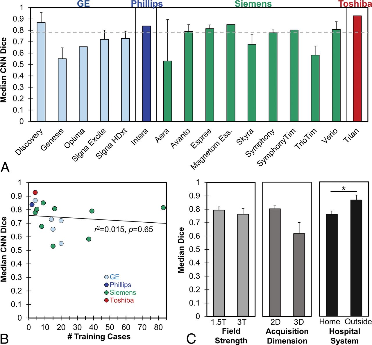
- Fig 5.
Performance of the CNN segmentation method according to technical characteristics. A, Median Dice scores on validation cases across different scanner models, grouped by MR imaging manufacturer. The dashed line indicates overall mean Dice score. There was no significant difference in Dice scores according to scanner model or manufacturer (P > .05 by 1-way ANOVA, see Results). B, Median Dice scores according to the number of training cases from that scanner model, with the best fit line. There is no significant correlation between the number of training cases and Dice scores (P > .05). C, Median Dice scores on validation cases grouped by field strength (left panel), acquisition dimension (middle panel), and hospital system where images were acquired (right panel). Error bars in all panels represent ± 1 standard error of the mean across cases. The asterisk denotes P < .05 for the 2-tailed t test among groups. See Table 1 for manufacturers' information. Ess indicates Essenza.


Tables
- Table 1:
Heterogeneous scanning parameters used for FLAIR sequences in training and validation samples, showing the number of study subjects in each categorya
Summary Training (n = 295) Validation (n = 92) Field strength 1.5T 230 (78.0%) 57 (62.0%) 3T 65 (22.0%) 35 (38.0%) Dimension 2D 287 (97.3%) 81 (88.0%) 3D 8 (2.7%) 11 (12.0%) Manufacturer/model GE Healthcareb Discovery MR750w 4 (1.4%) 3 (3.3%) Genesis Signa 20 (6.8%) 6 (6.5%) Optima MR450w 15 (5.1%) 1 (1.1%) Signa Excite 20 (6.8%) 7 (7.6%) Signa HDxt 14 (4.7%) 7 (7.6%) Phillipsc Intera 2 (0.7%) 1 (1.1%) Siemensd Magnetom Aera 15 (5.1%) 2 (2.2%) Avanto 39 (13.2%) 8 (8.7%) Magnetom Espree 83 (28.1%) 19 (20.1%) Magnetom Essenza 9 (3.1%) 1 (1.1%) Magnetom Skyra 8 (2.7%) 8 (8.7%) Magnetom Symphony 4 (1.4%) 3 (3.3%) Magnetom Symphony Tim 5 (1.7%) 1 (1.1%) Tim Trio 37 (12.5%) 11 (20.0%) Magnetom Verio 16 (5.4%) 13 (14.1%) Toshibae Titan 4 (1.4%) 1 (1.1%) TE (ms) Minimum 86 82 Median 136 136 Maximum 396 398 TR (ms) Minimum 5000 5000 Median 9000 9000 Maximum 12,000 12,000 - Table 2:
Summary measures of accuracy (Dice, voxelwise sensitivity, specificity, FDR, PPV/NPV) and comparisons of true and predicted lesion volumes by forecasting RMdSPE and Spearman correlation r of methodsa
Human CNN LST BIANCA Dice Median 0.805 0.789 0.562 0.410 SEM 0.017 0.022 0.026 0.027 Sensitivity (1-FNR) Median 0.800 0.767 0.599 0.556 SEM 0.017 0.025 0.026 0.020 Specificity (1-FPR) Median 0.999 0.999 0.999 0.997 SEM 0.000 0.000 0.000 0.000 PPV Median 0.824 0.769 0.690 0.335 SEM 0.018 0.018 0.030 0.034 NPV Median 0.999 0.999 0.999 0.999 SEM 0.000 0.000 0.001 0.001 RMdSPE 0.97% 1.38% 3.80% 6.56% Spearman r 0.991 0.985 0.862 0.655 Note:—PPV indicates positive predictive value; NPV, negative predictive value; FNR, false negative rate; FPR, false positive rate; SEM, standard error of the mean.
↵a Methods: Human, CNN, LST, and BIANCA.


Jump to section
Related Articles
Cited By...
- 3D Capsule Networks for Brain Image Segmentation
- 3D Capsule Networks for Brain Image Segmentation
- Development of Gestational Age-Based Fetal Brain and Intracranial Volume Reference Norms Using Deep Learning
- A Stacked Generalization of 3D Orthogonal Deep Learning Convolutional Neural Networks for Improved Detection of White Matter Hyperintensities in 3D FLAIR Images