
Article Figures & Data
Figures
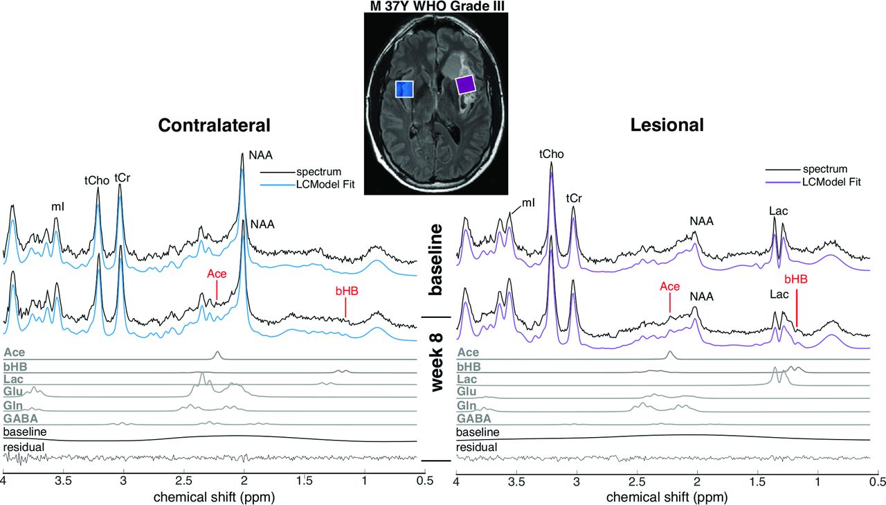
- Fig 1.
Representative MR spectra from 1 patient, together with spectral fitting results from the LCModel, at baseline and week 8. FLAIR MR imaging shows the voxel placement in the lesion in the left insular cortex and the corresponding voxel in the contralateral hemisphere. Individual fits of Ace and bHB at week 8 are also shown below the spectra, as well as those from Lac, glutamate (Glu), glutamine (Gln), and γ-aminobutyric acid (GABA). tCho indicates total Cho; tCr, total creatine; mI, myo-inositol.
- Fig 2.
Ketone body (left panel) and metabolite (right panel) concentrations as measured by MR spectroscopy at baseline and week 8 in the contralateral brain (A) and lesion (B) for all subjects. The asterisk indicates P ≤ .05; double asterisks, P ≤ .008 (Bonferroni-adjusted threshold for ketone bodies). Glx indicates glucose; tCho, total choline; tCr, total creatine; tNAA, total NAA; mI, myo-inositol.
- Fig 3.
Association between contralateral brain acetone levels estimated by MR spectroscopy and systemic measures of ketosis at week 8. Spearman rank correlation between acetone concentrations (IU error bars represent the ±95% confidence interval based on the LCModel Cramér-Rao values) in the contralateral brain at week 8 plotted against the urinary ketosis score (A) and fasting serum glucose levels (B). Urine ketones are defined as 1, trace (∼5 mg/dL); 2, small (∼15 mg/dL); 3, moderate (∼40 mg/dL); and 4, large (≥80 mg/dL).


Tables
Demographics (No.) (%) Age (mean) (SD) (yr) 49.2 (10.6) Male sex 6 (60%) WHO grade III 7 (70%) IV 3 (30%) Extent of resection Biopsy 2 (20%) Subtotal 3 (30%) Gross total 5 (50%) IDH1/2 mutational status IDH wild-type 4 (40%) IDH mutant 6 (60%) MGMT promoter methylation status Unmethylated 4 (40%) Methylated 4 (40%) Unknown 2 (20%) Concurrent TMZ (median) (range) (% completed) 100% (80%–100%) Adjuvant TMZ (median) (range) (No. of cycles) 6 (6–12) Note:—IDH indicates isocitrate dehydrogenase; MGMT, O6-methylguanine-DNA methyltransferase; TMZ, temozolomide.
- Table 2:
Ketone body concentrations as measured by MR spectroscopy before and after treatment with a ketogenic diet in both the lesion and contralateral braina
Baseline (IU) No. Week 8 (IU) No. P Value Ace Contralateral brain 0.04 ± 0.01 5 0.16 ± 0.04 9 .004b Lesion 0.06 ± 0.03 4 0.27 ± 0.06 9 .005b bHB Contralateral brain 0.09 ± 0.06 2 0.28 ± 0.08 6 .12 Lesion 0.07 ± 0.07 1 0.79 ± 0.32 5 .046c AcAc Contralateral brain 0.07 ± 0.04 3 0.06 ± 0.03 3 .76 Lesion 0.03 ± 0.03 1 0.02 ± 0.02 1 .72


