
Article Figures & Data
Figures
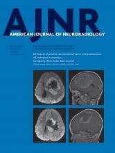
- Fig 1.
Example of plaque from 4 patients with symptomatic MCA plaque (A and C) and asymptomatic MCA plaque (B and D) and a histogram of 2 individual plaques (E). A and B, Scanned with a non-fat-suppressed T2 sequence. C and D, Scanned with a fat-suppressed T2 sequence. A and B, Plaque has a similar total volume but different signal features. E, Orange bars represents the signal distribution of A, which has more hyperintense signals seen as larger volume within high normalized plaque signal; blue bars represent the signal distribution of B, which has fewer hyperintense signals seen as larger volume within low normalized plaque signal. The plaque area (red dotted contour lines), vessel outer wall area (blue contour lines), and luminal area (yellow contour lines) are manually delineated on every cross-section containing plaque. Facial muscle signals are sampled by the pink box. The muscle signal of D was sampled in adjacent slices. Note the consistent image quality.
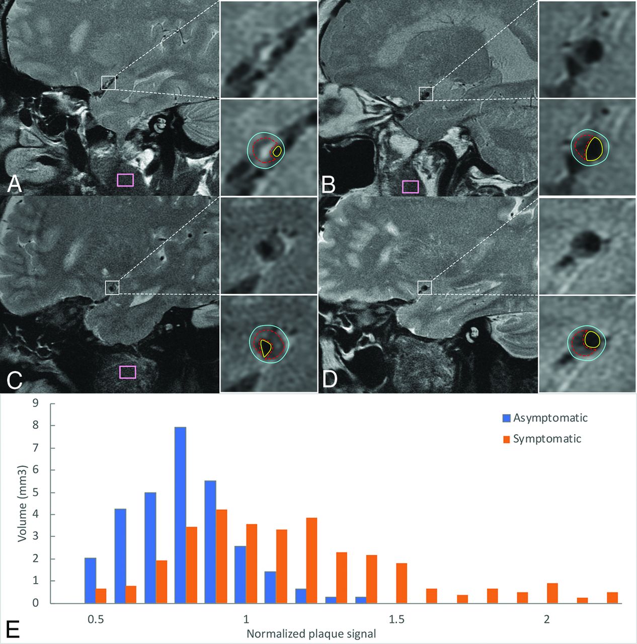
- Fig 2.
The summary of the median volume of normalized plaque signal in non-fat-suppressed and fat-suppressed T2 imaging. A, The median volume of symptomatic and asymptomatic MCA plaque in patients with non-fat-suppressed T2-weighted imaging. The difference between the 2 groups becomes more significant when normalized plaque signal is high. B, The median volume of symptomatic and asymptomatic MCA plaque in patients with fat-suppressed T2-weighted imaging. The difference between the 2 groups also becomes more significant when normalized plaque signal is high.


Tables
- Table 1:
Volume of NPS threshold, stenosis degree, and remodeling ratio in non-fat-suppressed imaginga
Non-Fat-Suppressed T2-Weighted Vessel Wall MR Imaging Symptomatic MCA Plaque (n = 47) Asymptomatic MCA Plaque (n = 26) AUC Stenosis degree (%) 72 ± 14 62 ± 12 0.692 Remodeling ratio 1.10 ± 0.20 1.03 ± 0.18 0.608 Volume of each NPS threshold, mm3 0.4∼0.5 0.1 (0–0.9) 0.4 (0–1.5) 0.408 0.5∼0.6 0.6 (0.1–2.2) 1.3 (0.3–3.3) 0.394 0.6∼0.7 1.2 (0.4–3.2) 2.5 (0.8–5.0) 0.415 0.7∼0.8 1.4 (0.9–4.2) 3.4 (1.3–6) 0.398 0.8∼0.9 2.4 (1.2–4.6) 2.4 (1.5–5.5) 0.482 0.9∼1.0 3.1 (1.4–4.6) 2.2 (0.8–3.3) 0.592 1.0∼1.1 2.8 (1.5–4.2) 1.9 (0.8–3.3) 0.615 1.1∼1.2 2.6 (1.7–3.8) 0.8 (0.5–2.2) 0.740 1.2∼1.3 1.9 (0.9–3.2) 0.5 (0.1–1.2) 0.797 1.3∼1.4 1.2 (0.3–2.8) 0.1 (0–0.5) 0.837 1.4∼1.5 1.2 (0.1–1.9) 0 (0–0.3) 0.798 1.5∼1.6 0.5 (0–1.2) 0 (0–0) 0.764 1.6∼1.7 0.1 (0–0.9) 0 (0–0) 0.731 1.7∼1.8 0 (0–0.6) 0 (0–0) 0.667 ≥0.9 15.1 (9.2–22.7) 7.0 (2.9–12.3) 0.760 ≥1.0 11.1 (6.3–18.8) 3.8 (1.5–8.2) 0.772 ≥1.1 8.8 (3.8–14.0) 2.2 (0.6–4.6) 0.795 ≥1.2 6.8 (1.5–10.8) 0.8 (0.1–2.2) 0.795 ≥1.3 4.0 (0.5–7.8) 0.3 (0–0.9) 0.815 ≥1.4 1.9 (0.1–5.2) 0 (0–0.4) 0.795 ≥1.5 0.6 (0–3.2) 0 (0–0) 0.772 ↵a Data are mean ± SD or median (interquartile range).
- Table 2:
Volume of NPS threshold, stenosis degree, and remodeling ratio in fat-suppressed imaginga
Fat-Suppressed T2-Weighted Vessel Wall MR Imaging Symptomatic MCA Plaque (n = 25) Asymptomatic MCA Plaque (n = 10) AUC Stenosis degree (%) 66 ± 13 56 ± 4 0.768 Remodeling ratio 1.16 ± 0.27 0.93 ± 0.09 0.800 Volume of each NPS threshold (mm3) 0.4∼0.5 0.3 (0.1–1.2) 0.8 (0.1–2.8) 0.412 0.5∼0.6 1.2 (0.5–1.9) 1.0 (0.4–4.9) 0.466 0.6∼0.7 1.7 (0.6–2.7) 1.7 (1.0–5.5) 0.426 0.7∼0.8 2.6 (1–3.1) 2.2 (1.0–3.5) 0.508 0.8∼0.9 2.4 (1.2–4.2) 2.0 (1.2–3.3) 0.578 0.9∼1.0 2.3 (1.2–4.5) 1.9 (0.4–2.8) 0.656 1.0∼1.1 2.0 (1.2–3.5) 0.8 (0–1.8) 0.748 1.1∼1.2 1.5 (0.6–2.4) 0.4 (0–0.5) 0.738 1.2∼1.3 1.2 (0.5–2.2) 0.5 (0–0.8) 0.750 1.3∼1.4 0.6 (0.3–1.3) 0 (0–0.3) 0.774 1.4∼1.5 0.5 (0.1–0.8) 0 (0–0.1) 0.752 1.5∼1.6 0.1 (0–0.8) 0 (0–0.3) 0.616 1.6∼1.7 0.1 (0–0.6) 0 (0–0) 0.676 1.7∼1.8 0 (0–0.5) 0 (0–0) 0.680 ≥0.9 10.0 (4.9–15.1) 3.1 (1.4–5.9) 0.764 ≥1.0 7.7 (3.6–12.2) 1.8 (0.3–3.6) 0.794 ≥1.1 3.8 (2.6–8.7) 1.2 (0–1.8) 0.770 ≥1.2 2.3 (1.3–6.3) 0.9 (0–1.4) 0.772 ≥1.3 1.4 (0.4–4.5) 0.3 (0–0.6) 0.764 ≥1.4 0.8 (0.1–3.2) 0 (0–0.4) 0.746 ≥1.5 0.3 (0–2.8) 0 (0–0.3) 0.650 ↵a Data are mean ± SD or median (interquartile range).
Factors Included in Logistic Regression Model OR (95% CI) AUC (95% CI) Model 1 Volume of NPS 1.3–1.4, per 1-mm3 increase 6.25 (1.90–20.57) 0.884 (0.822–0.945) Stenosis degree, per 10% increase 1.99 (1.24–3.19) Remodeling ratio, per 0.1 increase 1.50 (1.08–2.10) Fat-suppressed imaging 2.88 (0.93–8.94) Model 2 Volumetric summation of NPS no less than 1.2, per 1-mm3 increase 1.30 (1.06–1.58) 0.863 (0.791–0.935) Stenosis degree, per 10% increase 1.98 (1.26–3.10) Remodeling ratio, per 0.1 increase 1.45 (1.05–2.00) Fat-suppressed imaging 2.63 (0.89–7.81) Model 3a Stenosis degree, per 10% increase 2.43 (1.61–3.67) 0.806 (0.725–0.887) Remodeling ratio, per 0.1 increase 1.51 (1.11–2.06) Fat-suppressed imaging 2.03 (0.73–5.69) ↵a The AUC of model 3 is significantly lower than those of model 1 (P = .008) and model 2 (P = .027).


Jump to section
Related Articles
Cited By...
- No citing articles found.