Advertisement

Main menu

  • Home
  • Content
    • Current Issue
    • Accepted Manuscripts
    • Article Preview
    • Past Issue Archive
    • Video Articles
    • AJNR Case Collection
    • Case of the Week Archive
    • Case of the Month Archive
    • Classic Case Archive
  • Special Collections
    • AJNR Awards
    • Low-Field MRI
    • Alzheimer Disease
    • ASNR Foundation Special Collection
    • Photon-Counting CT
    • View All
  • Multimedia
    • AJNR Podcasts
    • AJNR SCANtastic
    • Trainee Corner
    • MRI Safety Corner
    • Imaging Protocols
  • For Authors
    • Submit a Manuscript
    • Submit a Video Article
    • Submit an eLetter to the Editor/Response
    • Manuscript Submission Guidelines
    • Statistical Tips
    • Fast Publishing of Accepted Manuscripts
    • Graphical Abstract Preparation
    • Imaging Protocol Submission
    • Author Policies
  • About Us
    • About AJNR
    • Editorial Board
    • Editorial Board Alumni
  • More
    • Become a Reviewer/Academy of Reviewers
    • Subscribers
    • Permissions
    • Alerts
    • Feedback
    • Advertisers
    • ASNR Home

User menu

  • Alerts
  • Log in

Search

  • Advanced search
American Journal of Neuroradiology

American Journal of Neuroradiology

ASHNR American Society of Functional Neuroradiology ASHNR American Society of Pediatric Neuroradiology ASSR
  • Alerts
  • Log in

Advanced Search

  • Home
  • Content
    • Current Issue
    • Accepted Manuscripts
    • Article Preview
    • Past Issue Archive
    • Video Articles
    • AJNR Case Collection
    • Case of the Week Archive
    • Case of the Month Archive
    • Classic Case Archive
  • Special Collections
    • AJNR Awards
    • Low-Field MRI
    • Alzheimer Disease
    • ASNR Foundation Special Collection
    • Photon-Counting CT
    • View All
  • Multimedia
    • AJNR Podcasts
    • AJNR SCANtastic
    • Trainee Corner
    • MRI Safety Corner
    • Imaging Protocols
  • For Authors
    • Submit a Manuscript
    • Submit a Video Article
    • Submit an eLetter to the Editor/Response
    • Manuscript Submission Guidelines
    • Statistical Tips
    • Fast Publishing of Accepted Manuscripts
    • Graphical Abstract Preparation
    • Imaging Protocol Submission
    • Author Policies
  • About Us
    • About AJNR
    • Editorial Board
    • Editorial Board Alumni
  • More
    • Become a Reviewer/Academy of Reviewers
    • Subscribers
    • Permissions
    • Alerts
    • Feedback
    • Advertisers
    • ASNR Home
  • Follow AJNR on Twitter
  • Visit AJNR on Facebook
  • Follow AJNR on Instagram
  • Join AJNR on LinkedIn
  • RSS Feeds

AJNR Awards, New Junior Editors, and more. Read the latest AJNR updates

Research ArticlePediatrics

Biometry of the Cerebellar Vermis and Brain Stem in Children: MR Imaging Reference Data from Measurements in 718 Children

C. Jandeaux, G. Kuchcinski, C. Ternynck, A. Riquet, X. Leclerc, J.-P. Pruvo and G. Soto-Ares
American Journal of Neuroradiology November 2019, 40 (11) 1835-1841; DOI: https://doi.org/10.3174/ajnr.A6257
C. Jandeaux
aFrom the Departments of Neuroradiology (C.J., G.K., X.L., J.-P.P., G.S.-A.)
  • Find this author on Google Scholar
  • Find this author on PubMed
  • Search for this author on this site
  • ORCID record for C. Jandeaux
G. Kuchcinski
aFrom the Departments of Neuroradiology (C.J., G.K., X.L., J.-P.P., G.S.-A.)
  • Find this author on Google Scholar
  • Find this author on PubMed
  • Search for this author on this site
  • ORCID record for G. Kuchcinski
C. Ternynck
bBiostatistics and Epidemiology (C.T.)
  • Find this author on Google Scholar
  • Find this author on PubMed
  • Search for this author on this site
  • ORCID record for C. Ternynck
A. Riquet
cNeuropediatrics (A.R.), Centre Hospitalier Universitaire Lille, Lille, France
  • Find this author on Google Scholar
  • Find this author on PubMed
  • Search for this author on this site
  • ORCID record for A. Riquet
X. Leclerc
aFrom the Departments of Neuroradiology (C.J., G.K., X.L., J.-P.P., G.S.-A.)
  • Find this author on Google Scholar
  • Find this author on PubMed
  • Search for this author on this site
  • ORCID record for X. Leclerc
J.-P. Pruvo
aFrom the Departments of Neuroradiology (C.J., G.K., X.L., J.-P.P., G.S.-A.)
  • Find this author on Google Scholar
  • Find this author on PubMed
  • Search for this author on this site
  • ORCID record for J.-P. Pruvo
G. Soto-Ares
aFrom the Departments of Neuroradiology (C.J., G.K., X.L., J.-P.P., G.S.-A.)
  • Find this author on Google Scholar
  • Find this author on PubMed
  • Search for this author on this site
  • ORCID record for G. Soto-Ares
  • Article
  • Figures & Data
  • Supplemental
  • Info & Metrics
  • Responses
  • References
  • PDF
Loading

Article Figures & Data

Figures

  • Tables
  • Fig 1.
    • Download figure
    • Open in new tab
    • Download powerpoint
    Fig 1.

    Illustration of the 2D measurements of the cerebellar vermis and the brain stem. H-V indicates the largest craniocaudal diameter of the vermis; APD-V, the largest anterior-posterior diameter of the vermis passing through the tip of the V4; APD-MP, perpendicular to the major axis of the brain stem passing through the midbrain-pons junction; APD-P, perpendicular to the major axis of the brain stem passing through the middle of the pons.
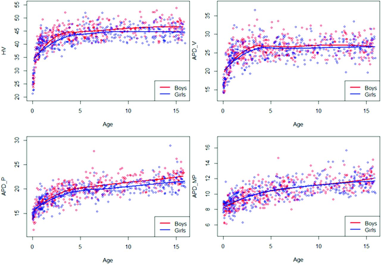
  • Fig 2.
    • Download figure
    • Open in new tab
    • Download powerpoint
    Fig 2.

    Smoothed curves of the different parameters (H-V, APD-V, APD-P, APD-MP) by age and sex.
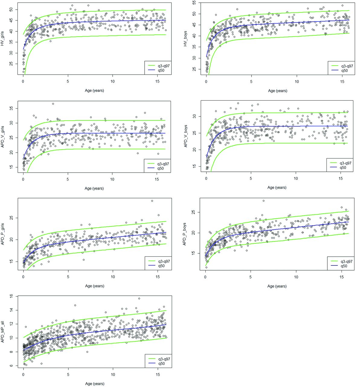
  • Fig 3.
    • Download figure
    • Open in new tab
    • Download powerpoint
    Fig 3.

    Reference intervals (third, 50th, 97th) for the different parameters (H-V, APD-V, APD-P, APD-MP). The values of APD-MP are similar in both sexes.

Tables

  • Figures
    • View popup
    Table 1:

    Distribution of the study population by age and sex (n = 718)

    Age Class (yr)Boys (n = 372)Girls (n = 346)All (n = 718)
    Younger than 15856114
    1–2373269
    2–3392867
    3–4312253
    4–5272350
    5–6202141
    6–7301848
    7–8171734
    8–991625
    9–10161228
    10–11101222
    11–12111425
    12–13111829
    13–14132033
    14–15171936
    15–16261844
    • View popup
    Table 2:

    Indications for brain MR imaging in the study population (n = 718)

    DiseasesPrevalence
    Epilepsy123 (17.2%)
    Supratentorial, suprasellar, or optic tumors65 (9%)
    Ocular globe pathology56 (7.7%)
    Suspicion of pituitary lesion (staturo-pondéral growth retardation, obesity, and so forth)55 (7.6%)
    Encephalitis, mastoiditis, meningitis, endocarditis, otitis, or empyema52 (7.3%)
    Extracerebral tumor or histiocytosis or blood disease40 (5.6%)
    Cranial trauma37 (5.2%)
    Spinal or medullary pathology, scoliosis, low back pain36 (5%)
    Supratentorial benign lesion (arachnoid cyst, pineal cyst, interhemispheric cyst, and so forth)32 (4.4%)
    Extracerebral or supratentorial vascular malformation28 (3.9%)
    Extracerebral venous-lymphatic malformation26 (3.6%)
    Isolated cervicofacial pathology (cervical mass, velar insufficiency, and so forth)21 (2.9%)
    Cyst or nasal root mass19 (2.6%)
    Headache15 (2.1%)
    Deafness10 (1.4%)
    Others103 (14.5%)
    • View popup
    Table 3:

    Equation for estimating reference values for the different parametersa

    Equations
    H-V girlsμ = 443 574.11 − 97 540.24Embedded Image − 231 497.89X3
    σ = 104 925.72 − 37 776.52X
    H-V boysμ = 707 969.47 − 45 502.94 log X − 538 257.55X3
    σ = 200 387.18 − 82 416.42X
    APD-V girlsμ = 585.75 − 336.12X3
    σ = 121.27
    APD-V boysμ = 1 083.91 − 689.41X3
    σ = 226.61
    APD-P girlsμ = 38.13 − 2 log X − 10.21X3
    σ = 4.09
    APD-P boysμ = 83.63 − 6.40 log X − 31.23X3
    σ = 12.09 − 4.71X
    APD-MPμ = 5.98 + 0.053X−0.05 − 1.31X
    σ = 0.48
    • ↵a With Embedded Image, where T denotes the age, T1 and Tn denote minimum and maximum ages, respectively, and ρ is a preselected constant for which a suitable value is .01. All measurements needed prior Box-Cox transformation. The values of APD-MP were similar in both sexes.

    • View popup
    Table 4:

    Inter- and intraobserver agreement for cerebellar vermis and brain stem parameters

    ParameterIntraobserver AgreementInterobserver Agreement
    Mean Bias95% Limits of AgreementICC (95% CI)Mean Bias95% Limits of AgreementICC (95% CI)
    H-V0.1560.869, 1.1810.994 (0.982–0.998)–0.302–2.129, 1.5250.981 (0.948–0.991)
    APD-V0.3321.831, 2.4940.941 (0.878–0.970)0.242–2.851, 3.3350.893 (0.799–0.948)
    APD-P0.100–1.161, 1.3610.916 (0.854–0.953)0.0881.704, 1.8800.846 (0.674–0.944)
    APD-MP0.052–0.729, 0.8330.988 (0.978–0.994)0.188–1.238, 1.6140.959 (0.918–0.978)
PreviousNext
Back to top

In this issue

American Journal of Neuroradiology: 40 (11)
American Journal of Neuroradiology
Vol. 40, Issue 11
1 Nov 2019
  • Table of Contents
  • Index by author
  • Complete Issue (PDF)
Advertisement
Print
Download PDF
Email Article

Thank you for your interest in spreading the word on American Journal of Neuroradiology.

NOTE: We only request your email address so that the person you are recommending the page to knows that you wanted them to see it, and that it is not junk mail. We do not capture any email address.

Enter multiple addresses on separate lines or separate them with commas.
Biometry of the Cerebellar Vermis and Brain Stem in Children: MR Imaging Reference Data from Measurements in 718 Children
(Your Name) has sent you a message from American Journal of Neuroradiology
(Your Name) thought you would like to see the American Journal of Neuroradiology web site.
CAPTCHA
This question is for testing whether or not you are a human visitor and to prevent automated spam submissions.
Cite this article
C. Jandeaux, G. Kuchcinski, C. Ternynck, A. Riquet, X. Leclerc, J.-P. Pruvo, G. Soto-Ares
Biometry of the Cerebellar Vermis and Brain Stem in Children: MR Imaging Reference Data from Measurements in 718 Children
American Journal of Neuroradiology Nov 2019, 40 (11) 1835-1841; DOI: 10.3174/ajnr.A6257

Citation Manager Formats

  • BibTeX
  • Bookends
  • EasyBib
  • EndNote (tagged)
  • EndNote 8 (xml)
  • Medlars
  • Mendeley
  • Papers
  • RefWorks Tagged
  • Ref Manager
  • RIS
  • Zotero
0 Responses
Respond to this article
Share
Bookmark this article
Biometry of the Cerebellar Vermis and Brain Stem in Children: MR Imaging Reference Data from Measurements in 718 Children
C. Jandeaux, G. Kuchcinski, C. Ternynck, A. Riquet, X. Leclerc, J.-P. Pruvo, G. Soto-Ares
American Journal of Neuroradiology Nov 2019, 40 (11) 1835-1841; DOI: 10.3174/ajnr.A6257
del.icio.us logo Twitter logo Facebook logo Mendeley logo
  • Tweet Widget
  • Facebook Like
  • Google Plus One
Purchase

Jump to section

  • Article
    • Abstract
    • ABBREVIATIONS:
    • MATERIALS AND METHODS
    • RESULTS
    • DISCUSSION
    • CONCLUSIONS
    • References
  • Figures & Data
  • Supplemental
  • Info & Metrics
  • Responses
  • References
  • PDF

Related Articles

  • PubMed
  • Google Scholar

Cited By...

  • Neuroradiologic, Clinical, and Genetic Characterization of Cerebellar Heterotopia: A Pediatric Multicentric Study
  • Imaging Findings and MRI Patterns in a Cohort of 18q Chromosomal Abnormalities
  • Neuroanatomical Features of NAA10- and NAA15-Related Neurodevelopmental Syndromes
  • Distinctive Brain Malformations in Zhu-Tokita-Takenouchi-Kim Syndrome
  • Refining the Neuroimaging Definition of the Dandy-Walker Phenotype
  • Brain Abnormalities in Patients with Germline Variants in H3F3: Novel Imaging Findings and Neurologic Symptoms Beyond Somatic Variants and Brain Tumors
  • Systematic Analysis of Brain MRI Findings in Adaptor Protein Complex 4-Associated Hereditary Spastic Paraplegia
  • BCL11A intellectual developmental disorder: defining the clinical spectrum and genotype-phenotype correlations
  • Crossref (11)
  • Google Scholar

This article has been cited by the following articles in journals that are participating in Crossref Cited-by Linking.

  • Refining the Neuroimaging Definition of the Dandy-Walker Phenotype
    M.T. Whitehead, M.J. Barkovich, J. Sidpra, C.A. Alves, D.M. Mirsky, Ö. Öztekin, D. Bhattacharya, L.T. Lucato, S. Sudhakar, A. Taranath, S. Andronikou, S.P. Prabhu, K.A. Aldinger, P. Haldipur, K.J. Millen, A.J. Barkovich, E. Boltshauser, W.B. Dobyns, K. Mankad
    American Journal of Neuroradiology 2022 43 10
  • Enhancing fetal alcohol spectrum disorders diagnosis with a classifier based on the intracerebellar gradient of volumetric undersizing
    Justine Fraize, Clara Fischer, Monique Elmaleh‐Bergès, Eliot Kerdreux, Anita Beggiato, Alexandra Ntorkou, Edouard Duchesnay, Dhaif Bekha, Odile Boespflug‐Tanguy, Richard Delorme, Lucie Hertz‐Pannier, David Germanaud
    Human Brain Mapping 2023 44 11
  • Brain Abnormalities in Patients with Germline Variants inH3F3: Novel Imaging Findings and Neurologic Symptoms Beyond Somatic Variants and Brain Tumors
    C.A.P.F. Alves, O. Sherbini, F. D’Arco, D. Steel, M.A. Kurian, F.C. Radio, G.B. Ferrero, D. Carli, M. Tartaglia, T.B. Balci, N.N. Powell-Hamilton, S.A. Schrier Vergano, H. Reutter, J. Hoefele, R. Günthner, E.R. Roeder, R.O. Littlejohn, D. Lessel, S. Lüttgen, C. Kentros, K. Anyane-Yeboa, C.B. Catarino, S. Mercimek-Andrews, J. Denecke, M.J. Lyons, T. Klopstock, E.J. Bhoj, L. Bryant, A. Vanderver
    American Journal of Neuroradiology 2022 43 7
  • Prenatal diagnosis of pontocerebellar hypoplasia with postnatal follow‐up
    Alienor Jaillard, Stéphanie Valence, Saskia Vande Perre, Ferdinand Dhombres, Delphine Héron, Thierry Billette de Villemeur, Boris Keren, Alexandra Afenjar, Leila Qebibo, Madeleine Harion, Geneviève Quenum‐Miraillet, Diana Rodriguez, Jean‐Marie Jouannic, Lydie Burglen, Catherine Garel
    Prenatal Diagnosis 2024 44 1
  • Undifferentiated psychosis or schizophrenia associated with vermis‐predominant cerebellar hypoplasia
    Alison C. Leslie, Mitchell P. Ward, William B. Dobyns
    American Journal of Medical Genetics Part A 2024 194 3
  • Corpus Callosum Length and Cerebellar Vermian Height in Fetal Growth Restriction
    Manesha Putra, Odessa P. Hamidi, Camille Driver, Emma E. Peek, Matthew A. Bolt, Diane Gumina, Shane A. Reeves, John C. Hobbins
    Fetal Diagnosis and Therapy 2024 51 3
  • Distinctive Brain Malformations in Zhu-Tokita-Takenouchi-Kim Syndrome
    B.J. Halliday, G. Baynam, L. Ewans, L. Greenhalgh, R.J. Leventer, D.T. Pilz, R. Sachdev, I.E. Scheffer, D.M. Markie, G. McGillivray, S.P. Robertson, S. Mandelstam
    American Journal of Neuroradiology 2022 43 11
  • Morphological characteristics of cerebellum, pons and thalamus in Reccurent isolated sleep paralysis – A pilot study
    Eva Miletínová, Monika Kliková, Amálie Dostalíková, Jitka Bušková
    Frontiers in Neuroanatomy 2024 18
  • Imaging Findings and MRI Patterns in a Cohort of 18q Chromosomal Abnormalities
    Prateek Malik, Helen Branson, Grace Yoon, Manohar Shroff, Susan Blaser, Pradeep Krishnan
    American Journal of Neuroradiology 2024 45 10
  • Neuroradiologic, Clinical, and Genetic Characterization of Cerebellar Heterotopia: A Pediatric Multicentric Study
    Ludovica Pasca, Filippo Arrigoni, Romina Romaniello, Maria Savina Severino, Davide Politano, Fulvio D’Abrusco, Jessica Garau, Valentina De Giorgis, Adriana Carpani, Sabrina Signorini, Simona Orcesi, Felice D’Arco, Enrico Alfei, Elisa Cattaneo, Elisa Rognone, Sara Uccella, Maria Teresa Divizia, Paolo Infantino, Enza Maria Valente, Renato Borgatti, Anna Pichiecchio
    American Journal of Neuroradiology 2025 46 1

More in this TOC Section

  • SyMRI & MR Fingerprinting in Brainstem Myelination
  • Comparison of Image Quality and Radiation Dose in Pediatric Temporal Bone CT Using Photon-Counting Detector CT and Energy-Integrating Detector CT
  • ASL in PEDS Nontraumatic Orbital Lesions
Show more Pediatrics

Similar Articles

Advertisement

Indexed Content

  • Current Issue
  • Accepted Manuscripts
  • Article Preview
  • Past Issues
  • Editorials
  • Editor's Choice
  • Fellows' Journal Club
  • Letters to the Editor
  • Video Articles

Cases

  • Case Collection
  • Archive - Case of the Week
  • Archive - Case of the Month
  • Archive - Classic Case

More from AJNR

  • Trainee Corner
  • Imaging Protocols
  • MRI Safety Corner

Multimedia

  • AJNR Podcasts
  • AJNR Scantastics

Resources

  • Turnaround Time
  • Submit a Manuscript
  • Submit a Video Article
  • Submit an eLetter to the Editor/Response
  • Manuscript Submission Guidelines
  • Statistical Tips
  • Fast Publishing of Accepted Manuscripts
  • Graphical Abstract Preparation
  • Imaging Protocol Submission
  • Evidence-Based Medicine Level Guide
  • Publishing Checklists
  • Author Policies
  • Become a Reviewer/Academy of Reviewers
  • News and Updates

About Us

  • About AJNR
  • Editorial Board
  • Editorial Board Alumni
  • Alerts
  • Permissions
  • Not an AJNR Subscriber? Join Now
  • Advertise with Us
  • Librarian Resources
  • Feedback
  • Terms and Conditions
  • AJNR Editorial Board Alumni

American Society of Neuroradiology

  • Not an ASNR Member? Join Now

© 2025 by the American Society of Neuroradiology All rights, including for text and data mining, AI training, and similar technologies, are reserved.
Print ISSN: 0195-6108 Online ISSN: 1936-959X

Powered by HighWire