Advertisement

Main menu

  • Home
  • Content
    • Current Issue
    • Accepted Manuscripts
    • Article Preview
    • Past Issue Archive
    • Video Articles
    • AJNR Case Collection
    • Case of the Week Archive
    • Case of the Month Archive
    • Classic Case Archive
  • Special Collections
    • AJNR Awards
    • Low-Field MRI
    • Alzheimer Disease
    • ASNR Foundation Special Collection
    • Photon-Counting CT
    • View All
  • Multimedia
    • AJNR Podcasts
    • AJNR SCANtastic
    • Trainee Corner
    • MRI Safety Corner
    • Imaging Protocols
  • For Authors
    • Submit a Manuscript
    • Submit a Video Article
    • Submit an eLetter to the Editor/Response
    • Manuscript Submission Guidelines
    • Statistical Tips
    • Fast Publishing of Accepted Manuscripts
    • Graphical Abstract Preparation
    • Imaging Protocol Submission
    • Author Policies
  • About Us
    • About AJNR
    • Editorial Board
    • Editorial Board Alumni
  • More
    • Become a Reviewer/Academy of Reviewers
    • Subscribers
    • Permissions
    • Alerts
    • Feedback
    • Advertisers
    • ASNR Home

User menu

  • Alerts
  • Log in

Search

  • Advanced search
American Journal of Neuroradiology

American Journal of Neuroradiology

ASHNR American Society of Functional Neuroradiology ASHNR American Society of Pediatric Neuroradiology ASSR
  • Alerts
  • Log in

Advanced Search

  • Home
  • Content
    • Current Issue
    • Accepted Manuscripts
    • Article Preview
    • Past Issue Archive
    • Video Articles
    • AJNR Case Collection
    • Case of the Week Archive
    • Case of the Month Archive
    • Classic Case Archive
  • Special Collections
    • AJNR Awards
    • Low-Field MRI
    • Alzheimer Disease
    • ASNR Foundation Special Collection
    • Photon-Counting CT
    • View All
  • Multimedia
    • AJNR Podcasts
    • AJNR SCANtastic
    • Trainee Corner
    • MRI Safety Corner
    • Imaging Protocols
  • For Authors
    • Submit a Manuscript
    • Submit a Video Article
    • Submit an eLetter to the Editor/Response
    • Manuscript Submission Guidelines
    • Statistical Tips
    • Fast Publishing of Accepted Manuscripts
    • Graphical Abstract Preparation
    • Imaging Protocol Submission
    • Author Policies
  • About Us
    • About AJNR
    • Editorial Board
    • Editorial Board Alumni
  • More
    • Become a Reviewer/Academy of Reviewers
    • Subscribers
    • Permissions
    • Alerts
    • Feedback
    • Advertisers
    • ASNR Home
  • Follow AJNR on Twitter
  • Visit AJNR on Facebook
  • Follow AJNR on Instagram
  • Join AJNR on LinkedIn
  • RSS Feeds

AJNR Awards, New Junior Editors, and more. Read the latest AJNR updates

Research ArticleAdult Brain
Open Access

Quantitative MRI of Perivascular Spaces at 3T for Early Diagnosis of Mild Cognitive Impairment

M. Niazi, M. Karaman, S. Das, X.J. Zhou, P. Yushkevich and K. Cai
American Journal of Neuroradiology September 2018, 39 (9) 1622-1628; DOI: https://doi.org/10.3174/ajnr.A5734
M. Niazi
aFrom the Department of Radiology (M.N., X.J.Z., K.C.)
cChicago College of Osteopathic Medicine (M.N.), Midwestern University, Downers Grove, Illinois
  • Find this author on Google Scholar
  • Find this author on PubMed
  • Search for this author on this site
  • ORCID record for M. Niazi
M. Karaman
bCenter for MR Research (M.K., X.J.Z., K.C.), College of Medicine, University of Illinois at Chicago, Chicago, Illinois
  • Find this author on Google Scholar
  • Find this author on PubMed
  • Search for this author on this site
  • ORCID record for M. Karaman
S. Das
dDepartment of Radiology (S.D., P.Y.), School of Medicine, University of Pennsylvania, Philadelphia, Pennsylvania.
  • Find this author on Google Scholar
  • Find this author on PubMed
  • Search for this author on this site
  • ORCID record for S. Das
X.J. Zhou
aFrom the Department of Radiology (M.N., X.J.Z., K.C.)
bCenter for MR Research (M.K., X.J.Z., K.C.), College of Medicine, University of Illinois at Chicago, Chicago, Illinois
  • Find this author on Google Scholar
  • Find this author on PubMed
  • Search for this author on this site
  • ORCID record for X.J. Zhou
P. Yushkevich
dDepartment of Radiology (S.D., P.Y.), School of Medicine, University of Pennsylvania, Philadelphia, Pennsylvania.
  • Find this author on Google Scholar
  • Find this author on PubMed
  • Search for this author on this site
  • ORCID record for P. Yushkevich
K. Cai
aFrom the Department of Radiology (M.N., X.J.Z., K.C.)
bCenter for MR Research (M.K., X.J.Z., K.C.), College of Medicine, University of Illinois at Chicago, Chicago, Illinois
  • Find this author on Google Scholar
  • Find this author on PubMed
  • Search for this author on this site
  • ORCID record for K. Cai
  • Article
  • Figures & Data
  • Info & Metrics
  • Responses
  • References
  • PDF
Loading

Article Figures & Data

Figures

  • Fig 1.
    • Download figure
    • Open in new tab
    • Download powerpoint
    Fig 1.

    Illustration of the EPVS segmentation process. A, A coronal MR brain image of a patient with aMCI. The original image fitted with a brain mask (B) and the original image fitted with a mask generated with first-order k-means clustering to remove the CSF (C). D, Contrast-enhanced image of the brain fitted with the mask generated with second-order k-means clustering to exclude the cortical gray matter. E, A mask for pixels with high and positive spatial gradient values obtained from a pixel-wise spatial gradient map. The brain stem was manually removed. F, EPVS selected on the basis of object size after removing the edges of the brain structures. G, Color-coded EPVS overlaid on the original brain image.

  • Fig 2.
    • Download figure
    • Open in new tab
    • Download powerpoint
    Fig 2.

    A side-by-side comparison of EPVS in a cognitively healthy control versus a patient with aMCI A, A coronal MR brain image of a cognitively healthy control. B, Segmented EPVS color-overlaid on the MR brain image of a cognitively healthy control. C, A coronal MR brain image of a patient with aMCI. D, Color-coded EPVS overlaid on the MR brain image of a patient with aMCI.

  • Fig 3.
    • Download figure
    • Open in new tab
    • Download powerpoint
    Fig 3.

    Representative multislice MIP projections of EPVS in the subcortical brain structures and the basal ganglia of a control and a subject with aMCI. A, MIP image of subcortical structures within the middle of the brain of a control subject. B, The MIP image of subcortical structures within the middle of the brain of a subject with aMCI. C, MIP image of the basal ganglia of a control subject. D, MIP image of the basal ganglia of a subject with aMCI.
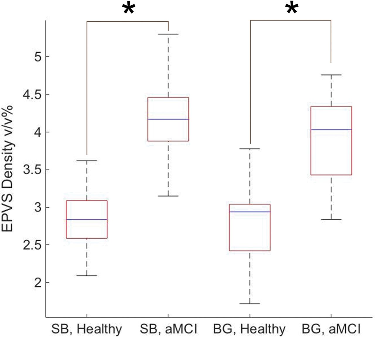
  • Fig 4.
    • Download figure
    • Open in new tab
    • Download powerpoint
    Fig 4.

    Summarized EPVS densities within the middle part of the subcortical brain (SB) and the basal ganglia (BG) of healthy controls and subjects with aMCI. The asterisk indicates P < .001.

  • Fig 5.
    • Download figure
    • Open in new tab
    • Download powerpoint
    Fig 5.

    The receiver operating characteristic curve for using EPVS as a differentiating biomarker between aMCI and cognitively healthy individuals. The area under the curve is 0.96, the 95% confidence interval is 0.89–1.00, and the standard error is 0.04. The cutoff point that gives the best sensitivity (92.86%) and the specificity (93.33%) is 3.35 v/v%.

PreviousNext
Back to top

In this issue

American Journal of Neuroradiology: 39 (9)
American Journal of Neuroradiology
Vol. 39, Issue 9
1 Sep 2018
  • Table of Contents
  • Index by author
  • Complete Issue (PDF)
Advertisement
Print
Download PDF
Email Article

Thank you for your interest in spreading the word on American Journal of Neuroradiology.

NOTE: We only request your email address so that the person you are recommending the page to knows that you wanted them to see it, and that it is not junk mail. We do not capture any email address.

Enter multiple addresses on separate lines or separate them with commas.
Quantitative MRI of Perivascular Spaces at 3T for Early Diagnosis of Mild Cognitive Impairment
(Your Name) has sent you a message from American Journal of Neuroradiology
(Your Name) thought you would like to see the American Journal of Neuroradiology web site.
CAPTCHA
This question is for testing whether or not you are a human visitor and to prevent automated spam submissions.
Cite this article
M. Niazi, M. Karaman, S. Das, X.J. Zhou, P. Yushkevich, K. Cai
Quantitative MRI of Perivascular Spaces at 3T for Early Diagnosis of Mild Cognitive Impairment
American Journal of Neuroradiology Sep 2018, 39 (9) 1622-1628; DOI: 10.3174/ajnr.A5734

Citation Manager Formats

  • BibTeX
  • Bookends
  • EasyBib
  • EndNote (tagged)
  • EndNote 8 (xml)
  • Medlars
  • Mendeley
  • Papers
  • RefWorks Tagged
  • Ref Manager
  • RIS
  • Zotero
0 Responses
Respond to this article
Share
Bookmark this article
Quantitative MRI of Perivascular Spaces at 3T for Early Diagnosis of Mild Cognitive Impairment
M. Niazi, M. Karaman, S. Das, X.J. Zhou, P. Yushkevich, K. Cai
American Journal of Neuroradiology Sep 2018, 39 (9) 1622-1628; DOI: 10.3174/ajnr.A5734
del.icio.us logo Twitter logo Facebook logo Mendeley logo
  • Tweet Widget
  • Facebook Like
  • Google Plus One
Purchase

Jump to section

  • Article
    • Abstract
    • ABBREVIATIONS:
    • Materials and Methods
    • Results
    • Discussion
    • Conclusions
    • Acknowledgments
    • Footnotes
    • References
  • Figures & Data
  • Info & Metrics
  • Responses
  • References
  • PDF

Related Articles

  • No related articles found.
  • PubMed
  • Google Scholar

Cited By...

  • A Systematic Review and Meta-Analysis of Automated Methods for Quantifying Enlarged Perivascular Spaces in the Brain
  • Lesion Volume in Relapsing Multiple Sclerosis is Associated with Perivascular Space Enlargement at the Level of the Basal Ganglia
  • Increasing age is independently associated with higher free water in non-active MS brain - A multi-compartment analysis using FAST-T2
  • Crossref (34)
  • Google Scholar

This article has been cited by the following articles in journals that are participating in Crossref Cited-by Linking.

  • Automated MRI-Based Deep Learning Model for Detection of Alzheimer’s Disease Process
    Wei Feng, Nicholas Van Halm-Lutterodt, Hao Tang, Andrew Mecum, Mohamed Kamal Mesregah, Yuan Ma, Haibin Li, Feng Zhang, Zhiyuan Wu, Erlin Yao, Xiuhua Guo
    International Journal of Neural Systems 2020 30 06
  • Optimal Mass Transport with Lagrangian Workflow Reveals Advective and Diffusion Driven Solute Transport in the Glymphatic System
    Sunil Koundal, Rena Elkin, Saad Nadeem, Yuechuan Xue, Stefan Constantinou, Simon Sanggaard, Xiaodan Liu, Brittany Monte, Feng Xu, William Van Nostrand, Maiken Nedergaard, Hedok Lee, Joanna Wardlaw, Helene Benveniste, Allen Tannenbaum
    Scientific Reports 2020 10 1
  • What is ‘Alzheimer&#039;s disease’? The neuropathological heterogeneity of clinically defined Alzheimer&#039;s dementia
    Rupal I. Mehta, Julie A. Schneider
    Current Opinion in Neurology 2021 34 2
  • The role of brain perivascular space burden in early-stage Parkinson’s disease
    Ting Shen, Yumei Yue, Shuai Zhao, Juanjuan Xie, Yanxing Chen, Jun Tian, Wen Lv, Chun-Yi Zac Lo, Yi-Cheng Hsu, Tobias Kober, Baorong Zhang, Hsin-Yi Lai
    npj Parkinson's Disease 2021 7 1
  • Quantification of visible Virchow–Robin spaces for detecting the functional status of the glymphatic system in children with newly diagnosed idiopathic generalized epilepsy
    Congcong Liu, Tafawa Habib, Mustafa Salimeen, Anuja Pradhan, Martha Singh, Miaomiao Wang, Fan Wu, Yuli Zhang, Lu Gao, Guanyu Yang, Xianjun Li, Jian Yang
    Seizure 2020 78
  • Glutamatergic neurotransmission: A potential pharmacotherapeutic target for the treatment of cognitive disorders
    Pratik Chakraborty, Abhijit Dey, Abilash Valsala Gopalakrishnan, Kumari Swati, Shreesh Ojha, Anand Prakash, Dhruv Kumar, Rashmi K. Ambasta, Niraj Kumar Jha, Saurabh Kumar Jha, Saikat Dewanjee
    Ageing Research Reviews 2023 85
  • A critical guide to the automated quantification of perivascular spaces in magnetic resonance imaging
    William Pham, Miranda Lynch, Gershon Spitz, Terence O’Brien, Lucy Vivash, Benjamin Sinclair, Meng Law
    Frontiers in Neuroscience 2022 16
  • Why Are Perivascular Spaces Important?
    Tatyana Shulyatnikova, Melvin R. Hayden
    Medicina 2023 59 5
  • Automated Methods for Detecting and Quantitation of Enlarged Perivascular spaces on MRI
    Jasmine Moses, Ben Sinclair, Meng Law, Terence J. O'Brien, Lucy Vivash
    Journal of Magnetic Resonance Imaging 2023 57 1
  • Quantity and Morphology of Perivascular Spaces: Associations With Vascular Risk Factors and Cerebral Small Vessel Disease
    Shuyue Wang, Peiyu Huang, Ruiting Zhang, Hui Hong, Yeerfan Jiaerken, Chunfeng Lian, Xinfeng Yu, Xiao Luo, Kaicheng Li, Qingze Zeng, Xiaopei Xu, Wenke Yu, Xiao Wu, Minming Zhang
    Journal of Magnetic Resonance Imaging 2021 54 4

More in this TOC Section

  • Diagnostic Neuroradiology of Monoclonal Antibodies
  • Clinical Outcomes After Chiari I Decompression
  • Segmentation of Brain Metastases with BLAST
Show more ADULT BRAIN

Similar Articles

Advertisement

Indexed Content

  • Current Issue
  • Accepted Manuscripts
  • Article Preview
  • Past Issues
  • Editorials
  • Editor's Choice
  • Fellows' Journal Club
  • Letters to the Editor
  • Video Articles

Cases

  • Case Collection
  • Archive - Case of the Week
  • Archive - Case of the Month
  • Archive - Classic Case

More from AJNR

  • Trainee Corner
  • Imaging Protocols
  • MRI Safety Corner
  • Book Reviews

Multimedia

  • AJNR Podcasts
  • AJNR Scantastics

Resources

  • Turnaround Time
  • Submit a Manuscript
  • Submit a Video Article
  • Submit an eLetter to the Editor/Response
  • Manuscript Submission Guidelines
  • Statistical Tips
  • Fast Publishing of Accepted Manuscripts
  • Graphical Abstract Preparation
  • Imaging Protocol Submission
  • Evidence-Based Medicine Level Guide
  • Publishing Checklists
  • Author Policies
  • Become a Reviewer/Academy of Reviewers
  • News and Updates

About Us

  • About AJNR
  • Editorial Board
  • Editorial Board Alumni
  • Alerts
  • Permissions
  • Not an AJNR Subscriber? Join Now
  • Advertise with Us
  • Librarian Resources
  • Feedback
  • Terms and Conditions
  • AJNR Editorial Board Alumni

American Society of Neuroradiology

  • Not an ASNR Member? Join Now

© 2025 by the American Society of Neuroradiology All rights, including for text and data mining, AI training, and similar technologies, are reserved.
Print ISSN: 0195-6108 Online ISSN: 1936-959X

Powered by HighWire