Advertisement

Main menu

  • Home
  • Content
    • Current Issue
    • Accepted Manuscripts
    • Article Preview
    • Past Issue Archive
    • Video Articles
    • AJNR Case Collection
    • Case of the Week Archive
    • Case of the Month Archive
    • Classic Case Archive
  • Special Collections
    • Advancing NeuroMRI with High-Relaxivity Contrast Agents
    • AJNR Awards
    • Low-Field MRI
    • Alzheimer Disease
    • ASNR Foundation Special Collection
    • Photon-Counting CT
    • View All
  • Multimedia
    • AJNR Podcasts
    • AJNR SCANtastic
    • Trainee Corner
    • MRI Safety Corner
    • Imaging Protocols
  • For Authors
    • Submit a Manuscript
    • Submit a Video Article
    • Submit an eLetter to the Editor/Response
    • Manuscript Submission Guidelines
    • Statistical Tips
    • Fast Publishing of Accepted Manuscripts
    • Graphical Abstract Preparation
    • Imaging Protocol Submission
    • Author Policies
  • About Us
    • About AJNR
    • Editorial Board
    • Editorial Board Alumni
  • More
    • Become a Reviewer/Academy of Reviewers
    • Subscribers
    • Permissions
    • Alerts
    • Feedback
    • Advertisers
    • ASNR Home

User menu

  • Alerts
  • Log in

Search

  • Advanced search
American Journal of Neuroradiology

American Journal of Neuroradiology

ASHNR American Society of Functional Neuroradiology ASHNR American Society of Pediatric Neuroradiology ASSR
  • Alerts
  • Log in

Advanced Search

  • Home
  • Content
    • Current Issue
    • Accepted Manuscripts
    • Article Preview
    • Past Issue Archive
    • Video Articles
    • AJNR Case Collection
    • Case of the Week Archive
    • Case of the Month Archive
    • Classic Case Archive
  • Special Collections
    • Advancing NeuroMRI with High-Relaxivity Contrast Agents
    • AJNR Awards
    • Low-Field MRI
    • Alzheimer Disease
    • ASNR Foundation Special Collection
    • Photon-Counting CT
    • View All
  • Multimedia
    • AJNR Podcasts
    • AJNR SCANtastic
    • Trainee Corner
    • MRI Safety Corner
    • Imaging Protocols
  • For Authors
    • Submit a Manuscript
    • Submit a Video Article
    • Submit an eLetter to the Editor/Response
    • Manuscript Submission Guidelines
    • Statistical Tips
    • Fast Publishing of Accepted Manuscripts
    • Graphical Abstract Preparation
    • Imaging Protocol Submission
    • Author Policies
  • About Us
    • About AJNR
    • Editorial Board
    • Editorial Board Alumni
  • More
    • Become a Reviewer/Academy of Reviewers
    • Subscribers
    • Permissions
    • Alerts
    • Feedback
    • Advertisers
    • ASNR Home
  • Follow AJNR on Twitter
  • Visit AJNR on Facebook
  • Follow AJNR on Instagram
  • Join AJNR on LinkedIn
  • RSS Feeds

AJNR Awards, New Junior Editors, and more. Read the latest AJNR updates


Improved Turnaround Times | Median time to first decision: 12 days

Research ArticleAdult Brain
Open Access

Hybrid 3D/2D Convolutional Neural Network for Hemorrhage Evaluation on Head CT

P.D. Chang, E. Kuoy, J. Grinband, B.D. Weinberg, M. Thompson, R. Homo, J. Chen, H. Abcede, M. Shafie, L. Sugrue, C.G. Filippi, M.-Y. Su, W. Yu, C. Hess and D. Chow
American Journal of Neuroradiology September 2018, 39 (9) 1609-1616; DOI: https://doi.org/10.3174/ajnr.A5742
P.D. Chang
aFrom the Departments of Radiology (P.D.C., E.K., M.T., R.H., M.-Y.S., D.C.)
dDepartments of Radiology (P.D.C., L.S., C.H.), University of California, San Francisco, California
  • Find this author on Google Scholar
  • Find this author on PubMed
  • Search for this author on this site
  • ORCID record for P.D. Chang
E. Kuoy
aFrom the Departments of Radiology (P.D.C., E.K., M.T., R.H., M.-Y.S., D.C.)
  • Find this author on Google Scholar
  • Find this author on PubMed
  • Search for this author on this site
  • ORCID record for E. Kuoy
J. Grinband
eDepartment of Radiology (J.G.), Columbia University, New York, New York
  • Find this author on Google Scholar
  • Find this author on PubMed
  • Search for this author on this site
  • ORCID record for J. Grinband
B.D. Weinberg
fDepartment of Radiology (B.D.W.), Emory University School of Medicine, Atlanta, Georgia
  • Find this author on Google Scholar
  • Find this author on PubMed
  • Search for this author on this site
  • ORCID record for B.D. Weinberg
M. Thompson
aFrom the Departments of Radiology (P.D.C., E.K., M.T., R.H., M.-Y.S., D.C.)
  • Find this author on Google Scholar
  • Find this author on PubMed
  • Search for this author on this site
  • ORCID record for M. Thompson
R. Homo
aFrom the Departments of Radiology (P.D.C., E.K., M.T., R.H., M.-Y.S., D.C.)
  • Find this author on Google Scholar
  • Find this author on PubMed
  • Search for this author on this site
  • ORCID record for R. Homo
J. Chen
bNeurosurgery (J.C.)
  • Find this author on Google Scholar
  • Find this author on PubMed
  • Search for this author on this site
  • ORCID record for J. Chen
H. Abcede
cNeurology (H.A., M.S., W.Y.), University of California Irvine
  • Find this author on Google Scholar
  • Find this author on PubMed
  • Search for this author on this site
  • ORCID record for H. Abcede
M. Shafie
cNeurology (H.A., M.S., W.Y.), University of California Irvine
  • Find this author on Google Scholar
  • Find this author on PubMed
  • Search for this author on this site
  • ORCID record for M. Shafie
L. Sugrue
dDepartments of Radiology (P.D.C., L.S., C.H.), University of California, San Francisco, California
  • Find this author on Google Scholar
  • Find this author on PubMed
  • Search for this author on this site
  • ORCID record for L. Sugrue
C.G. Filippi
gDepartment of Radiology (C.G.F.), North Shore University Hospital, Long Island, New York.
  • Find this author on Google Scholar
  • Find this author on PubMed
  • Search for this author on this site
  • ORCID record for C.G. Filippi
M.-Y. Su
aFrom the Departments of Radiology (P.D.C., E.K., M.T., R.H., M.-Y.S., D.C.)
  • Find this author on Google Scholar
  • Find this author on PubMed
  • Search for this author on this site
  • ORCID record for M.-Y. Su
W. Yu
cNeurology (H.A., M.S., W.Y.), University of California Irvine
  • Find this author on Google Scholar
  • Find this author on PubMed
  • Search for this author on this site
  • ORCID record for W. Yu
C. Hess
dDepartments of Radiology (P.D.C., L.S., C.H.), University of California, San Francisco, California
  • Find this author on Google Scholar
  • Find this author on PubMed
  • Search for this author on this site
  • ORCID record for C. Hess
D. Chow
aFrom the Departments of Radiology (P.D.C., E.K., M.T., R.H., M.-Y.S., D.C.)
  • Find this author on Google Scholar
  • Find this author on PubMed
  • Search for this author on this site
  • ORCID record for D. Chow
  • Article
  • Figures & Data
  • Info & Metrics
  • Responses
  • References
  • PDF
Loading

Article Figures & Data

Figures

  • Tables
  • Fig 1.
    • Download figure
    • Open in new tab
    • Download powerpoint
    Fig 1.

    Overview of the mask R-CNN approach. Mask R-CNN architectures provide a flexible and efficient framework for parallel evaluation of region proposal (attention), object detection (classification), and instance segmentation. A, Preconfigured bounding boxes at various shapes and resolutions are tested for the presence of a potential abnormality. B, The highest ranking bounding boxes are identified and used to generate region proposals that focus algorithm attention. C, Composite region proposals are pruned using nonmaximum suppression and are used as input into a classifier to determine the presence or absence of hemorrhage. D, Segmentation masks are generated for cases positive for hemorrhage.
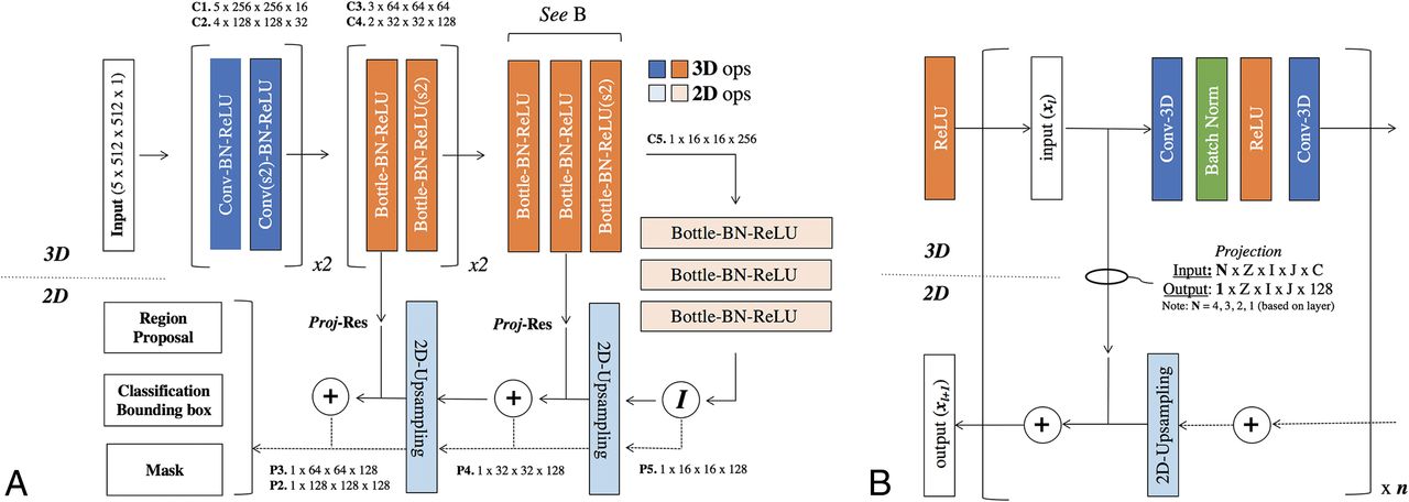
  • Fig 2.
    • Download figure
    • Open in new tab
    • Download powerpoint
    Fig 2.

    Convolutional neural network architecture. A, Hybrid 3D-contracting (bottom-up) and 2D-expanding (top-down) fully convolutional feature-pyramid network architecture used for the mask R-CNN backbone. The architecture incorporates both traditional 3 × 3 filters (blue) as well as bottleneck 1 × 1–3 × 3–1 × 1 modules (orange). The contracting arm is composed of 3D operations and convolutional kernels. Subsampling in the x- and y-directions is implemented via 1 × 2 × 2 strided convolutions (marked by s2). Subsampling in the z-direction is mediated by a 2 × 1 x 1 convolutional kernel with valid padding. The expanding arm is composed entirely of 2D operations. B, Connections between the contracting and expanding arms are facilitated by residual addition operations between corresponding layers. 3D layers in the contracting arm are mapped to 2D layers in the expanding arm by projection operations, which are designed both to match in the input (N) and output (1) z-dimension shape in addition to input (C) and output (128) feature map sizes. Ops indicates operations; Conv, convolutions; BN-ReLU, Batch Normalization Rectified Linear Unit; Proj-Res, Projection-Residual; Z, Z-axis; I, In plane axis; J, In plane axis.

  • Fig 3.
    • Download figure
    • Open in new tab
    • Download powerpoint
    Fig 3.

    Sample network predictions: true-positives. Network predictions by the algorithm include bounding-box region proposals for potential areas of abnormality (to focus algorithm attention) and final network predictions, including confidence of results. Correctly identified areas of hemorrhage (green) include subtle abnormalities representing subarachnoid (A), subdural (B and C), and intraparenchymal (D) hemorrhage. Correctly identified areas of excluded hemorrhage often include common mimics for blood on NCCT, including thickening/high density along the falx (A, C, and D) and beam-hardening along the periphery (B).

  • Fig 4.
    • Download figure
    • Open in new tab
    • Download powerpoint
    Fig 4.

    Sample network predictions: false-positives and false-negatives. Network predictions by the algorithm include bounding-box region proposals for potential areas of abnormality (to focus algorithm attention) and final network predictions including confidence of results. False-positive predictions for hemorrhage (purple) often include areas of motion artifacts and/or posterior fossa beam-hardening (A) or high-density mimics such as cortical calcification (C). False-negative predictions for excluded hemorrhage often include small volume abnormalities with relatively lower density, resulting in decreased conspicuity. Examples include subtle subarachnoid hemorrhage along the posterior right frontal lobe (B) and right inferior parietal lobe (D).

Tables

  • Figures
    • View popup
    Table 1:

    Distribution of hemorrhages by type and sizea

    SizeIPHEDH/SDHSAH
    ValidTestValidTestValidTest
    Large1921318819859
    Medium8887915533
    Small631494526
    Punctate15130343
    Total358233193822421
    • ↵a Large, medium, small, and punctate hemorrhages were defined as >25, 5–25, 0.01–5.0, and <0.01 mL, respectively.

    • View popup
    Table 2:

    Balanced dataset performance statistics stratified by hemorrhage type and sizea

    SizeAccuracyAUCSensitivitySpecificityPPVNPV
    ValidTestValidTestValidTestValidTestValidTestValidTest
    All ICHs0.9840.9720.9910.9890.9710.9510.9750.9730.9750.9720.9710.952
        Large0.9990.9970.9990.9991.0001.0000.9750.9730.9750.9731.0001.000
        Medium0.9920.9770.9950.9820.9860.9620.9750.9730.9750.9720.9860.962
        Small0.9650.9060.9720.9870.9330.8180.9750.9730.9740.9680.9360.843
        Punctate0.8830.8720.8950.9030.7690.7500.9750.9730.9680.9650.8090.796
    IPH0.9920.9970.9960.9990.9861.0000.9750.9730.9750.9730.9861.000
        Large0.9990.9970.9990.9991.0001.0000.9750.9730.9750.9731.0001.000
        Medium0.9990.9970.9990.9991.0001.0000.9750.9730.9750.9731.0001.000
        Small0.9830.9970.9990.9990.9681.0000.9750.9730.9740.9730.9681.000
        Punctate0.8990.9970.9210.9990.8001.0000.9750.9730.9690.9730.8301.000
    EDH/SDH0.9860.9700.9890.9740.9750.9470.9750.9730.9750.9720.9750.949
        Large0.9990.9970.9990.9991.0001.0000.9750.9730.9750.9731.0001.000
        Medium0.9800.9630.9830.9710.9620.9330.9750.9730.9740.9710.9630.936
        Small0.9580.8720.9680.8820.9180.7500.9750.9730.9730.9650.9230.796
        Punctate0.832NA0.857NA0.667NA0.9750.9730.963NA0.745NA
    SAH0.9700.9490.9720.9530.9420.9050.9750.9730.9740.9710.9440.911
        Large0.9990.9970.9990.9991.0001.0000.9750.9730.9750.9731.0001.000
        Medium0.9990.9970.9990.9991.0001.0000.9750.9730.9750.9731.0001.000
        Small0.9500.9130.9600.9280.9040.8330.9750.9730.9730.9680.9100.854
        Punctate0.8810.8300.8910.8330.7650.6670.9750.9730.9680.9610.8060.745
    • Note:—AUC indicates area under the curve; NA, not applicable; PPV, positive predictive value; NPV, negative predictive value.

    • ↵a Large, medium, small, and punctate hemorrhages were defined as >25, 5–25, 0.01–5.0, and <0.01 mL, respectively.

PreviousNext
Back to top

In this issue

American Journal of Neuroradiology: 39 (9)
American Journal of Neuroradiology
Vol. 39, Issue 9
1 Sep 2018
  • Table of Contents
  • Index by author
  • Complete Issue (PDF)
Advertisement
Print
Download PDF
Email Article

Thank you for your interest in spreading the word on American Journal of Neuroradiology.

NOTE: We only request your email address so that the person you are recommending the page to knows that you wanted them to see it, and that it is not junk mail. We do not capture any email address.

Enter multiple addresses on separate lines or separate them with commas.
Hybrid 3D/2D Convolutional Neural Network for Hemorrhage Evaluation on Head CT
(Your Name) has sent you a message from American Journal of Neuroradiology
(Your Name) thought you would like to see the American Journal of Neuroradiology web site.
CAPTCHA
This question is for testing whether or not you are a human visitor and to prevent automated spam submissions.
Cite this article
P.D. Chang, E. Kuoy, J. Grinband, B.D. Weinberg, M. Thompson, R. Homo, J. Chen, H. Abcede, M. Shafie, L. Sugrue, C.G. Filippi, M.-Y. Su, W. Yu, C. Hess, D. Chow
Hybrid 3D/2D Convolutional Neural Network for Hemorrhage Evaluation on Head CT
American Journal of Neuroradiology Sep 2018, 39 (9) 1609-1616; DOI: 10.3174/ajnr.A5742

Citation Manager Formats

  • BibTeX
  • Bookends
  • EasyBib
  • EndNote (tagged)
  • EndNote 8 (xml)
  • Medlars
  • Mendeley
  • Papers
  • RefWorks Tagged
  • Ref Manager
  • RIS
  • Zotero
0 Responses
Respond to this article
Share
Bookmark this article
Hybrid 3D/2D Convolutional Neural Network for Hemorrhage Evaluation on Head CT
P.D. Chang, E. Kuoy, J. Grinband, B.D. Weinberg, M. Thompson, R. Homo, J. Chen, H. Abcede, M. Shafie, L. Sugrue, C.G. Filippi, M.-Y. Su, W. Yu, C. Hess, D. Chow
American Journal of Neuroradiology Sep 2018, 39 (9) 1609-1616; DOI: 10.3174/ajnr.A5742
del.icio.us logo Twitter logo Facebook logo Mendeley logo
  • Tweet Widget
  • Facebook Like
  • Google Plus One
Purchase

Jump to section

  • Article
    • Abstract
    • ABBREVIATIONS:
    • Materials and Methods
    • Results
    • Discussion
    • Conclusions
    • Footnotes
    • References
  • Figures & Data
  • Info & Metrics
  • Responses
  • References
  • PDF

Related Articles

  • No related articles found.
  • PubMed
  • Google Scholar

Cited By...

  • Deep Learning-Based ASPECTS Algorithm Enhances Reader Performance and Reduces Interpretation Time
  • Application of deep learning models for detection of subdural hematoma: a systematic review and meta-analysis
  • Predicting vasospasm risk using first presentation aneurysmal subarachnoid haemorrhage volume: a semi-automated CT image segmentation analysis in ITK-SNAP
  • Labeling Noncontrast Head CT Reports for Common Findings Using Natural Language Processing
  • Artificial Intelligence Assessment of Renal Scarring (AIRS Study)
  • Review of deep learning algorithms for the automatic detection of intracranial hemorrhages on computed tomography head imaging
  • Automated Cerebral Hemorrhage Detection Using RAPID
  • Artificial Intelligence and Acute Stroke Imaging
  • 3D Deep Neural Network Segmentation of Intracerebral Hemorrhage: Development and Validation for Clinical Trials
  • Artificial Intelligence in Neuroradiology: Current Status and Future Directions
  • Expert-level detection of acute intracranial hemorrhage on head computed tomography using deep learning
  • Convolutional Neural Network for Automated FLAIR Lesion Segmentation on Clinical Brain MR Imaging
  • Towards Reproducible Results: Validating CT Hemorrhage-Detection Algorithms on Standard Datasets
  • Crossref
  • Google Scholar

This article has not yet been cited by articles in journals that are participating in Crossref Cited-by Linking.

More in this TOC Section

  • Diagnostic Neuroradiology of Monoclonal Antibodies
  • Clinical Outcomes After Chiari I Decompression
  • Segmentation of Brain Metastases with BLAST
Show more Adult Brain

Similar Articles

Advertisement

Indexed Content

  • Current Issue
  • Accepted Manuscripts
  • Article Preview
  • Past Issues
  • Editorials
  • Editor's Choice
  • Fellows' Journal Club
  • Letters to the Editor
  • Video Articles

Cases

  • Case Collection
  • Archive - Case of the Week
  • Archive - Case of the Month
  • Archive - Classic Case

More from AJNR

  • Trainee Corner
  • Imaging Protocols
  • MRI Safety Corner
  • Book Reviews

Multimedia

  • AJNR Podcasts
  • AJNR Scantastics

Resources

  • Turnaround Time
  • Submit a Manuscript
  • Submit a Video Article
  • Submit an eLetter to the Editor/Response
  • Manuscript Submission Guidelines
  • Statistical Tips
  • Fast Publishing of Accepted Manuscripts
  • Graphical Abstract Preparation
  • Imaging Protocol Submission
  • Evidence-Based Medicine Level Guide
  • Publishing Checklists
  • Author Policies
  • Become a Reviewer/Academy of Reviewers
  • News and Updates

About Us

  • About AJNR
  • Editorial Board
  • Editorial Board Alumni
  • Alerts
  • Permissions
  • Not an AJNR Subscriber? Join Now
  • Advertise with Us
  • Librarian Resources
  • Feedback
  • Terms and Conditions
  • AJNR Editorial Board Alumni

American Society of Neuroradiology

  • Not an ASNR Member? Join Now

© 2025 by the American Society of Neuroradiology All rights, including for text and data mining, AI training, and similar technologies, are reserved.
Print ISSN: 0195-6108 Online ISSN: 1936-959X

Powered by HighWire