
Article Figures & Data
Figures
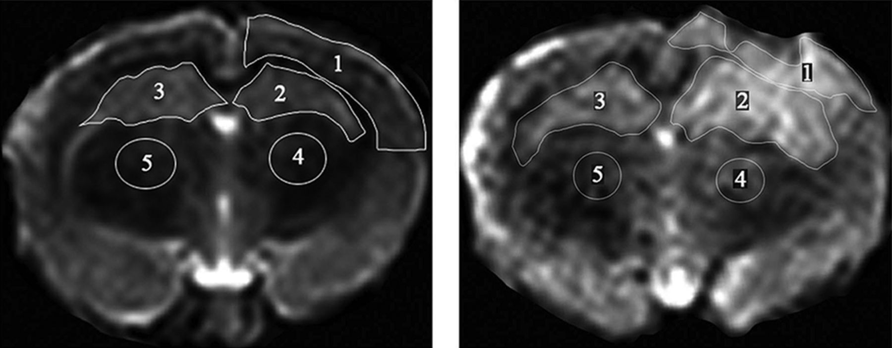
- Fig 1.
T2-weighted MR imaging displaying the manually outlined ROI. Left, Sham group ROIs: all ROIs correspond to ROIs as identified in the TBI group. Right, TBI group ROIs: ROI 1, ipsilateral cortex; ROI 2, ipsilateral hippocampus; ROI 3, contralateral hippocampus; ROI 4, ipsilateral thalamus; and ROI 5, contralateral thalamus.
- Fig 2.
MR imaging from a representative animal in the PTE, non-PTE, and sham groups. The Ktrans values of the PTE and non-PTE groups were higher than those of the sham operation group. In addition, the Ktrans values of the corresponding ROI in the PTE group were higher than those in the non-PTE group.
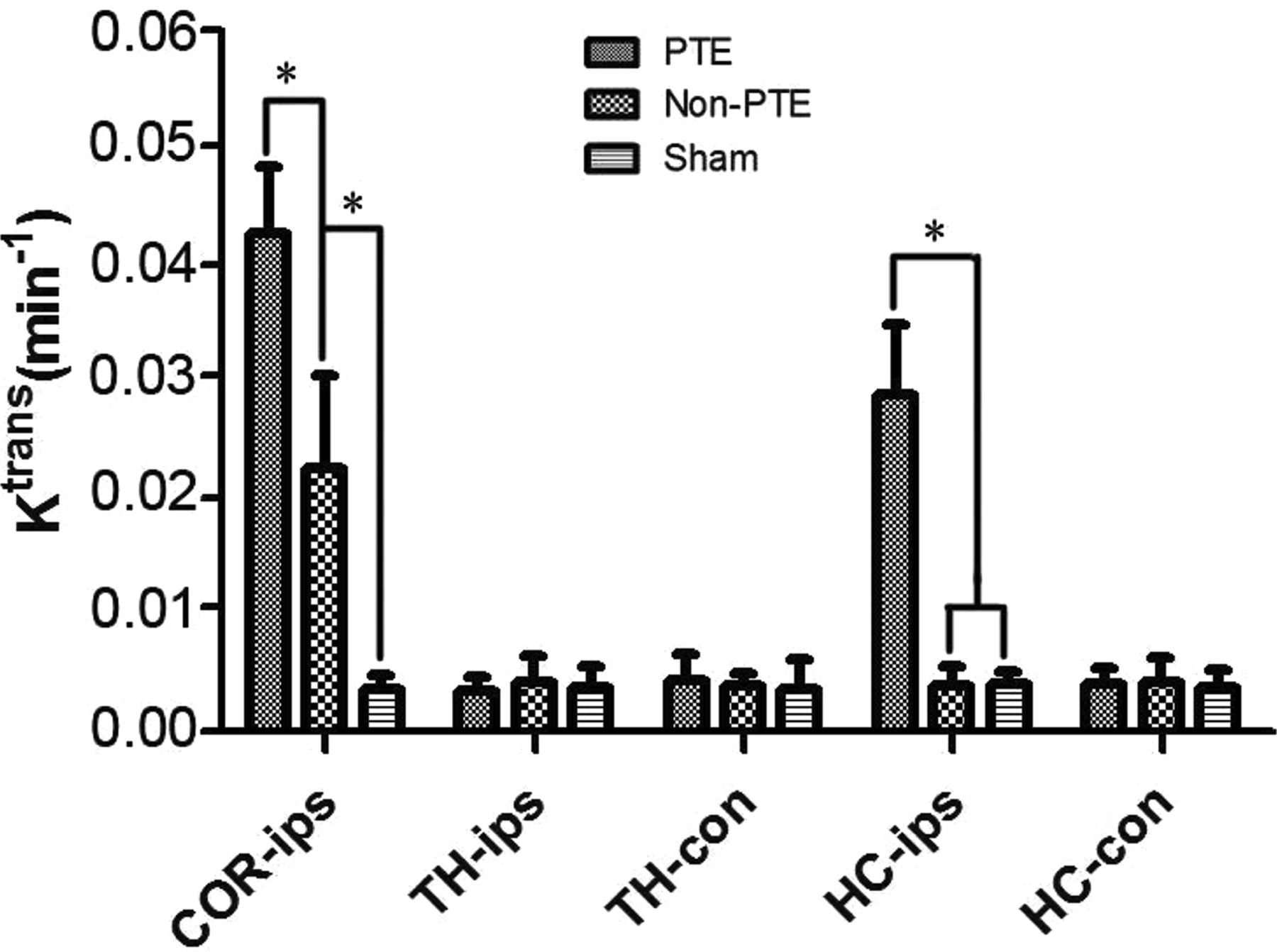
- Fig 3.
Comparison of Ktrans in all ROIs among all study groups. The asterisk indicates comparison between the 2 groups, P < .01; COR-ips, ipsilateral cortex; TH-ips, ipsilateral thalamus; TH-con, contralateral thalamus; HC-ips, ipsilateral hippocampus; HC-con, contralateral hippocampus.
- Fig 4.
Comparison of MK in all ROIs among all study groups. The asterisk indicates comparison between the 2 groups, P < .01; COR-ips, ipsilateral cortex; TH-ips, ipsilateral thalamus; TH-con, contralateral thalamus; HC-ips, ipsilateral hippocampus; HC-con, contralateral hippocampus.
- Fig 5.
Comparison of the number of cell bodies in the contralateral hippocampus: 1) sham group (100×). 2) Non-PTE group (100×). 3) PTE group (100×). The number of cell bodies in the contralateral hippocampus in the PTE group was lower than that in the non-PTE and sham groups. The number of cell bodies in the non-PTE group was also lower than that in the sham group.


Jump to section
Related Articles
Cited By...
- No citing articles found.