Advertisement

Main menu

  • Home
  • Content
    • Current Issue
    • Accepted Manuscripts
    • Article Preview
    • Past Issue Archive
    • Video Articles
    • AJNR Case Collection
    • Case of the Week Archive
    • Case of the Month Archive
    • Classic Case Archive
  • Special Collections
    • AJNR Awards
    • Low-Field MRI
    • Alzheimer Disease
    • ASNR Foundation Special Collection
    • Photon-Counting CT
    • View All
  • Multimedia
    • AJNR Podcasts
    • AJNR SCANtastic
    • Trainee Corner
    • MRI Safety Corner
    • Imaging Protocols
  • For Authors
    • Submit a Manuscript
    • Submit a Video Article
    • Submit an eLetter to the Editor/Response
    • Manuscript Submission Guidelines
    • Statistical Tips
    • Fast Publishing of Accepted Manuscripts
    • Graphical Abstract Preparation
    • Imaging Protocol Submission
    • Author Policies
  • About Us
    • About AJNR
    • Editorial Board
    • Editorial Board Alumni
  • More
    • Become a Reviewer/Academy of Reviewers
    • Subscribers
    • Permissions
    • Alerts
    • Feedback
    • Advertisers
    • ASNR Home

User menu

  • Alerts
  • Log in

Search

  • Advanced search
American Journal of Neuroradiology

American Journal of Neuroradiology

ASHNR American Society of Functional Neuroradiology ASHNR American Society of Pediatric Neuroradiology ASSR
  • Alerts
  • Log in

Advanced Search

  • Home
  • Content
    • Current Issue
    • Accepted Manuscripts
    • Article Preview
    • Past Issue Archive
    • Video Articles
    • AJNR Case Collection
    • Case of the Week Archive
    • Case of the Month Archive
    • Classic Case Archive
  • Special Collections
    • AJNR Awards
    • Low-Field MRI
    • Alzheimer Disease
    • ASNR Foundation Special Collection
    • Photon-Counting CT
    • View All
  • Multimedia
    • AJNR Podcasts
    • AJNR SCANtastic
    • Trainee Corner
    • MRI Safety Corner
    • Imaging Protocols
  • For Authors
    • Submit a Manuscript
    • Submit a Video Article
    • Submit an eLetter to the Editor/Response
    • Manuscript Submission Guidelines
    • Statistical Tips
    • Fast Publishing of Accepted Manuscripts
    • Graphical Abstract Preparation
    • Imaging Protocol Submission
    • Author Policies
  • About Us
    • About AJNR
    • Editorial Board
    • Editorial Board Alumni
  • More
    • Become a Reviewer/Academy of Reviewers
    • Subscribers
    • Permissions
    • Alerts
    • Feedback
    • Advertisers
    • ASNR Home
  • Follow AJNR on Twitter
  • Visit AJNR on Facebook
  • Follow AJNR on Instagram
  • Join AJNR on LinkedIn
  • RSS Feeds

AJNR Awards, New Junior Editors, and more. Read the latest AJNR updates

Research ArticleHead and Neck Imaging

MRI with DWI for the Detection of Posttreatment Head and Neck Squamous Cell Carcinoma: Why Morphologic MRI Criteria Matter

A. Ailianou, P. Mundada, T. De Perrot, M. Pusztaszieri, P.-A. Poletti and M. Becker
American Journal of Neuroradiology April 2018, 39 (4) 748-755; DOI: https://doi.org/10.3174/ajnr.A5548
A. Ailianou
aFrom the Division of Radiology (A.A., P.M., T.D.P., P.-A.P., M.B.), Department of Imaging and Medical Informatics
  • Find this author on Google Scholar
  • Find this author on PubMed
  • Search for this author on this site
  • ORCID record for A. Ailianou
P. Mundada
aFrom the Division of Radiology (A.A., P.M., T.D.P., P.-A.P., M.B.), Department of Imaging and Medical Informatics
  • Find this author on Google Scholar
  • Find this author on PubMed
  • Search for this author on this site
  • ORCID record for P. Mundada
T. De Perrot
aFrom the Division of Radiology (A.A., P.M., T.D.P., P.-A.P., M.B.), Department of Imaging and Medical Informatics
  • Find this author on Google Scholar
  • Find this author on PubMed
  • Search for this author on this site
  • ORCID record for T. De Perrot
M. Pusztaszieri
bDivision of Clinical Pathology (M.P.), Department of Laboratory and Genetics, Geneva University Hospitals, University of Geneva, Geneva, Switzerland.
  • Find this author on Google Scholar
  • Find this author on PubMed
  • Search for this author on this site
  • ORCID record for M. Pusztaszieri
P.-A. Poletti
aFrom the Division of Radiology (A.A., P.M., T.D.P., P.-A.P., M.B.), Department of Imaging and Medical Informatics
  • Find this author on Google Scholar
  • Find this author on PubMed
  • Search for this author on this site
  • ORCID record for P.-A. Poletti
M. Becker
aFrom the Division of Radiology (A.A., P.M., T.D.P., P.-A.P., M.B.), Department of Imaging and Medical Informatics
  • Find this author on Google Scholar
  • Find this author on PubMed
  • Search for this author on this site
  • ORCID record for M. Becker
  • Article
  • Figures & Data
  • Supplemental
  • Info & Metrics
  • Responses
  • References
  • PDF
Loading

Article Figures & Data

Figures

  • Tables
  • Fig 1.
    • Download figure
    • Open in new tab
    • Download powerpoint
    Fig 1.

    DWIMRI obtained 23 months after RTH for laryngeal squamous cell carcinoma and increasing hoarseness. Axial T2 (A), T1 (B), and contrast-enhanced T1 (C) show an oval lesion (arrows) in the left false cord and left paraglottic space with intermediate signal intensity on T2, low signal intensity on T1, and moderate contrast enhancement highly suspicious for rHNSCC. The b=1000 image (D) shows high signal intensity in the lesion. ADC map (E) reveals low signal intensity compatible with restricted diffusion (arrow), further suggesting recurrence (ADCmean = 0.798 × 10⁻³ mm2/s). Endoscopic biopsy confirmed rHNSCC. F, Histology (H&E, original magnification ×100) shows squamous cell carcinoma with areas of densely packed and loosely packed squamous cells of variable size with keratin pearls (asterisk).

  • Fig 2.
    • Download figure
    • Open in new tab
    • Download powerpoint
    Fig 2.

    DWIMRI obtained 13 months after RTH and neck dissection for squamous cell carcinoma of the larynx and oropharynx. The patient had massive weight loss, malnutrition, and recurrent aspiration pneumonia. Endoscopy showed intact mucosa and fixed vocal cords bilaterally. Axial T2 (A), T1 (B), and contrast-enhanced T1 (C) show a triangular lesion (arrows) in the left true vocal cord with very low signal intensity on T2, low signal intensity on T1, and faint contrast enhancement suggesting post-RTH late fibrosis. In contrast, the right vocal cord (dashed arrows) displays high signal intensity on T2, low signal on T1, and enhancement. Findings on the right were interpreted as suggesting inflammatory edema. The b=1000 image (D) and ADC map (E) reveal no restricted diffusion in the right vocal cord (ADCmean = 1.643 × 10⁻³ mm2/s) and restricted diffusion with low ADC in the left vocal cord (ADCmean = 1.006 × 10⁻³ mm2/s). Because the nonfunctional larynx was the cause of malnutrition and recurrent aspiration pneumonia, laryngectomy was performed. F, Corresponding whole-organ histologic slice (H&E) shows extensive muscle fibrosis on the left (arrows) and inflammatory edema with denervation on the right (dashed arrows).

  • Fig 3.
    • Download figure
    • Open in new tab
    • Download powerpoint
    Fig 3.

    DWIMRI obtained 3 months after RTH and bucopharyngectomy for squamous cell carcinoma of the retromolar trigone. The patient had right reflex otalgia and progressing trismus. Endoscopy could not be performed. Axial T2 (A) and coronal STIR (B) images show a triangular, elongated, strongly hypointense lesion (arrows) on the right. There was no contrast enhancement (not shown). The diagnosis of benign post-RTH late fibrosis was made. The b=1000 image (C) reveals low signal. ADC map (D) likewise shows low signal (ADCmean = 0.731 × 10⁻³ mm2/s). Follow-up at 38 months (not shown) showed no recurrence but progressive scar retraction on MRI.
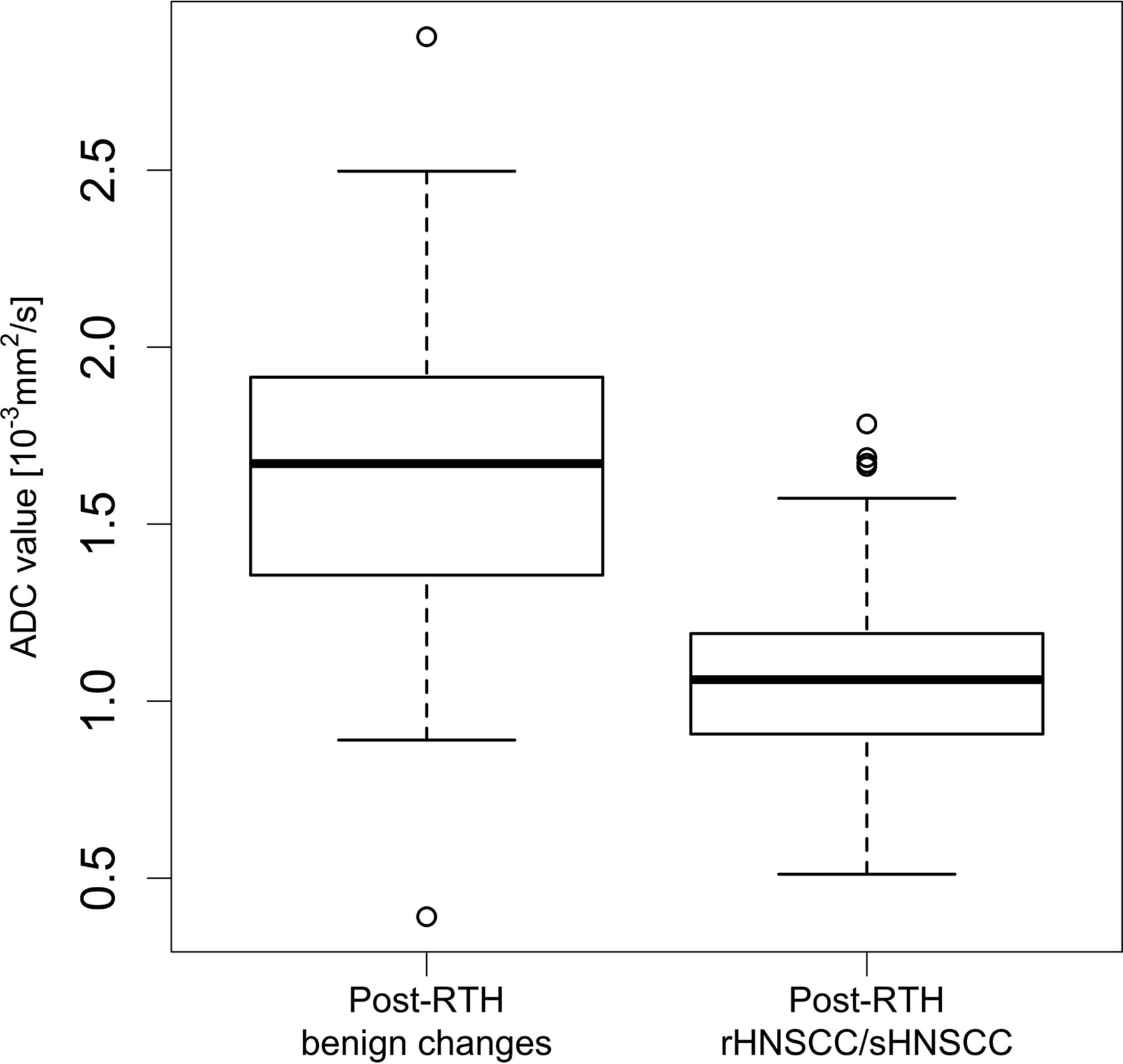
  • Fig 4.
    • Download figure
    • Open in new tab
    • Download powerpoint
    Fig 4.

    Box-and-whisker plots of ADCmean values in patients with post-RTH changes and post-RTH tumors. The horizontal lines represent the median values, and the bottom and the top of the box represent the 25th and 75th quartiles, respectively. Median ADCmean (25th–75th quartiles) for rHNSCC/sHNSCC = 1.061 (0.907–1.191) × 10−3 mm2/s. Median ADCmean (25th–75th quartiles) for post-RTH changes (late fibrosis and inflammatory edema together) = 1.671 (1.3355–1.915) × 10−3 mm2/s.

  • Fig 5.
    • Download figure
    • Open in new tab
    • Download powerpoint
    Fig 5.

    Box-and-whisker plots of ADCmean values in patients with post-RTH inflammatory edema, late fibrosis, and post-RTH HNSCCs. The horizontal lines represent the median values; the bottom and the top of the box represent the 25th and 75th quartiles, respectively. Median ADCmean (25th–75th quartiles) for rHNSCC/sHNSCC = 1.061 (0.907–1.191) × 10−3 mm2/s. Median ADCmean (25th–75th quartiles) for post-RTH inflammatory edema = 1.764 (1.575–1.938) × 10−3 mm2/s. Median ADCmean (25th–75th quartiles) for late fibrosis/mature scar post-RTH = 1.068 (0.939–1.152) × 10−3 mm2/s. There was no statistically significant difference between ADCmean in late fibrosis and rHNSCC/sHNSCC (P > .05). However, there was a significant difference between ADCmean in inflammatory post-RTH edema and late fibrosis (P < .05).
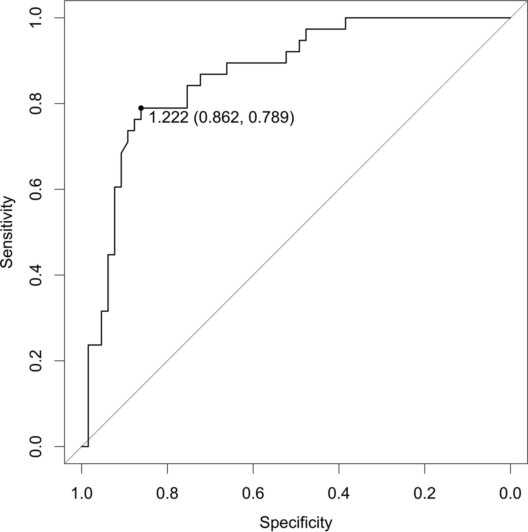
  • Fig 6.
    • Download figure
    • Open in new tab
    • Download powerpoint
    Fig 6.

    Receiver operating characteristic curve for the quantitative analysis of ADCmean values showing the area under the curve of 0.8678. A threshold of ADC = 1.222 × 10⁻³ mm2/s was found (see description in the text). This threshold yielded a sensitivity of 78.9%, a specificity of 86.2%, and an accuracy of 83.5%.

Tables

  • Figures
    • View popup
    Table 1:

    Patient and tumor characteristics

    Primary HNSCCrHNSCC and sHNSSC after Treatment
    Total No. of patients100
    Total No. of tumors10338a
    Female (No.) (%)22 (22%)
    Male (No.) (%)78 (78%)
    Mean age (yr)59.3 ± 11.361.5 ± 11.1
    Treatment modalities in pHNSCC (No. of patients)NA
        RTH alone52
        Operation followed by RTH48
        Median interval (quartile 1–quartile 3) between end of RTH and rHNSCC/sHNSCC (mo)NA14 (4.5–51)
    Tumor location (No. of tumor sites) (%)
        Nasopharynx7 (6.8%)1 (2.6%)
        Oral cavity22 (21.3%)13 (34.2%)
        Oropharynx38 (36.9%)8 (21.1%)
        Hypopharynx12 (11.6%)5 (13.1%)
        Larynx19 (18.5%)6 (15.8%)
        Paranasal sinuses4 (3.9%)2 (5.3%)
        Base of the skull0 (0%)3 (7.9%)
        Unknown primary tumor1 (1%)0 (0%)
    T classificationb
        Tx1 (1%)0 (0%)
        Tis1 (1%)1 (2.6%)
        T114 (13.6%)3 (7.9%)
        T230 (29.1%)7 (18.4%)
        T325 (24.3%)6 (15.8%)
        T432 (31.0%)21 (55.3%)
    N classificationb
        N031 (30.1%)26 (68.4%)
        N120 (19.4%)5 (13.2%)
        N245 (43.7%)6 (15.8%)
        N36 (5.8%)1 (2.6%)
        Nx1 (1%)0 (0%)
    M classificationb
        Mx7 (6.8%)0 (0%)
        M094 (91.3%)36 (94.7%)
        M12 (1.9%)2 (5.3%)
    • Note:—NA indicates not applicable.

    • ↵a Thirty-three rHNSCCs and 5 sHNSCCs.

    • ↵b Tumor, Node, Metastasis (TNM) classification according to Union for International Cancer Control 2016.28

    • View popup
    Table 2:

    Diagnostic performance of morphologic MRI alone, quantitative DWI alone, qualitative DWIMRI, and quantitative DWIMRI with ADCmean < 1.22 × 10⁻³ mm2/s

    Morphologic MRIQuantitative DWI with ADCmean < 1.22Morphologic MRI with Qualitative DWIMorphologic MRI with Quantitative DWI (ADCmean < 1.22)
    TP (No.)30303435
    TN (No.)52565962
    FP (No.)13963
    FN (No.)8843
    Sensitivity (%)a78.978.989.492.1
        (95% CI)(65.9–91.9)(65.9–91.9)(79.7–99.2)(83.5–100.0)
    Specificity (%)b80.086.190.895.4
        (95% CI)(70.3–89.7)(74.8–93.1)(83.7–97.8)(90.3–100.0)
    PPV (%)69.776.985.092.1
        (95% CI)(56.1–83.5)(60.3–88.3)(73.9–96.1)(83.5–100.0)
    NPV (%)86.687.593.695.4
        (95% CI)(78.1–95.3)(76.3–94.1)(87.6–99.6)(90.2–100.0)
    LR+c3.945.709.6919.9
        (95% CI)(2.36–6.59)(3.04–10.68)(4.48–20.9)(6.58–60.5)
    LR−d0.260.240.110.08
        (95% CI)(0.14–0.49)(0.13–0.45)(0.04–0.29)(0.03–0.24)
    • Note:—TP indicates true-positive; TN, true-negative; FP, false-positive; FN, false-negative; PPV, positive predictive value; NPV, negative predictive value.

    • ↵a Comparison of sensitivities: MRI vs DWI: P = 1; MRI vs qualitative DWIMRI: P = .10; MRI vs quantitative DWIMRI: P = .025; qualitative DWIMRI vs quantitative DWIMRI: P = .31; DWI vs quantitative DWIMRI: P = .05.

    • ↵b Comparison of specificities: MRI vs DWI: P = .34; MRI vs qualitative DWIMRI: P = .05; MRI vs quantitative DWIMRI: P = .004; qualitative DWIMRI vs quantitative DWIMRI: P = .18; DWI vs quantitative DWIMRI: P = .03.

    • ↵c Comparison of LR+: MRI vs DWI: P = .36; MRI vs qualitative DWIMRI: P = .03; MRI vs quantitative DWIMRI: P = .004; qualitative DWIMRI vs quantitative DWIMRI: P = .17; DWI vs quantitative DWIMRI: P = .02.

    • ↵d Comparison of LR−: MRI vs DWI: P = .85; MRI vs qualitative DWIMRI: P = .06; MRI vs quantitative DWIMRI: P = .01; qualitative DWIMRI vs quantitative DWIMRI: P = .24; DWI vs quantitative DWIMRI: P = .04.

PreviousNext
Back to top

In this issue

American Journal of Neuroradiology: 39 (4)
American Journal of Neuroradiology
Vol. 39, Issue 4
1 Apr 2018
  • Table of Contents
  • Index by author
  • Complete Issue (PDF)
Advertisement
Print
Download PDF
Email Article

Thank you for your interest in spreading the word on American Journal of Neuroradiology.

NOTE: We only request your email address so that the person you are recommending the page to knows that you wanted them to see it, and that it is not junk mail. We do not capture any email address.

Enter multiple addresses on separate lines or separate them with commas.
MRI with DWI for the Detection of Posttreatment Head and Neck Squamous Cell Carcinoma: Why Morphologic MRI Criteria Matter
(Your Name) has sent you a message from American Journal of Neuroradiology
(Your Name) thought you would like to see the American Journal of Neuroradiology web site.
CAPTCHA
This question is for testing whether or not you are a human visitor and to prevent automated spam submissions.
Cite this article
A. Ailianou, P. Mundada, T. De Perrot, M. Pusztaszieri, P.-A. Poletti, M. Becker
MRI with DWI for the Detection of Posttreatment Head and Neck Squamous Cell Carcinoma: Why Morphologic MRI Criteria Matter
American Journal of Neuroradiology Apr 2018, 39 (4) 748-755; DOI: 10.3174/ajnr.A5548

Citation Manager Formats

  • BibTeX
  • Bookends
  • EasyBib
  • EndNote (tagged)
  • EndNote 8 (xml)
  • Medlars
  • Mendeley
  • Papers
  • RefWorks Tagged
  • Ref Manager
  • RIS
  • Zotero
0 Responses
Respond to this article
Share
Bookmark this article
MRI with DWI for the Detection of Posttreatment Head and Neck Squamous Cell Carcinoma: Why Morphologic MRI Criteria Matter
A. Ailianou, P. Mundada, T. De Perrot, M. Pusztaszieri, P.-A. Poletti, M. Becker
American Journal of Neuroradiology Apr 2018, 39 (4) 748-755; DOI: 10.3174/ajnr.A5548
del.icio.us logo Twitter logo Facebook logo Mendeley logo
  • Tweet Widget
  • Facebook Like
  • Google Plus One
Purchase

Jump to section

  • Article
    • Abstract
    • ABBREVIATIONS:
    • Materials and Methods
    • Results
    • Discussion
    • Conclusions
    • References
  • Figures & Data
  • Supplemental
  • Info & Metrics
  • Responses
  • References
  • PDF

Related Articles

  • PubMed
  • Google Scholar

Cited By...

  • Normalized Parameters of Dynamic Contrast-Enhanced Perfusion MRI and DWI-ADC for Differentiation between Posttreatment Changes and Recurrence in Head and Neck Cancer
  • Adding MR Diffusion Imaging and T2 Signal Intensity to Neck Imaging Reporting and Data System Categories 2 and 3 in Primary Sites of Postsurgical Oral Cavity Carcinoma Provides Incremental Diagnostic Value
  • ADC for Differentiation between Posttreatment Changes and Recurrence in Head and Neck Cancer: A Systematic Review and Meta-analysis
  • MRI Posttreatment Surveillance for Head and Neck Squamous Cell Carcinoma: Proposed MR NI-RADS Criteria
  • Detection of Local Recurrence in Patients with Head and Neck Squamous Cell Carcinoma Using Voxel-Based Color Maps of Initial and Final Area under the Curve Values Derived from DCE-MRI
  • Crossref (33)
  • Google Scholar

This article has been cited by the following articles in journals that are participating in Crossref Cited-by Linking.

  • Recent advances in MRI of the head and neck, skull base and cranial nerves: new and evolving sequences, analyses and clinical applications
    Philip Touska, Steve E. J. Connor
    The British Journal of Radiology 2019 92 1104
  • Apparent diffusion coefficient (ADC) histogram analysis: differentiation of benign from malignant parotid gland tumors using readout-segmented diffusion-weighted imaging
    Zanxia Zhang, Chengru Song, Yong Zhang, Baohong Wen, Jinxia Zhu, Jingliang Cheng
    Dentomaxillofacial Radiology 2019 48 7
  • MR Imaging of Laryngeal and Hypopharyngeal Cancer
    Minerva Becker, Yann Monnier, Claudio de Vito
    Magnetic Resonance Imaging Clinics of North America 2022 30 1
  • Normalized Parameters of Dynamic Contrast-Enhanced Perfusion MRI and DWI-ADC for Differentiation between Posttreatment Changes and Recurrence in Head and Neck Cancer
    A. Baba, R. Kurokawa, E. Rawie, M. Kurokawa, Y. Ota, A. Srinivasan
    American Journal of Neuroradiology 2022 43 8
  • Prediction of Response to Neoadjuvant Chemoradiotherapy by MRI-Based Machine Learning Texture Analysis in Rectal Cancer Patients
    Sajad P. Shayesteh, Afsaneh Alikhassi, Farshid Farhan, Reza Gahletaki, Masume Soltanabadi, Peiman Haddad, Ahmad Bitarafan-Rajabi
    Journal of Gastrointestinal Cancer 2020 51 2
  • Detection of Local Recurrence in Patients with Head and Neck Squamous Cell Carcinoma Using Voxel-Based Color Maps of Initial and Final Area under the Curve Values Derived from DCE-MRI
    J.Y. Lee, K.L. Cheng, J.H. Lee, Y.J. Choi, H.W. Kim, Y.S. Sung, S.R. Chung, K.H. Ryu, M.S. Chung, S.Y. Kim, S.-W. Lee, J.H. Baek
    American Journal of Neuroradiology 2019 40 8
  • MRI Posttreatment Surveillance for Head and Neck Squamous Cell Carcinoma: Proposed MR NI-RADS Criteria
    M.M. Ashour, E.A.F. Darwish, R.M. Fahiem, T.T. Abdelaziz
    American Journal of Neuroradiology 2021 42 6
  • ADC for Differentiation between Posttreatment Changes and Recurrence in Head and Neck Cancer: A Systematic Review and Meta-analysis
    A. Baba, R. Kurokawa, M. Kurokawa, O. Hassan, Y. Ota, A. Srinivasan
    American Journal of Neuroradiology 2022 43 3
  • Extra-nodal extension in head and neck cancer: how radiologists can help staging and treatment planning
    Takashi Hiyama, Hirofumi Kuno, Takahiko Nagaki, Kotaro Sekiya, Shioto Oda, Satoshi Fujii, Ryuichi Hayashi, Tatsushi Kobayashi
    Japanese Journal of Radiology 2020 38 6
  • The ability of post-chemoradiotherapy DWI ADCmean and 18F-FDG SUVmax to predict treatment outcomes in head and neck cancer: impact of human papilloma virus oropharyngeal cancer status
    S. Connor, C. Sit, M. Anjari, M. Lei, T. Guerrero-Urbano, T. Szyszko, G. Cook, P. Bassett, V. Goh
    Journal of Cancer Research and Clinical Oncology 2021 147 8

More in this TOC Section

  • ASL Sensitivity for Head and Neck Paraganglioma
  • Post SRS Peritumoral Hyperintense Signal of VSs
  • Contrast Enhanced Pituitary CISS/FIESTA
Show more Head and Neck Imaging

Similar Articles

Advertisement

Indexed Content

  • Current Issue
  • Accepted Manuscripts
  • Article Preview
  • Past Issues
  • Editorials
  • Editor's Choice
  • Fellows' Journal Club
  • Letters to the Editor
  • Video Articles

Cases

  • Case Collection
  • Archive - Case of the Week
  • Archive - Case of the Month
  • Archive - Classic Case

More from AJNR

  • Trainee Corner
  • Imaging Protocols
  • MRI Safety Corner
  • Book Reviews

Multimedia

  • AJNR Podcasts
  • AJNR Scantastics

Resources

  • Turnaround Time
  • Submit a Manuscript
  • Submit a Video Article
  • Submit an eLetter to the Editor/Response
  • Manuscript Submission Guidelines
  • Statistical Tips
  • Fast Publishing of Accepted Manuscripts
  • Graphical Abstract Preparation
  • Imaging Protocol Submission
  • Evidence-Based Medicine Level Guide
  • Publishing Checklists
  • Author Policies
  • Become a Reviewer/Academy of Reviewers
  • News and Updates

About Us

  • About AJNR
  • Editorial Board
  • Editorial Board Alumni
  • Alerts
  • Permissions
  • Not an AJNR Subscriber? Join Now
  • Advertise with Us
  • Librarian Resources
  • Feedback
  • Terms and Conditions
  • AJNR Editorial Board Alumni

American Society of Neuroradiology

  • Not an ASNR Member? Join Now

© 2025 by the American Society of Neuroradiology All rights, including for text and data mining, AI training, and similar technologies, are reserved.
Print ISSN: 0195-6108 Online ISSN: 1936-959X

Powered by HighWire