
Article Figures & Data
Figures
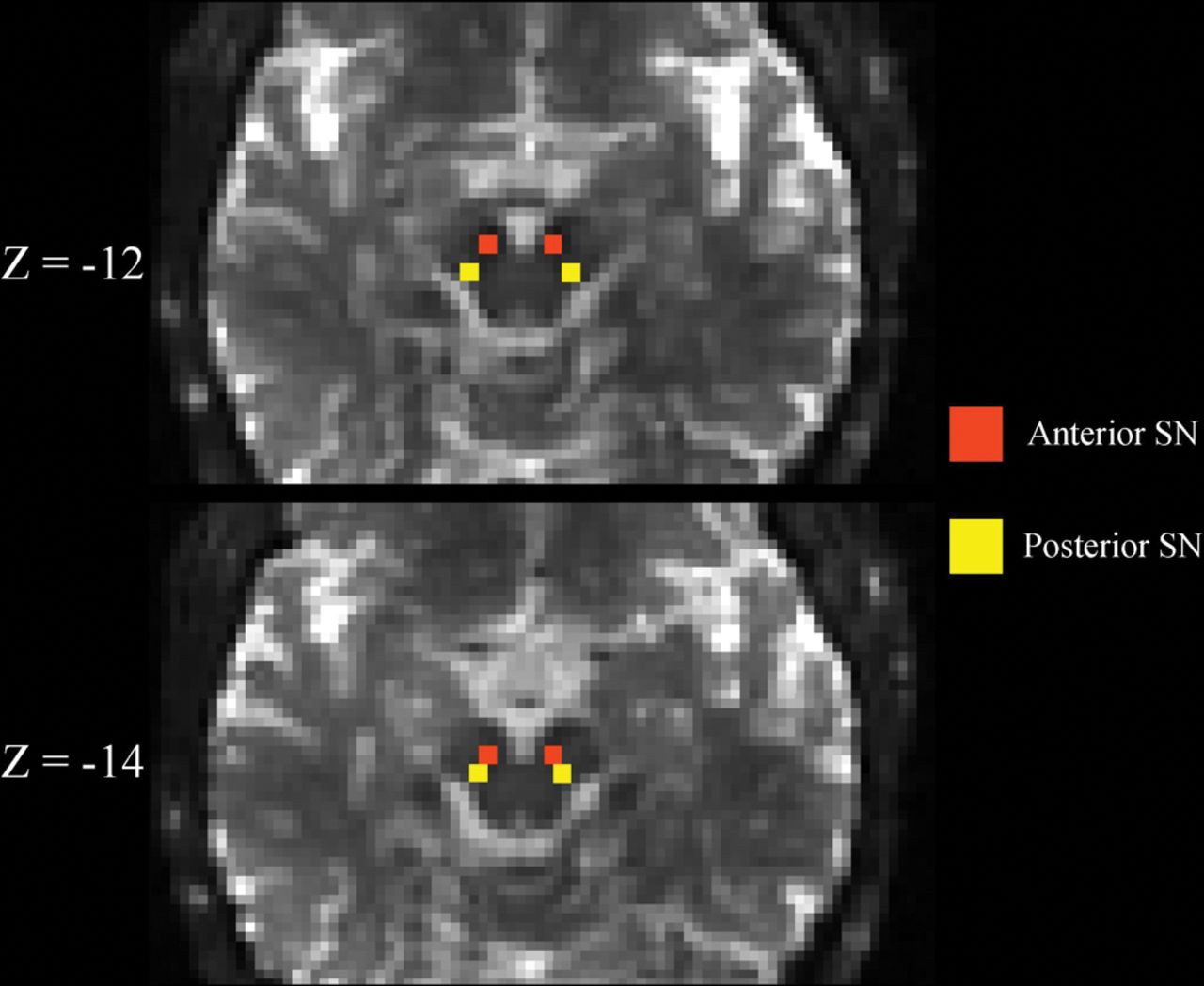
- Fig 1.
Substantia nigra ROIs. Representative b=0 s/mm2 images linearly registered to the MNI atlas are shown demonstrating the placement of the substantia nigra ROIs. Z-coordinates refer to the slice shown in MNI space. See the “Materials and Methods” section for a more detailed description of how ROIs were placed.
- Fig 2.
Longitudinal changes in SN FW in patients with later-stage IPD and HCs. aSN and pSN FW group and group × time interaction intergroup differences were P < .001, = 0.021, and P = .014, = 0.651, respectively. Follow-up was approximately 3 years from baseline. Error bars are ± 1 standard error of mean.
- Fig 3.
Change in the in bi-temporal lobe white matter FW-corrected mean diffusivity of subjects with IPD and corresponding changes in the MMSE and MoCA scores over 3 years. R2 indicates coefficient of determination.


Tables
HC (n = 19) IPD (n = 19) Between Groups BL F/U BL F/U P Value within BL P Value within F/U Age (yr) 56.5 (10.1) 59.6 (10.3) 59.8 (8.4) 63.2 (8.4) .276 .251 Male sex (No.) (%) 5 (26) – 12 (63) – .022 – Disease duration (yr) – – 7.1 (5.1) 10.4 (5.3) – – Time to F/U (mo) – 36.2 (5.4) – 43.8 (7.8) – .001 MMSE score 29.4 (0.9) 29.5 (0.9) 29.4 (1.0) 27.6 (4.8) .586 .862 MoCA score 27.3 (2.4) 27.9 (2.1) 24.4 (4.2) 24.3 (6.9) .095 .046 CDR score (mean, median) (IQR) 0, 0 (0–0) 0.1, 0 (0–0) 0.2, 0 (0–0.5) 0.3, 0 (0–0.5) .053 .317 UPDRS-III score 0.8 (1.2) 1.3 (2.3) 18.8 (7.3) 22.1 (9.2) <.001 <.001 H&Y (mean, median) (IQR) 0, 0 (0–0) 0, 0 (0) 1.9, 2 (1–2.5) 2.4, 2.5 (2–3) <.001 <.001 S&E score 100 (0) 100 (0) 91.3 (9.3) 79.5 (21.8) .001 .001 GDS score 1.9 (2.9) 1.2 (1.9) 2.3 (2.1) 5.2 (7.9) .615 .044 Note:—H&Y indicates Hoehn and Yahr scale; S&E, Schwab and England Activities of Daily Living Scale; GDS, Geriatric Depression Scale; IQR, interquartile range; BL, baseline; F/U, follow up; CDR, Clinical Dementia Rating; UPDRS III, Unified Parkinson's Disease Rating Scale, Part III.
↵a Results are presented as mean (SD), unless otherwise noted.
HC (n = 19) IPD (n = 19) Group Effect Time Effect Interaction Baseline Follow-Up Baseline Follow-Up aSN FW .2301 (.0243) .2361 (.0351) .2713 (.0535) .2989 (.0364) <.001 .112 .021 pSN FW .2221 (.0349) .2452 (.0367) .2691 (.0513) .2955 (.0441) .014 .246 .651 ↵a Values are mean (SD). Group, time, and group × time interaction effect values are P values.


