Advertisement

Main menu

  • Home
  • Content
    • Current Issue
    • Accepted Manuscripts
    • Article Preview
    • Past Issue Archive
    • Video Articles
    • AJNR Case Collection
    • Case of the Week Archive
    • Case of the Month Archive
    • Classic Case Archive
  • Special Collections
    • AJNR Awards
    • Low-Field MRI
    • Alzheimer Disease
    • ASNR Foundation Special Collection
    • Photon-Counting CT
    • View All
  • Multimedia
    • AJNR Podcasts
    • AJNR SCANtastic
    • Trainee Corner
    • MRI Safety Corner
    • Imaging Protocols
  • For Authors
    • Submit a Manuscript
    • Submit a Video Article
    • Submit an eLetter to the Editor/Response
    • Manuscript Submission Guidelines
    • Statistical Tips
    • Fast Publishing of Accepted Manuscripts
    • Graphical Abstract Preparation
    • Imaging Protocol Submission
    • Author Policies
  • About Us
    • About AJNR
    • Editorial Board
    • Editorial Board Alumni
  • More
    • Become a Reviewer/Academy of Reviewers
    • Subscribers
    • Permissions
    • Alerts
    • Feedback
    • Advertisers
    • ASNR Home

User menu

  • Alerts
  • Log in

Search

  • Advanced search
American Journal of Neuroradiology

American Journal of Neuroradiology

ASHNR American Society of Functional Neuroradiology ASHNR American Society of Pediatric Neuroradiology ASSR
  • Alerts
  • Log in

Advanced Search

  • Home
  • Content
    • Current Issue
    • Accepted Manuscripts
    • Article Preview
    • Past Issue Archive
    • Video Articles
    • AJNR Case Collection
    • Case of the Week Archive
    • Case of the Month Archive
    • Classic Case Archive
  • Special Collections
    • AJNR Awards
    • Low-Field MRI
    • Alzheimer Disease
    • ASNR Foundation Special Collection
    • Photon-Counting CT
    • View All
  • Multimedia
    • AJNR Podcasts
    • AJNR SCANtastic
    • Trainee Corner
    • MRI Safety Corner
    • Imaging Protocols
  • For Authors
    • Submit a Manuscript
    • Submit a Video Article
    • Submit an eLetter to the Editor/Response
    • Manuscript Submission Guidelines
    • Statistical Tips
    • Fast Publishing of Accepted Manuscripts
    • Graphical Abstract Preparation
    • Imaging Protocol Submission
    • Author Policies
  • About Us
    • About AJNR
    • Editorial Board
    • Editorial Board Alumni
  • More
    • Become a Reviewer/Academy of Reviewers
    • Subscribers
    • Permissions
    • Alerts
    • Feedback
    • Advertisers
    • ASNR Home
  • Follow AJNR on Twitter
  • Visit AJNR on Facebook
  • Follow AJNR on Instagram
  • Join AJNR on LinkedIn
  • RSS Feeds

AJNR Awards, New Junior Editors, and more. Read the latest AJNR updates

Research ArticleAdult Brain
Open Access

Pressure Mapping and Hemodynamic Assessment of Intracranial Dural Sinuses and Dural Arteriovenous Fistulas with 4D Flow MRI

L.A. Rivera-Rivera, K.M. Johnson, P.A. Turski and O. Wieben
American Journal of Neuroradiology March 2018, 39 (3) 485-487; DOI: https://doi.org/10.3174/ajnr.A5494
L.A. Rivera-Rivera
aFrom the Departments of Medical Physics (L.A.R.-R., K.M.J., P.A.T., O.W.)
  • Find this author on Google Scholar
  • Find this author on PubMed
  • Search for this author on this site
  • ORCID record for L.A. Rivera-Rivera
K.M. Johnson
aFrom the Departments of Medical Physics (L.A.R.-R., K.M.J., P.A.T., O.W.)
bRadiology (K.M.J., P.A.T., O.W.), University of Wisconsin School of Medicine and Public Health, Madison, Wisconsin.
  • Find this author on Google Scholar
  • Find this author on PubMed
  • Search for this author on this site
  • ORCID record for K.M. Johnson
P.A. Turski
aFrom the Departments of Medical Physics (L.A.R.-R., K.M.J., P.A.T., O.W.)
bRadiology (K.M.J., P.A.T., O.W.), University of Wisconsin School of Medicine and Public Health, Madison, Wisconsin.
  • Find this author on Google Scholar
  • Find this author on PubMed
  • Search for this author on this site
  • ORCID record for P.A. Turski
O. Wieben
aFrom the Departments of Medical Physics (L.A.R.-R., K.M.J., P.A.T., O.W.)
bRadiology (K.M.J., P.A.T., O.W.), University of Wisconsin School of Medicine and Public Health, Madison, Wisconsin.
  • Find this author on Google Scholar
  • Find this author on PubMed
  • Search for this author on this site
  • ORCID record for O. Wieben
  • Article
  • Figures & Data
  • Supplemental
  • Info & Metrics
  • Responses
  • References
  • PDF
Loading

Abstract

SUMMARY: The feasibility of 4D flow MR imaging to visualize flow patterns and generate relative pressure maps in the dural venous sinus in healthy subjects (n = 60) and patients with dural arteriovenous fistulas (n = 7) was investigated. Dural venous drainage was classified based on torcular Herophili anatomy by using 4D flow MR imaging–derived angiograms and magnitude images. Subjects were scanned in a 3T clinical MR imaging system. 4D flow MR imaging enabled noninvasive characterization of dural sinus anatomy and mapping of relative pressure differences.

ABBREVIATIONS:

DAVF
dural arteriovenous fistula
TS
transverse sinus

Venous hypertension is thought to be implicated in dural arteriovenous fistulas (DAVFs) with aggressive presentation.1,2 We investigated the use of 4D flow MR imaging for the noninvasive assessment of DAVFs3 through measuring the vascular velocity vector field. These data can be processed for the analysis of the spatial and temporal distributions of flow and pressure gradients,4⇓–6 including dural venous sinus pressure and flow patterns. The purpose of this study was to compare 4D flow MR imaging–derived hemodynamics and relative pressure maps in the dural venous drainage of patients with DAVF and healthy subjects. In addition, we report on the anatomic variations found in the dural venous drainage.

Materials and Methods

Seven patients (age range, 33–72 years; mean age, 52 years; 2 women) diagnosed with unilateral DAVFs affecting the transverse/sigmoid sinus and 60 healthy subjects (age range, 45–75 years; mean age, 64 years; 32 women) were scanned by using a 3T clinical MR imaging system (MR750; GE Healthcare, Milwaukee, Wisconsin) with an 8-channel head coil. Phase-contrast MR imaging data with 3-directional velocity encoding were acquired with a radially undersampled 4D flow MR imaging sequence, phase contrast with vastly undersampled isotropic projection (PC-VIPR),7,8 and the following imaging parameters: velocity encoding = 80 cm/s for control patients (TR, 7.4 ms; TE, 2.7 ms) and 100 cm/s for patients with DAVFs (TR, 7.8 ms; TE, 2.5 ms); FOV = 22 × 22 × 16 cm3; 0.7 mm isotropic resolution; 14,000 projection angles; scan time = ∼7 minutes; flip angle α = 10°; and receiver bandwidth = ±83 kHz. Magnitude, velocity data, and PC angiograms were all generated with off-line reconstruction. Background phase correction of velocities was performed in Matlab (MathWorks, Natick, Massachusetts), and vessel segmentation was performed semiautomatically (Mimics; Materialise, Leuven, Belgium) from the phase-contrast angiograms. Pressure maps were also calculated in Matlab by using previously validated methods.5,6 This approach solves the Navier-Stokes equation by assuming blood is an incompressible Newtonian fluid (density = 1060 kg/m3; viscosity = 3.2 cP). Visualization of pressure differences and flow quantification were carried out in EnSight (CEI, Apex, North Carolina). Flow and area were quantified from cut planes in each transverse sinus (TS), left and right, placed 25 mm from the torcular Herophili, and pressure gradients were quantified as the pressure difference at 25 mm and 50 mm from the torcular Herophili. These measurements were performed by a Ph.D. candidate with 5 years of MR imaging postprocessing experience (L.A.R.-R.). The anatomy of the dural sinuses was classified according to Gökçe et al9 based on torcular Herophili anatomy. A senior neuroradiologist (P.A.T., >30 years of experience) inspected the images to determine anatomic classification.

Statistics were assessed by using ANOVA followed by post hoc analysis with the Tukey-Kramer method. Data normality was assessed with quantile-quantile plots. Analysis was performed in Matlab. P < .05 was set as the threshold for statistical significance.

Results

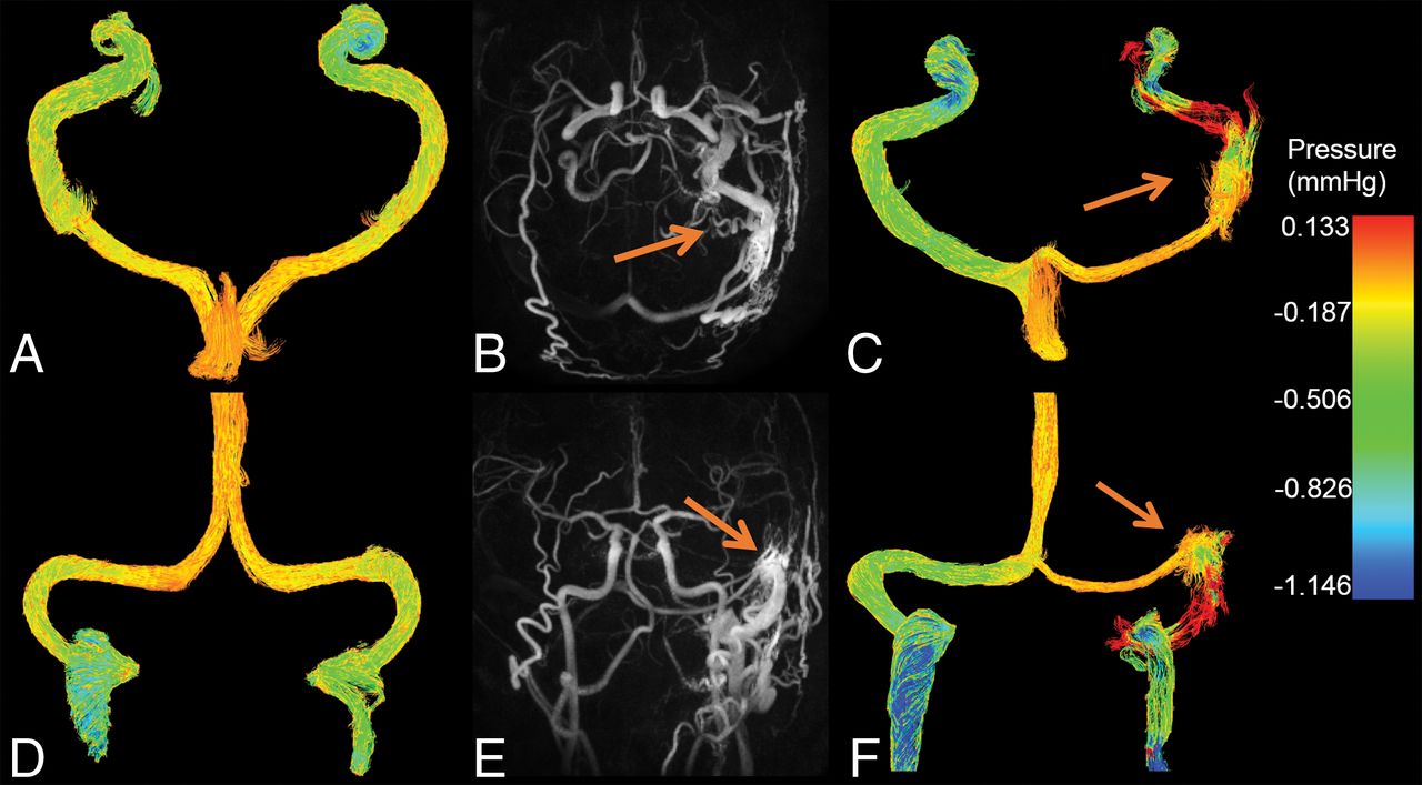
Representative color pressure maps in a healthy subject and a patient with DAVF are shown in Fig 1. For the healthy subject (Fig 1A, -D), blood pressure decreased downstream with lower pressure in the TS than in the superior sagittal sinus. However, the patient with DAVF (Fig 1C, -F) shows a pressure increase in the TS affected by the fistula and a decrease on the TS contralateral to the DAVF. Another patient pressure map and flow streamlines can be found in On-line Figs 1 and 2. The streamlines move retrograde from the site affected by the fistula into the right TS.

Fig 1.
  • Download figure
  • Open in new tab
  • Download powerpoint
Fig 1.

Color pressure maps (mm Hg) in a healthy subject in the axial (A) and coronal (D) planes as well as for a patient (C and F) with a type IIa DAVF affecting the left transverse and sigmoid sinus (arrows). MIP images in the axial (B) and coronal (E) plane show the site of arteriovenous shunting (arrows) where there is increased pressure seen on the axial (C) and coronal (F) pressure maps.

The anatomic variations in the dural sinus drainage were catalogued as follows: for healthy subjects, there were 10 type I, 35 type II, and 15 type III variations (On-line Fig 3 and On-line Table 1). For patients with DAVF, there were 5 type I and 2 type II variations. Flow and cross-sectional area measurements in healthy subjects are summarized in On-line Table 2.

Flow and pressure drops in the TS for all subjects are shown in Fig 2. For patients with DAVF, pressure increased along the TS ipsilateral to the fistula with a median of 0.055 ± 0.130 mm Hg, whereas pressure decreased in the TS contralateral to the fistula with a median of −0.367 ± 0.205 mm Hg (P = .109). In healthy subjects, the pressure decreased very similarly in both TSs, with values of −0.105 ± 0.033 mm Hg and −0.101 ± 0.043 mm Hg, respectively (P = .994). In patients with DAVF, flow in the TS affected by the fistula was 40 ± 129 mL/min, and flow in the TS contralateral to the fistula was 328 ± 134 mL/min (P = .046). In healthy subjects, independent of anatomy type, flow was significantly larger in the right TS (262 ± 83 mL/min) compared with the left TS (121 ± 56 mL/min; P < .001).

Fig 2.
  • Download figure
  • Open in new tab
  • Download powerpoint
Fig 2.

Boxplots showing the pressure change (A) and blood flow rates (B) along the TS in the patients with DAVF (n = 7) and healthy subjects (n = 60). The pressure drops were computed from the difference in pressure in 2 cut planes placed on each TS. The cut planes were placed 25 mm and 50 mm from the torcular Herophili. The median pressure drop in the ipsilateral and contralateral TS to the DAVF were 0.055 ± 0.130 mm Hg and −0.367 ± 0.205 mm Hg, respectively (P = .109). In the healthy subjects, the pressure drops were −0.105 ± 0.033 mm Hg in the left TS and −0.101 ± 0.043 mm Hg in the right TS (P = .994). The median blood flow rates in the patients with DAVF show a value of 40 ± 129 mL/min in the TS affected by the fistula and, in the TS contralateral to the fistula, equal to 328 ± 134 mL/min (P = .046). The median flow in healthy subjects including all anatomy types was significantly larger in the right TS compared with the left TS, with values of 262 ± 83 mL/min and 121 ± 56 mL/min, respectively (P < .001).

Discussion

Initially, we characterized dural venous sinus pressure in a population of 60 healthy subjects, establishing a reference frame for the interpretation of dural sinus pathology. The main findings of this study are an increase in pressure in the TS ipsilateral to the DAVF compared with a pressure drop in the contralateral TS. In healthy subjects, the pressure dropped similarly in the left and right TS. These findings suggest asymmetric pressure drops can potentially be used to identify patients with DAVFs, in whom the TS displaying an increase in pressure or smaller pressure drop represents the site affected by the DAVF. During conventional angiography, dural sinus pressure gradients are frequently measured invasively by navigating a coaxial microcatheter to the draining dural sinus. This preliminary study demonstrates that it is feasible to characterize relative pressure in DAVFs noninvasively by using 4D flow MR imaging.

Retrograde dural/cortical venous drainage is a risk factor for intracranial hemorrhage in patients with DAVFs.1 Consequently, hemodynamic assessment of DAVFs is important for an accurate characterization of severity. Blood flow directionality was successfully assessed for both patients with DAVF and healthy subjects. During this study, 1 patient DAVF classification was changed. Initially, the fistula was classified as type I; after demonstrating retrograde flow in the 4D flow MR imaging, the fistula was reclassified as Type IIa in the Cognard system. The technique described in this study can be implemented to explore other pathologies such as venous stenosis, which is often associated with idiopathic intracranial hypertension and pulsatile tinnitus.10

There are a number of limitations in this study. First, for group comparisons, we used time-average data because cardiac data were not recorded for all but 1 patient with DAVF. Second, the MR imaging technique we used has some potential inaccuracies in pressure mapping in the presence of turbulent flow, which requires more advanced pressure solvers.11 The pressure maps have not been validated against a criterion standard, which will be addressed in a follow-up study with invasive conventional angiography measures that navigate a coaxial microcatheter to the draining dural sinus. This study will also contain more patients with DAVFs.

Conclusions

Noninvasive pressure mapping in the dural sinuses is feasible with 4D flow MR imaging. In addition, 4D flow MR imaging allows blood flow directionality assessment along the dural sinuses, which can help clinicians detect retrograde flow in DAVFs.

Acknowledgments

One of the main contributors to this publication, Zachary J. Clark, MD, passed away suddenly and tragically before this manuscript was completed. Due to ICJME (international committee of medical journal editors) authorship requirements that all authors have the opportunity to review revisions and proofs prior to publication, Dr. Clark could not be included in the authors list. Zach greatly contributed to this work and will be dearly missed.

Footnotes

  • Disclosures: Kevin M. Johnson—RELATED: Grants/Grants Pending: National Institutes of Health*; UNRELATED: Consultancy: Vertex Pharmaceuticals; Other: GE Healthcare, Comments: the University of Wisconsin-Madison receives research support from GE Healthcare*. Patrick A. Turski—RELATED: Grants/Grants Pending: National Institutes of Health, Comments: RO1 Accelerated Neurovascular MRA*. Oliver Wieben—UNRELATED: Other: GE Healthcare, Comments: research agreement*. *Money paid to the institution.

  • This work was supported by National Institute on Aging grant P50-AG033514, National Institute of Neurological Disorders and Stroke grant R01NS066982, National Heart, Lung, and Blood Institute grant R01HL072260, National Institute of General Medical Sciences grant R25GM083252, the American Society of Neuroradiology Alzheimer's Grant Award, and GE Healthcare.

  • Paper previously presented at: Annual Meeting and Exhibition of the International Society for Magnetic Resonance in Medicine, April 22–27, 2017; Honolulu, Hawaii, and Annual Meeting of the American Society of Neuroradiology, April 22–27, 2017; Long Beach, California.

Indicates open access to non-subscribers at www.ajnr.org

References

  1. 1.↵
    1. Gandhi D,
    2. Chen J,
    3. Pearl M, et al
    . Intracranial dural arteriovenous fistulas: classification, imaging findings, and treatment. AJNR Am J Neuroradiol 2012;33:1007–13 doi:10.3174/ajnr.A2798 pmid:22241393
    Abstract/FREE Full Text
  2. 2.↵
    1. Bernstein RA,
    2. Gress DR
    . Dural arteriovenous fistulas and the neurology of venous hypertension. Semin Cerebrovasc Dis Stroke 2004;4:168–75 doi:10.1053/j.scds.2005.01.003
    CrossRef
  3. 3.↵
    1. Markl M,
    2. Frydrychowicz A,
    3. Kozerke S, et al
    . 4D flow MRI. J Magn Reson Imaging 2012;36:1015–36 doi:10.1002/jmri.23632 pmid:23090914
    CrossRefPubMed
  4. 4.↵
    1. Tyszka JM,
    2. Laidlaw DH,
    3. Asa JW, et al
    . Three-dimensional, time-resolved (4D) relative pressure mapping using magnetic resonance imaging. J Magn Reson Imaging 2000;12:321–29 doi:10.1002/1522-2586(200008)12:2<321::AID-JMRI15>3.0.CO%3B2-2 pmid:10931596
    CrossRefPubMed
  5. 5.↵
    1. Moftakhar R,
    2. Aagaard-Kienitz B,
    3. Johnson K, et al
    . Noninvasive measurement of intra-aneurysmal pressure and flow pattern using phase contrast with vastly undersampled isotropic projection imaging. AJNR Am J Neuroradiol 2007;28:1710–14 doi:10.3174/ajnr.A0648 pmid:17885239
    Abstract/FREE Full Text
  6. 6.↵
    1. Bley TA,
    2. Johnson KM,
    3. François CJ, et al
    . Noninvasive assessment of transstenotic pressure gradients in porcine renal artery stenoses by using vastly undersampled phase-contrast MR angiography. Radiology 2011;261:266–73 doi:10.1148/radiol.11101175 pmid:21813739
    CrossRefPubMed
  7. 7.↵
    1. Gu T,
    2. Korosec FR,
    3. Block WF, et al
    . PC VIPR: a high-speed 3D phase-contrast method for flow quantification and high-resolution angiography. AJNR Am J Neuroradiol 2005;26:743–49 pmid:15814915
    Abstract/FREE Full Text
  8. 8.↵
    1. Johnson KM,
    2. Lum DP,
    3. Turski PA, et al
    . Improved 3D phase contrast MRI with off-resonance corrected dual echo VIPR. Magn Reson Med 2008;60:1329–36 doi:10.1002/mrm.21763 pmid:19025882
    CrossRefPubMed
  9. 9.↵
    1. Gökçe E,
    2. Pınarbaşılı T,
    3. Acu B, et al
    . Torcular Herophili classification and evaluation of dural venous sinus variations using digital subtraction angiography and magnetic resonance venographies. Surg Radiol Anat 2014;36:527–36 doi:10.1007/s00276-013-1223-0 pmid:24154635
    CrossRefPubMed
  10. 10.↵
    1. Lansley JA,
    2. Tucker W,
    3. Eriksen MR, et al
    . Sigmoid sinus diverticulum, dehiscence, and venous sinus stenosis: potential causes of pulsatile tinnitus in patients with idiopathic intracranial hypertension? AJNR Am J Neuroradiol 2017;38:1783–88 doi:10.3174/ajnr.A5277 pmid:28705815
    Abstract/FREE Full Text
  11. 11.↵
    1. Ha H,
    2. Lantz J,
    3. Ziegler M, et al
    . Estimating the irreversible pressure drop across a stenosis by quantifying turbulence production using 4D flow MRI. Sci Rep 2017;7:46618 doi:10.1038/srep46618 pmid:28425452
    CrossRefPubMed
  • Received July 7, 2017.
  • Accepted after revision October 26, 2017.
  • © 2018 by American Journal of Neuroradiology
View Abstract
PreviousNext
Back to top

In this issue

American Journal of Neuroradiology: 39 (3)
American Journal of Neuroradiology
Vol. 39, Issue 3
1 Mar 2018
  • Table of Contents
  • Index by author
  • Complete Issue (PDF)
Advertisement
Print
Download PDF
Email Article

Thank you for your interest in spreading the word on American Journal of Neuroradiology.

NOTE: We only request your email address so that the person you are recommending the page to knows that you wanted them to see it, and that it is not junk mail. We do not capture any email address.

Enter multiple addresses on separate lines or separate them with commas.
Pressure Mapping and Hemodynamic Assessment of Intracranial Dural Sinuses and Dural Arteriovenous Fistulas with 4D Flow MRI
(Your Name) has sent you a message from American Journal of Neuroradiology
(Your Name) thought you would like to see the American Journal of Neuroradiology web site.
CAPTCHA
This question is for testing whether or not you are a human visitor and to prevent automated spam submissions.
Cite this article
L.A. Rivera-Rivera, K.M. Johnson, P.A. Turski, O. Wieben
Pressure Mapping and Hemodynamic Assessment of Intracranial Dural Sinuses and Dural Arteriovenous Fistulas with 4D Flow MRI
American Journal of Neuroradiology Mar 2018, 39 (3) 485-487; DOI: 10.3174/ajnr.A5494

Citation Manager Formats

  • BibTeX
  • Bookends
  • EasyBib
  • EndNote (tagged)
  • EndNote 8 (xml)
  • Medlars
  • Mendeley
  • Papers
  • RefWorks Tagged
  • Ref Manager
  • RIS
  • Zotero
0 Responses
Respond to this article
Share
Bookmark this article
Pressure Mapping and Hemodynamic Assessment of Intracranial Dural Sinuses and Dural Arteriovenous Fistulas with 4D Flow MRI
L.A. Rivera-Rivera, K.M. Johnson, P.A. Turski, O. Wieben
American Journal of Neuroradiology Mar 2018, 39 (3) 485-487; DOI: 10.3174/ajnr.A5494
del.icio.us logo Twitter logo Facebook logo Mendeley logo
  • Tweet Widget
  • Facebook Like
  • Google Plus One
Purchase

Jump to section

  • Article
    • Abstract
    • ABBREVIATIONS:
    • Materials and Methods
    • Results
    • Discussion
    • Conclusions
    • Acknowledgments
    • Footnotes
    • References
  • Figures & Data
  • Supplemental
  • Info & Metrics
  • Responses
  • References
  • PDF

Related Articles

  • PubMed
  • Google Scholar

Cited By...

  • Utilisation of advanced MRI techniques to understand neurovascular complications of PHACE syndrome: a case of arterial stenosis and dissection
  • Crossref (19)
  • Google Scholar

This article has been cited by the following articles in journals that are participating in Crossref Cited-by Linking.

  • 4D Flow with MRI
    Gilles Soulat, Patrick McCarthy, Michael Markl
    Annual Review of Biomedical Engineering 2020 22 1
  • Standardized Evaluation of Cerebral Arteriovenous Malformations Using Flow Distribution Network Graphs and Dual‐venc 4D Flow MRI
    Maria Aristova, Alireza Vali, Sameer A. Ansari, Ali Shaibani, Tord D. Alden, Michael C. Hurley, Babak S. Jahromi, Matthew B. Potts, Michael Markl, Susanne Schnell
    Journal of Magnetic Resonance Imaging 2019 50 6
  • Pressure drop mapping using 4D flow MRI in patients with bicuspid aortic valve disease: A novel marker of valvular obstruction
    Ali Fatehi Hassanabad, Fiona Burns, Michael S. Bristow, Carmen Lydell, Andrew G. Howarth, Bobak Heydari, Xuexin Gao, Paul W.M. Fedak, James A. White, Julio Garcia
    Magnetic Resonance Imaging 2020 65
  • 4D flow MRI hemodynamic biomarkers for cerebrovascular diseases
    Anders Wåhlin, Anders Eklund, Jan Malm
    Journal of Internal Medicine 2022 291 2
  • Noninvasive quantification of cerebrovascular pressure changes using 4D Flow MRI
    David Marlevi, Jonas Schollenberger, Maria Aristova, Edward Ferdian, Yue Ma, Alistair A. Young, Elazer R. Edelman, Susanne Schnell, C. Alberto Figueroa, David A. Nordsletten
    Magnetic Resonance in Medicine 2021 86 6
  • Estimation of venous sinus pressure drop in patients with idiopathic intracranial hypertension using 4D-flow MRI
    Yupeng Zhang, Chao Ma, Shikai Liang, Changxuan Li, Haoyu Zhu, Zhiye Li, Zhongrong Miao, Xu Tong, Kehui Dong, Chuhan Jiang, Binbin Sui, Dapeng Mo
    European Radiology 2022 33 4
  • Serial Quantitative and Qualitative Measurements of Flow in Vein of Galen Malformations Using 4-Dimensional Flow Magnetic Resonance Imaging (Phase Contrast Vastly undersampled Isotropic PRojection)
    Yiping Li, Raheel Ahmed, Leonardo A. Rivera-Rivera, James A. Stadler, Patrick Turski, Beverly Aagaard-Kienitz
    World Neurosurgery 2019 126
  • Clinical outcomes of adjunct sinus stenting in dural arteriovenous fistulas
    Yen-Heng Lin, Chung-Wei Lee, Hon-Man Liu
    Clinical Neuroradiology 2022 32 2
  • Utilisation of advanced MRI techniques to understand neurovascular complications of PHACE syndrome: a case of arterial stenosis and dissection
    Laura B Eisenmenger, Leonardo A Rivera-Rivera, Kevin M Johnson, Beth A Drolet
    BMJ Case Reports 2020 13 9
  • 4D flow imaging with 2D‐selective excitation
    Clarissa Wink, Giulio Ferrazzi, Jean Pierre Bassenge, Sebastian Flassbeck, Simon Schmidt, Tobias Schaeffter, Sebastian Schmitter
    Magnetic Resonance in Medicine 2019 82 3

More in this TOC Section

  • Diagnostic Neuroradiology of Monoclonal Antibodies
  • Clinical Outcomes After Chiari I Decompression
  • Segmentation of Brain Metastases with BLAST
Show more Adult Brain

Similar Articles

Advertisement

Indexed Content

  • Current Issue
  • Accepted Manuscripts
  • Article Preview
  • Past Issues
  • Editorials
  • Editor's Choice
  • Fellows' Journal Club
  • Letters to the Editor
  • Video Articles

Cases

  • Case Collection
  • Archive - Case of the Week
  • Archive - Case of the Month
  • Archive - Classic Case

More from AJNR

  • Trainee Corner
  • Imaging Protocols
  • MRI Safety Corner
  • Book Reviews

Multimedia

  • AJNR Podcasts
  • AJNR Scantastics

Resources

  • Turnaround Time
  • Submit a Manuscript
  • Submit a Video Article
  • Submit an eLetter to the Editor/Response
  • Manuscript Submission Guidelines
  • Statistical Tips
  • Fast Publishing of Accepted Manuscripts
  • Graphical Abstract Preparation
  • Imaging Protocol Submission
  • Evidence-Based Medicine Level Guide
  • Publishing Checklists
  • Author Policies
  • Become a Reviewer/Academy of Reviewers
  • News and Updates

About Us

  • About AJNR
  • Editorial Board
  • Editorial Board Alumni
  • Alerts
  • Permissions
  • Not an AJNR Subscriber? Join Now
  • Advertise with Us
  • Librarian Resources
  • Feedback
  • Terms and Conditions
  • AJNR Editorial Board Alumni

American Society of Neuroradiology

  • Not an ASNR Member? Join Now

© 2025 by the American Society of Neuroradiology All rights, including for text and data mining, AI training, and similar technologies, are reserved.
Print ISSN: 0195-6108 Online ISSN: 1936-959X

Powered by HighWire