Advertisement

Main menu

  • Home
  • Content
    • Current Issue
    • Accepted Manuscripts
    • Article Preview
    • Past Issue Archive
    • Video Articles
    • AJNR Case Collection
    • Case of the Week Archive
    • Case of the Month Archive
    • Classic Case Archive
  • Special Collections
    • Advancing NeuroMRI with High-Relaxivity Contrast Agents
    • AJNR Awards
    • Low-Field MRI
    • Alzheimer Disease
    • ASNR Foundation Special Collection
    • Photon-Counting CT
    • View All
  • Multimedia
    • AJNR Podcasts
    • AJNR SCANtastic
    • Trainee Corner
    • MRI Safety Corner
    • Imaging Protocols
  • For Authors
    • Submit a Manuscript
    • Submit a Video Article
    • Submit an eLetter to the Editor/Response
    • Manuscript Submission Guidelines
    • Statistical Tips
    • Fast Publishing of Accepted Manuscripts
    • Graphical Abstract Preparation
    • Imaging Protocol Submission
    • Author Policies
  • About Us
    • About AJNR
    • Editorial Board
    • Editorial Board Alumni
  • More
    • Become a Reviewer/Academy of Reviewers
    • Subscribers
    • Permissions
    • Alerts
    • Feedback
    • Advertisers
    • ASNR Home

User menu

  • Alerts
  • Log in

Search

  • Advanced search
American Journal of Neuroradiology

American Journal of Neuroradiology

ASHNR American Society of Functional Neuroradiology ASHNR American Society of Pediatric Neuroradiology ASSR
  • Alerts
  • Log in

Advanced Search

  • Home
  • Content
    • Current Issue
    • Accepted Manuscripts
    • Article Preview
    • Past Issue Archive
    • Video Articles
    • AJNR Case Collection
    • Case of the Week Archive
    • Case of the Month Archive
    • Classic Case Archive
  • Special Collections
    • Advancing NeuroMRI with High-Relaxivity Contrast Agents
    • AJNR Awards
    • Low-Field MRI
    • Alzheimer Disease
    • ASNR Foundation Special Collection
    • Photon-Counting CT
    • View All
  • Multimedia
    • AJNR Podcasts
    • AJNR SCANtastic
    • Trainee Corner
    • MRI Safety Corner
    • Imaging Protocols
  • For Authors
    • Submit a Manuscript
    • Submit a Video Article
    • Submit an eLetter to the Editor/Response
    • Manuscript Submission Guidelines
    • Statistical Tips
    • Fast Publishing of Accepted Manuscripts
    • Graphical Abstract Preparation
    • Imaging Protocol Submission
    • Author Policies
  • About Us
    • About AJNR
    • Editorial Board
    • Editorial Board Alumni
  • More
    • Become a Reviewer/Academy of Reviewers
    • Subscribers
    • Permissions
    • Alerts
    • Feedback
    • Advertisers
    • ASNR Home
  • Follow AJNR on Twitter
  • Visit AJNR on Facebook
  • Follow AJNR on Instagram
  • Join AJNR on LinkedIn
  • RSS Feeds

AJNR Awards, New Junior Editors, and more. Read the latest AJNR updates


Improved Turnaround Times | Median time to first decision: 12 days

Research ArticleAdult Brain

Natural History of Endoscopic Third Ventriculostomy in Adults: Serial Evaluation with High-Resolution CISS

M. Trelles, A.K. Ahmed, C.H. Mitchell, I. Josue-Torres, D. Rigamonti and A.M. Blitz
American Journal of Neuroradiology December 2018, 39 (12) 2231-2236; DOI: https://doi.org/10.3174/ajnr.A5861
M. Trelles
aFrom the Departments of Radiology and Radiological Sciences (M.T., A.M.B.)
  • Find this author on Google Scholar
  • Find this author on PubMed
  • Search for this author on this site
  • ORCID record for M. Trelles
A.K. Ahmed
bNeurosurgery (A.K.A., I.J.-T., D.R.)
  • Find this author on Google Scholar
  • Find this author on PubMed
  • Search for this author on this site
  • ORCID record for A.K. Ahmed
C.H. Mitchell
cNeurology (C.H.M.), Johns Hopkins University School of Medicine, Baltimore, Maryland.
  • Find this author on Google Scholar
  • Find this author on PubMed
  • Search for this author on this site
  • ORCID record for C.H. Mitchell
I. Josue-Torres
bNeurosurgery (A.K.A., I.J.-T., D.R.)
  • Find this author on Google Scholar
  • Find this author on PubMed
  • Search for this author on this site
  • ORCID record for I. Josue-Torres
D. Rigamonti
bNeurosurgery (A.K.A., I.J.-T., D.R.)
  • Find this author on Google Scholar
  • Find this author on PubMed
  • Search for this author on this site
  • ORCID record for D. Rigamonti
A.M. Blitz
aFrom the Departments of Radiology and Radiological Sciences (M.T., A.M.B.)
  • Find this author on Google Scholar
  • Find this author on PubMed
  • Search for this author on this site
  • ORCID record for A.M. Blitz
  • Article
  • Figures & Data
  • Info & Metrics
  • Responses
  • References
  • PDF
Loading

Article Figures & Data

Figures

  • Tables
  • Fig 1.
    • Download figure
    • Open in new tab
    • Download powerpoint
    Fig 1.

    Representative sagittal CISS image and coronal MPR reconstruction of a third ventriculostomy defect with anteroposterior and third ventriculostomy defect size measurement.

  • Fig 2.
    • Download figure
    • Open in new tab
    • Download powerpoint
    Fig 2.

    Defect size area across time after surgery.
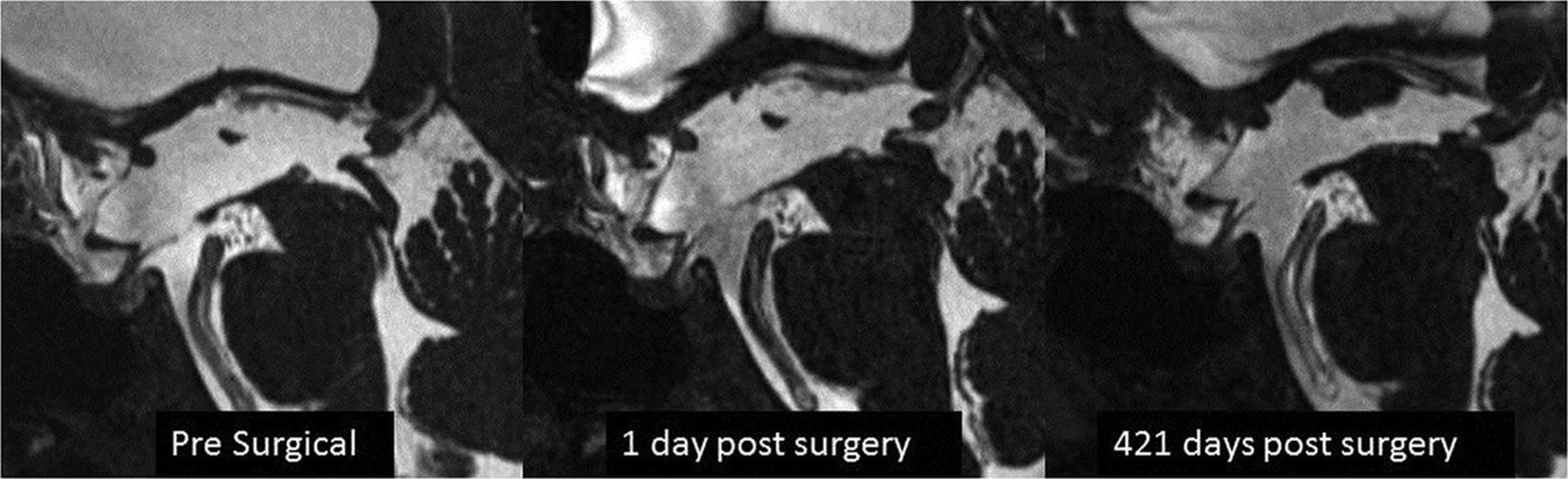
  • Fig 3.
    • Download figure
    • Open in new tab
    • Download powerpoint
    Fig 3.

    Representative case showing presurgical imaging, first follow-up at 1 day postsurgery, and final follow-up at 421 days postsurgery showing an increase in the ETV defect size.

  • Fig 4.
    • Download figure
    • Open in new tab
    • Download powerpoint
    Fig 4.

    Average increase in the size of the defect across time.

  • Fig 5.
    • Download figure
    • Open in new tab
    • Download powerpoint
    Fig 5.

    Maximum defect size and age at the operation.

Tables

  • Figures
    • View popup
    Table 1:

    Demographic information

    Demographics%No.
    Sex
        Male44%15
        Female56%19
    Mean age (range) (yr)54 (19–76)
    • View popup
    Table 2:

    Site of obstruction to the flow of CSF

    Location of Obstruction%No.
    Cerebral aqueduct82%28
    Fourth ventricle outflow3%1
    Prepontine cistern9%3
    Unknown3%1
    Other3%1
    34
    • View popup
    Table 3:

    Average defect size ± 95% confidence interval and t test for the NICO Myriad device at last follow-up

    NICO Myriad Device Used
    YesNoP Value
    Average defect size at LFU (mean)28.21 ± 7.4811.25 ± 6.80<.05
    • Note:—LFU indicates last follow-up.

    • View popup
    Table 4:

    Presenting symptoms

    NICO Myriad UsedNo NICO MyriadP Value
    Headache60%75%>.05
    Dizziness26.7%30%>.05
    Nausea26.7%35%>.05
    Vision deficit40%45%>.05
    Gait abnormality53.3%50%>.05
    Urinary incontinence46.7%35%>.05
    Cognitive dysfunction53.3%60%>.05
    • View popup
    Table 5:

    Clinical outcomes at 6 months and last follow-up after ETVa

    ImprovedSame as BaselineWorse Compared with BaselineP Value
    NICO Myriad UsedNo NICO MyriadNICO Myriad UsedNo NICO MyriadNICO Myriad UsedNo NICO Myriad
    Headache
        6 Mo88.9%80%11.1%13.3%0%6.7%>.05
        LFU77.8%53.3%22.2%33.3%0%13.3%>.05
    Dizziness
        6 Mo100%83.3%0%16.7%0%0%>.05
        LFU100%83.3%0%16.7%0%0%>.05
    Nausea
        6 Mo75%75%25%25%0%0%>.05
        LFU50%75%50%25%0%0%>.05
    Vision deficit
        6 Mo83.3%77.8%16.7%22.2%0%0%>.05
        LFU83.3%77.8%16.7%22.2%0%0%>.05
    Gait abnormality
        6 Mo100%100%0%0%0%0%>.05
        LFU100%70%0%10%0%20%>.05
    Urinary incontinence
        6 Mo100%100%0%0%0%0%>.05
        LFU85.7%85.7%0%0%14.3%14.3%>.05
    Cognitive dysfunction
        6 Mo87.5%70%12.5%30%0%0%>.05
        LFU87.5%70%12.5%20%0%10%>.05
    • Note:—LFU indicates last follow-up.

    • ↵a Outcomes are given among those with preoperative symptoms.

PreviousNext
Back to top

In this issue

American Journal of Neuroradiology: 39 (12)
American Journal of Neuroradiology
Vol. 39, Issue 12
1 Dec 2018
  • Table of Contents
  • Index by author
  • Complete Issue (PDF)
Advertisement
Print
Download PDF
Email Article

Thank you for your interest in spreading the word on American Journal of Neuroradiology.

NOTE: We only request your email address so that the person you are recommending the page to knows that you wanted them to see it, and that it is not junk mail. We do not capture any email address.

Enter multiple addresses on separate lines or separate them with commas.
Natural History of Endoscopic Third Ventriculostomy in Adults: Serial Evaluation with High-Resolution CISS
(Your Name) has sent you a message from American Journal of Neuroradiology
(Your Name) thought you would like to see the American Journal of Neuroradiology web site.
CAPTCHA
This question is for testing whether or not you are a human visitor and to prevent automated spam submissions.
Cite this article
M. Trelles, A.K. Ahmed, C.H. Mitchell, I. Josue-Torres, D. Rigamonti, A.M. Blitz
Natural History of Endoscopic Third Ventriculostomy in Adults: Serial Evaluation with High-Resolution CISS
American Journal of Neuroradiology Dec 2018, 39 (12) 2231-2236; DOI: 10.3174/ajnr.A5861

Citation Manager Formats

  • BibTeX
  • Bookends
  • EasyBib
  • EndNote (tagged)
  • EndNote 8 (xml)
  • Medlars
  • Mendeley
  • Papers
  • RefWorks Tagged
  • Ref Manager
  • RIS
  • Zotero
0 Responses
Respond to this article
Share
Bookmark this article
Natural History of Endoscopic Third Ventriculostomy in Adults: Serial Evaluation with High-Resolution CISS
M. Trelles, A.K. Ahmed, C.H. Mitchell, I. Josue-Torres, D. Rigamonti, A.M. Blitz
American Journal of Neuroradiology Dec 2018, 39 (12) 2231-2236; DOI: 10.3174/ajnr.A5861
del.icio.us logo Twitter logo Facebook logo Mendeley logo
  • Tweet Widget
  • Facebook Like
  • Google Plus One
Purchase

Jump to section

  • Article
    • Abstract
    • ABBREVIATION:
    • Materials and Methods
    • Results
    • Discussion
    • Conclusions
    • Footnotes
    • References
  • Figures & Data
  • Info & Metrics
  • Responses
  • References
  • PDF

Related Articles

  • No related articles found.
  • PubMed
  • Google Scholar

Cited By...

  • No citing articles found.
  • Crossref
  • Google Scholar

This article has not yet been cited by articles in journals that are participating in Crossref Cited-by Linking.

More in this TOC Section

  • Diagnostic Neuroradiology of Monoclonal Antibodies
  • Clinical Outcomes After Chiari I Decompression
  • Segmentation of Brain Metastases with BLAST
Show more Adult Brain

Similar Articles

Advertisement

Indexed Content

  • Current Issue
  • Accepted Manuscripts
  • Article Preview
  • Past Issues
  • Editorials
  • Editor's Choice
  • Fellows' Journal Club
  • Letters to the Editor
  • Video Articles

Cases

  • Case Collection
  • Archive - Case of the Week
  • Archive - Case of the Month
  • Archive - Classic Case

More from AJNR

  • Trainee Corner
  • Imaging Protocols
  • MRI Safety Corner
  • Book Reviews

Multimedia

  • AJNR Podcasts
  • AJNR Scantastics

Resources

  • Turnaround Time
  • Submit a Manuscript
  • Submit a Video Article
  • Submit an eLetter to the Editor/Response
  • Manuscript Submission Guidelines
  • Statistical Tips
  • Fast Publishing of Accepted Manuscripts
  • Graphical Abstract Preparation
  • Imaging Protocol Submission
  • Evidence-Based Medicine Level Guide
  • Publishing Checklists
  • Author Policies
  • Become a Reviewer/Academy of Reviewers
  • News and Updates

About Us

  • About AJNR
  • Editorial Board
  • Editorial Board Alumni
  • Alerts
  • Permissions
  • Not an AJNR Subscriber? Join Now
  • Advertise with Us
  • Librarian Resources
  • Feedback
  • Terms and Conditions
  • AJNR Editorial Board Alumni

American Society of Neuroradiology

  • Not an ASNR Member? Join Now

© 2025 by the American Society of Neuroradiology All rights, including for text and data mining, AI training, and similar technologies, are reserved.
Print ISSN: 0195-6108 Online ISSN: 1936-959X

Powered by HighWire