Advertisement

Main menu

  • Home
  • Content
    • Current Issue
    • Accepted Manuscripts
    • Article Preview
    • Past Issue Archive
    • Video Articles
    • AJNR Case Collection
    • Case of the Week Archive
    • Case of the Month Archive
    • Classic Case Archive
  • Special Collections
    • AJNR Awards
    • Low-Field MRI
    • Alzheimer Disease
    • ASNR Foundation Special Collection
    • Photon-Counting CT
    • View All
  • Multimedia
    • AJNR Podcasts
    • AJNR SCANtastic
    • Trainee Corner
    • MRI Safety Corner
    • Imaging Protocols
  • For Authors
    • Submit a Manuscript
    • Submit a Video Article
    • Submit an eLetter to the Editor/Response
    • Manuscript Submission Guidelines
    • Statistical Tips
    • Fast Publishing of Accepted Manuscripts
    • Graphical Abstract Preparation
    • Imaging Protocol Submission
    • Author Policies
  • About Us
    • About AJNR
    • Editorial Board
    • Editorial Board Alumni
  • More
    • Become a Reviewer/Academy of Reviewers
    • Subscribers
    • Permissions
    • Alerts
    • Feedback
    • Advertisers
    • ASNR Home

User menu

  • Alerts
  • Log in

Search

  • Advanced search
American Journal of Neuroradiology

American Journal of Neuroradiology

ASHNR American Society of Functional Neuroradiology ASHNR American Society of Pediatric Neuroradiology ASSR
  • Alerts
  • Log in

Advanced Search

  • Home
  • Content
    • Current Issue
    • Accepted Manuscripts
    • Article Preview
    • Past Issue Archive
    • Video Articles
    • AJNR Case Collection
    • Case of the Week Archive
    • Case of the Month Archive
    • Classic Case Archive
  • Special Collections
    • AJNR Awards
    • Low-Field MRI
    • Alzheimer Disease
    • ASNR Foundation Special Collection
    • Photon-Counting CT
    • View All
  • Multimedia
    • AJNR Podcasts
    • AJNR SCANtastic
    • Trainee Corner
    • MRI Safety Corner
    • Imaging Protocols
  • For Authors
    • Submit a Manuscript
    • Submit a Video Article
    • Submit an eLetter to the Editor/Response
    • Manuscript Submission Guidelines
    • Statistical Tips
    • Fast Publishing of Accepted Manuscripts
    • Graphical Abstract Preparation
    • Imaging Protocol Submission
    • Author Policies
  • About Us
    • About AJNR
    • Editorial Board
    • Editorial Board Alumni
  • More
    • Become a Reviewer/Academy of Reviewers
    • Subscribers
    • Permissions
    • Alerts
    • Feedback
    • Advertisers
    • ASNR Home
  • Follow AJNR on Twitter
  • Visit AJNR on Facebook
  • Follow AJNR on Instagram
  • Join AJNR on LinkedIn
  • RSS Feeds

AJNR Awards, New Junior Editors, and more. Read the latest AJNR updates

Research ArticleExtracranial Vascular
Open Access

Lower Arterial Cross-Sectional Area of Carotid and Vertebral Arteries and Higher Frequency of Secondary Neck Vessels Are Associated with Multiple Sclerosis

P. Belov, D. Jakimovski, J. Krawiecki, C. Magnano, J. Hagemeier, L. Pelizzari, B. Weinstock-Guttman and R. Zivadinov
American Journal of Neuroradiology January 2018, 39 (1) 123-130; DOI: https://doi.org/10.3174/ajnr.A5469
P. Belov
aFrom the Buffalo Neuroimaging Analysis Center, Department of Neurology (P.B., D.J., J.K., C.M., J.H., R.Z.)
  • Find this author on Google Scholar
  • Find this author on PubMed
  • Search for this author on this site
  • ORCID record for P. Belov
D. Jakimovski
aFrom the Buffalo Neuroimaging Analysis Center, Department of Neurology (P.B., D.J., J.K., C.M., J.H., R.Z.)
  • Find this author on Google Scholar
  • Find this author on PubMed
  • Search for this author on this site
  • ORCID record for D. Jakimovski
J. Krawiecki
aFrom the Buffalo Neuroimaging Analysis Center, Department of Neurology (P.B., D.J., J.K., C.M., J.H., R.Z.)
  • Find this author on Google Scholar
  • Find this author on PubMed
  • Search for this author on this site
  • ORCID record for J. Krawiecki
C. Magnano
aFrom the Buffalo Neuroimaging Analysis Center, Department of Neurology (P.B., D.J., J.K., C.M., J.H., R.Z.)
  • Find this author on Google Scholar
  • Find this author on PubMed
  • Search for this author on this site
  • ORCID record for C. Magnano
J. Hagemeier
aFrom the Buffalo Neuroimaging Analysis Center, Department of Neurology (P.B., D.J., J.K., C.M., J.H., R.Z.)
  • Find this author on Google Scholar
  • Find this author on PubMed
  • Search for this author on this site
  • ORCID record for J. Hagemeier
L. Pelizzari
dDepartment of Electronics, Information and Bioengineering (L.P.), Politecnico di Milano, Milan, Italy
eFondazione Don Carlo Gnocchi Organizzazione Non Lucrativa di Utilita' Sociae (L.P.), Milan, Italy.
  • Find this author on Google Scholar
  • Find this author on PubMed
  • Search for this author on this site
  • ORCID record for L. Pelizzari
B. Weinstock-Guttman
bJacobs MS Center, Department of Neurology (B.W.-G.)
  • Find this author on Google Scholar
  • Find this author on PubMed
  • Search for this author on this site
  • ORCID record for B. Weinstock-Guttman
R. Zivadinov
aFrom the Buffalo Neuroimaging Analysis Center, Department of Neurology (P.B., D.J., J.K., C.M., J.H., R.Z.)
cCenter for Biomedical Imaging at Clinical Translational Science Institute (R.Z.), University at Buffalo, State University of New York, Buffalo, New York
  • Find this author on Google Scholar
  • Find this author on PubMed
  • Search for this author on this site
  • ORCID record for R. Zivadinov
  • Article
  • Figures & Data
  • Supplemental
  • Info & Metrics
  • Responses
  • References
  • PDF
Loading

Article Figures & Data

Figures

  • Tables
  • Fig 1.
    • Download figure
    • Open in new tab
    • Download powerpoint
    Fig 1.

    Radar plot representation of the neck vessel differences between patients with multiple sclerosis and healthy controls. Significant correlations are in boldface.
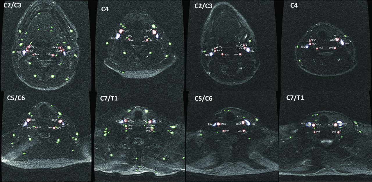
  • Fig 2.
    • Download figure
    • Open in new tab
    • Download powerpoint
    Fig 2.

    Comparison of main and secondary vessel number and cross-sectional area on a 2D-MRV sequence at 4 cervical levels in patients with multiple sclerosis (4 corresponding panels on the left) and age- and sex-matched healthy controls (4 corresponding panels on the right). VV indicates vertebral vein; L, left; R, right. Green color represents the secondary vessels, red color represents the CCA, ICA, EAC, and VA, while blue represents the IJV.

Tables

  • Figures
    • View popup
    Table 1:

    Demographic and clinical characteristics of patients with multiple sclerosis (n = 193) and healthy controls (n = 193)a

    MS (n = 193)HC (n = 193)P Value
    Female (No.) (%)130 (67.4)130 (67.4)1.000
    Age (mean) (SD) (yr)42.2 (13.9)42.9 (17.5).676
    BMI (mean) (SD)26.8 (5.8)26.8 (5.7).94
    Disease duration (mean) (SD) (yr)12.0 (9.4)NA–
    EDSS (median) (range)2 (0.0–6.5)NA–
    Smoking history (No.) (%)73 (46.8)58 (32.4).005b
    Heart disease (No.) (%)30 (19.7)20 (12.4).053
    Hypertension (No.) (%)38 (25.3)19 (11.3).001b
    • Note:—EDSS indicates Expanded Disability Status Scale; IQR, interquartile range; NA, not applicable.

    • ↵a χ2 and Student t test were used for comparing variables between groups.

    • ↵b An α level of .05 was considered significant.

    • View popup
    Table 2:

    Correlations of arterial, venous, and secondary neck vessel frequency and the cross-sectional area with age and BMI in study groupsa

    AgeBody Mass Index
    MS (n = 193)HC (n = 193)MS (n = 193)HC (n = 193)
    R ValueP ValueR ValueP ValueR ValueP ValueR ValueP Value
    Arterial CSA (mm2)
        C2/3−0.156.031b−0.163.023b0.130.0850.059.449
        C40.075.2980.045.5340.037.626−0.017.830
        C5/60.103.1560.122.0920.136.0730.095.224
        C7/T1−0.049.502−0.003.9680.085.2660.047.548
    IJV CSA (mm2)
        C2/30.257<.001b0.150.370.098.1980.083.287
        C40.255<.001b0.177.14b0.146.0540.139.075
        C5/60.196.006b0.252<.001b0.339<.001b0.231.003b
        C7/T10.187.009b0.327<.001b0.375<.001b0.344<.001b
    Secondary CSA (mm2)
        C2/30.039.594−0.060.404−0.192.011b−0.074.347
        C40.112.122−0.040.584−0.229.002b−0.121.121
        C5/60.047.5180.028.696−0.214.004b−0.064.417
        C7/T10.061.398−0.049.498−0.078.307−0.065.405
    No. of vessels
        C2/30.013.860−0.149.039b−0.204.007b−0.153.050b
        C4−0.460.527−0.211.003b−0.292<.001b−0.316<.001b
        C5/6−0.076.294−0.141.050b−0.333<.001b−0.251.001b
        C7/T1−0.060.406−0.155.031b−0.180.017b−0.174.025b
    • ↵a Spearman ranked correlations between age/BMI and the corresponding vascular variable were used.

    • ↵b An α level of .05 was considered significant.

    • View popup
    Table 3:

    Arterial, venous, and secondary neck vessel frequency and the cross-sectional area in the study groupsa

    Primary Vessel (CSA) (mm2)
    Arterial and VenousArterial (VAs)
    MS (n = 193)HC (n = 193)P ValueMS (n = 193)HC (n = 193)P Value
    Arterial (CCA/ICA/ECA)
        C2/C355.1 (16.4)60.9 (17.9).030b20.1 (4.4)21.8 (5.8).02b
        C460.8 (15.7)63.4 (16.3).22918.6 (4.2)20.3 (5.0).012b
        C5/C650.1 (10.1)53.9 (12.5).026b18.1 (6.9)19.3 (4.7).341
        C7/T147.6 (9.8)52 (9.9).005b16.3 (4.5)18.4 (5.9).006b
    Venous (IJVs)
        C2/C364.9 (27.4)66.0 (31.6).621
        C486.9 (35.9)91.1 (41.0).140
        C5/C692.3 (57.1)97.4 (60.2).418
        C7/T1113.3 (67.0)117.9 (79.3).790
    • ↵a Analysis of covariance adjusted for age and BMI, smoking history, heart disease, and hypertension was used. In the ANCOVA for frequency of vessels, ranked variables were used.

    • ↵b An α level of .05 was considered significant.

    • View popup
    Table 4:

    Secondary neck vessel frequency and the cross-sectional area in the study groupsa

    No. of VesselsCSA (mm2)
    MS (n = 193)HC (n = 193)P ValueMS (n = 193)HC (n = 193)P Value
    Secondary vessels
        C2/C312.9 (5.4)10 (4.2)<.001b92.1 (40.6)81.6 (35.5).016b
        C49.1 (4.2)7.5 (3.3)<.001b71.0 (33.7)65.3 (28.7).022b
        C5/C67.8 (3.9)6.8 (3.4).012b61.9 (32.2)57.2 (28.2).028b
        C7/T18.8 (4.9)6 (3.5)<.001b71.1 (40.5)56.7 (32.5)<.001b
    • ↵a Analysis of covariance adjusted for age and BMI, smoking history, heart disease, and hypertension was used. In the ANCOVA for frequency of vessels, ranked variables were used.

    • ↵b An α level of .05 was considered significant.

PreviousNext
Back to top

In this issue

American Journal of Neuroradiology: 39 (1)
American Journal of Neuroradiology
Vol. 39, Issue 1
1 Jan 2018
  • Table of Contents
  • Index by author
  • Complete Issue (PDF)
Advertisement
Print
Download PDF
Email Article

Thank you for your interest in spreading the word on American Journal of Neuroradiology.

NOTE: We only request your email address so that the person you are recommending the page to knows that you wanted them to see it, and that it is not junk mail. We do not capture any email address.

Enter multiple addresses on separate lines or separate them with commas.
Lower Arterial Cross-Sectional Area of Carotid and Vertebral Arteries and Higher Frequency of Secondary Neck Vessels Are Associated with Multiple Sclerosis
(Your Name) has sent you a message from American Journal of Neuroradiology
(Your Name) thought you would like to see the American Journal of Neuroradiology web site.
CAPTCHA
This question is for testing whether or not you are a human visitor and to prevent automated spam submissions.
Cite this article
P. Belov, D. Jakimovski, J. Krawiecki, C. Magnano, J. Hagemeier, L. Pelizzari, B. Weinstock-Guttman, R. Zivadinov
Lower Arterial Cross-Sectional Area of Carotid and Vertebral Arteries and Higher Frequency of Secondary Neck Vessels Are Associated with Multiple Sclerosis
American Journal of Neuroradiology Jan 2018, 39 (1) 123-130; DOI: 10.3174/ajnr.A5469

Citation Manager Formats

  • BibTeX
  • Bookends
  • EasyBib
  • EndNote (tagged)
  • EndNote 8 (xml)
  • Medlars
  • Mendeley
  • Papers
  • RefWorks Tagged
  • Ref Manager
  • RIS
  • Zotero
0 Responses
Respond to this article
Share
Bookmark this article
Lower Arterial Cross-Sectional Area of Carotid and Vertebral Arteries and Higher Frequency of Secondary Neck Vessels Are Associated with Multiple Sclerosis
P. Belov, D. Jakimovski, J. Krawiecki, C. Magnano, J. Hagemeier, L. Pelizzari, B. Weinstock-Guttman, R. Zivadinov
American Journal of Neuroradiology Jan 2018, 39 (1) 123-130; DOI: 10.3174/ajnr.A5469
del.icio.us logo Twitter logo Facebook logo Mendeley logo
  • Tweet Widget
  • Facebook Like
  • Google Plus One
Purchase

Jump to section

  • Article
    • Abstract
    • ABBREVIATIONS:
    • Materials and Methods
    • Results
    • Discussion
    • Conclusions
    • Footnotes
    • References
  • Figures & Data
  • Supplemental
  • Info & Metrics
  • Responses
  • References
  • PDF

Related Articles

  • PubMed
  • Google Scholar

Cited By...

  • Disability in Multiple Sclerosis is Associated with Vascular Factors: An Ultrasound study
  • Five-Year Longitudinal Study of Neck Vessel Cross-Sectional Area in Multiple Sclerosis
  • Crossref
  • Google Scholar

This article has not yet been cited by articles in journals that are participating in Crossref Cited-by Linking.

More in this TOC Section

Extracranial Vascular

  • Proximal Vertebral Artery Variants and Embryology
  • High-Risk Plaque Features in Carotid MRI
  • Nonstenotic Carotid Plaques and Stroke Review
Show more Extracranial Vascular

Adult Brain

  • Diagnostic Neuroradiology of Monoclonal Antibodies
  • Segmentation of Brain Metastases with BLAST
  • Cerebral ADC Changes in Fabry Disease
Show more Adult Brain

Similar Articles

Advertisement

Indexed Content

  • Current Issue
  • Accepted Manuscripts
  • Article Preview
  • Past Issues
  • Editorials
  • Editor's Choice
  • Fellows' Journal Club
  • Letters to the Editor
  • Video Articles

Cases

  • Case Collection
  • Archive - Case of the Week
  • Archive - Case of the Month
  • Archive - Classic Case

More from AJNR

  • Trainee Corner
  • Imaging Protocols
  • MRI Safety Corner
  • Book Reviews

Multimedia

  • AJNR Podcasts
  • AJNR Scantastics

Resources

  • Turnaround Time
  • Submit a Manuscript
  • Submit a Video Article
  • Submit an eLetter to the Editor/Response
  • Manuscript Submission Guidelines
  • Statistical Tips
  • Fast Publishing of Accepted Manuscripts
  • Graphical Abstract Preparation
  • Imaging Protocol Submission
  • Evidence-Based Medicine Level Guide
  • Publishing Checklists
  • Author Policies
  • Become a Reviewer/Academy of Reviewers
  • News and Updates

About Us

  • About AJNR
  • Editorial Board
  • Editorial Board Alumni
  • Alerts
  • Permissions
  • Not an AJNR Subscriber? Join Now
  • Advertise with Us
  • Librarian Resources
  • Feedback
  • Terms and Conditions
  • AJNR Editorial Board Alumni

American Society of Neuroradiology

  • Not an ASNR Member? Join Now

© 2025 by the American Society of Neuroradiology All rights, including for text and data mining, AI training, and similar technologies, are reserved.
Print ISSN: 0195-6108 Online ISSN: 1936-959X

Powered by HighWire