Advertisement

Main menu

  • Home
  • Content
    • Current Issue
    • Accepted Manuscripts
    • Article Preview
    • Past Issue Archive
    • Video Articles
    • AJNR Case Collection
    • Case of the Week Archive
    • Case of the Month Archive
    • Classic Case Archive
  • Special Collections
    • AJNR Awards
    • Low-Field MRI
    • Alzheimer Disease
    • ASNR Foundation Special Collection
    • Photon-Counting CT
    • View All
  • Multimedia
    • AJNR Podcasts
    • AJNR SCANtastic
    • Trainee Corner
    • MRI Safety Corner
    • Imaging Protocols
  • For Authors
    • Submit a Manuscript
    • Submit a Video Article
    • Submit an eLetter to the Editor/Response
    • Manuscript Submission Guidelines
    • Statistical Tips
    • Fast Publishing of Accepted Manuscripts
    • Graphical Abstract Preparation
    • Imaging Protocol Submission
    • Author Policies
  • About Us
    • About AJNR
    • Editorial Board
    • Editorial Board Alumni
  • More
    • Become a Reviewer/Academy of Reviewers
    • Subscribers
    • Permissions
    • Alerts
    • Feedback
    • Advertisers
    • ASNR Home

User menu

  • Alerts
  • Log in

Search

  • Advanced search
American Journal of Neuroradiology

American Journal of Neuroradiology

ASHNR American Society of Functional Neuroradiology ASHNR American Society of Pediatric Neuroradiology ASSR
  • Alerts
  • Log in

Advanced Search

  • Home
  • Content
    • Current Issue
    • Accepted Manuscripts
    • Article Preview
    • Past Issue Archive
    • Video Articles
    • AJNR Case Collection
    • Case of the Week Archive
    • Case of the Month Archive
    • Classic Case Archive
  • Special Collections
    • AJNR Awards
    • Low-Field MRI
    • Alzheimer Disease
    • ASNR Foundation Special Collection
    • Photon-Counting CT
    • View All
  • Multimedia
    • AJNR Podcasts
    • AJNR SCANtastic
    • Trainee Corner
    • MRI Safety Corner
    • Imaging Protocols
  • For Authors
    • Submit a Manuscript
    • Submit a Video Article
    • Submit an eLetter to the Editor/Response
    • Manuscript Submission Guidelines
    • Statistical Tips
    • Fast Publishing of Accepted Manuscripts
    • Graphical Abstract Preparation
    • Imaging Protocol Submission
    • Author Policies
  • About Us
    • About AJNR
    • Editorial Board
    • Editorial Board Alumni
  • More
    • Become a Reviewer/Academy of Reviewers
    • Subscribers
    • Permissions
    • Alerts
    • Feedback
    • Advertisers
    • ASNR Home
  • Follow AJNR on Twitter
  • Visit AJNR on Facebook
  • Follow AJNR on Instagram
  • Join AJNR on LinkedIn
  • RSS Feeds

AJNR Awards, New Junior Editors, and more. Read the latest AJNR updates

Research ArticleADULT BRAIN
Open Access

Retention of Gadolinium-Based Contrast Agents in Multiple Sclerosis: Retrospective Analysis of an 18-Year Longitudinal Study

Y. Forslin, S. Shams, F. Hashim, P. Aspelin, G. Bergendal, J. Martola, S. Fredrikson, M. Kristoffersen-Wiberg and T. Granberg
American Journal of Neuroradiology July 2017, 38 (7) 1311-1316; DOI: https://doi.org/10.3174/ajnr.A5211
Y. Forslin
aFrom the Departments of Clinical Science Intervention and Technology (Y.F., S.S., F.H., P.A., G.B., J.M., M.K.-W., T.G.)
bRadiology (Y.F., S.S., F.H., P.A., G.B., J.M., M.K.-W., T.G.)
  • Find this author on Google Scholar
  • Find this author on PubMed
  • Search for this author on this site
  • ORCID record for Y. Forslin
S. Shams
aFrom the Departments of Clinical Science Intervention and Technology (Y.F., S.S., F.H., P.A., G.B., J.M., M.K.-W., T.G.)
bRadiology (Y.F., S.S., F.H., P.A., G.B., J.M., M.K.-W., T.G.)
  • Find this author on Google Scholar
  • Find this author on PubMed
  • Search for this author on this site
  • ORCID record for S. Shams
F. Hashim
aFrom the Departments of Clinical Science Intervention and Technology (Y.F., S.S., F.H., P.A., G.B., J.M., M.K.-W., T.G.)
bRadiology (Y.F., S.S., F.H., P.A., G.B., J.M., M.K.-W., T.G.)
  • Find this author on Google Scholar
  • Find this author on PubMed
  • Search for this author on this site
  • ORCID record for F. Hashim
P. Aspelin
aFrom the Departments of Clinical Science Intervention and Technology (Y.F., S.S., F.H., P.A., G.B., J.M., M.K.-W., T.G.)
bRadiology (Y.F., S.S., F.H., P.A., G.B., J.M., M.K.-W., T.G.)
  • Find this author on Google Scholar
  • Find this author on PubMed
  • Search for this author on this site
  • ORCID record for P. Aspelin
G. Bergendal
aFrom the Departments of Clinical Science Intervention and Technology (Y.F., S.S., F.H., P.A., G.B., J.M., M.K.-W., T.G.)
bRadiology (Y.F., S.S., F.H., P.A., G.B., J.M., M.K.-W., T.G.)
cNeurology (G.B., S.F.)
  • Find this author on Google Scholar
  • Find this author on PubMed
  • Search for this author on this site
  • ORCID record for G. Bergendal
J. Martola
aFrom the Departments of Clinical Science Intervention and Technology (Y.F., S.S., F.H., P.A., G.B., J.M., M.K.-W., T.G.)
bRadiology (Y.F., S.S., F.H., P.A., G.B., J.M., M.K.-W., T.G.)
  • Find this author on Google Scholar
  • Find this author on PubMed
  • Search for this author on this site
  • ORCID record for J. Martola
S. Fredrikson
cNeurology (G.B., S.F.)
dClinical Neuroscience (S.F.), Karolinska Institutet, Stockholm, Sweden.
  • Find this author on Google Scholar
  • Find this author on PubMed
  • Search for this author on this site
  • ORCID record for S. Fredrikson
M. Kristoffersen-Wiberg
aFrom the Departments of Clinical Science Intervention and Technology (Y.F., S.S., F.H., P.A., G.B., J.M., M.K.-W., T.G.)
bRadiology (Y.F., S.S., F.H., P.A., G.B., J.M., M.K.-W., T.G.)
  • Find this author on Google Scholar
  • Find this author on PubMed
  • Search for this author on this site
  • ORCID record for M. Kristoffersen-Wiberg
T. Granberg
aFrom the Departments of Clinical Science Intervention and Technology (Y.F., S.S., F.H., P.A., G.B., J.M., M.K.-W., T.G.)
bRadiology (Y.F., S.S., F.H., P.A., G.B., J.M., M.K.-W., T.G.)
  • Find this author on Google Scholar
  • Find this author on PubMed
  • Search for this author on this site
  • ORCID record for T. Granberg
  • Article
  • Figures & Data
  • Supplemental
  • Info & Metrics
  • Responses
  • References
  • PDF
Loading

Abstract

BACKGROUND AND PURPOSE: Gadolinium-based contrast agents have been associated with lasting high T1-weighted signal intensity in the dentate nucleus and globus pallidus, with histopathologically confirmed gadolinium retention. We aimed to longitudinally investigate the relationship of multiple gadolinium-based contrast agent administrations to the Signal Intensity Index in the dentate nucleus and globus pallidus and any associations with cognitive function in multiple sclerosis.

MATERIALS AND METHODS: The Signal Intensity Index in the dentate nucleus and globus pallidus was retrospectively evaluated on T1-weighted MR imaging in an 18-year longitudinal cohort study of 23 patients with MS receiving multiple gadolinium-based contrast agent administrations and 23 healthy age- and sex-matched controls. Participants also underwent comprehensive neuropsychological testing.

RESULTS: Patients with MS had a higher Signal Intensity Index in the dentate nucleus (P < .001), but not in the globus pallidus (P = .19), compared with non-gadolinium-based contrast agent–exposed healthy controls by an unpaired t test. Increasing numbers of gadolinium-based contrast agent administrations were associated with an increased Signal Intensity Index in the dentate nucleus (β = 0.45, P < .001) and globus pallidus (β = 0.60, P < .001). This association remained stable with corrections for the age, disease duration, and physical disability for both the dentate nucleus (β = 0.43, P = .001) and globus pallidus (β = 0.58, P < .001). An increased Signal Intensity Index in the dentate nucleus among patients with MS was associated with lower verbal fluency scores, which remained significant after correction for several aspects of disease severity (β = −0.40 P = .013).

CONCLUSIONS: Our data corroborate previous reports of lasting gadolinium retention in brain tissues. An increased Signal Intensity Index in the dentate nucleus and globus pallidus was associated with lower verbal fluency, which does not prove causality but encourages further studies on cognition and gadolinium-based contrast agent administration.

ABBREVIATIONS:

DN
dentate nucleus
EDSS
Expanded Disability Status Scale
GBCA
gadolinium-based contrast agent
GP
globus pallidus
SII
Signal Intensity Index

Gadolinium-based contrast agents (GBCAs) are an important aid in MR imaging diagnostics for improving the detection and characterization of pathologic processes. Approximately 30 million contrast-enhanced MRIs are performed every year, with an estimated 300 million administered thus far.1 In multiple sclerosis, GBCAs play a key role in detecting disease activity. Gadolinium has paramagnetic properties that shorten the T1 relaxation in tissues, resulting in an increased signal on T1-weighted imaging. Because gadolinium (Gd3+) is toxic, it is chelated to stabilize the compound and mitigate these effects.2 Macrocyclic GBCAs are kinetically more stable than linear GBCAs with less dissociation of gadolinium, while nonionic linear GBCAs are the least stable.3 Dissociation of gadolinium has been implicated in the development of nephrogenic systemic fibrosis, which is typically seen in patients with impaired renal function.2 This implication has led to the recommendation to primarily use macrocyclic GBCAs in patients with renal impairment.4

We know from earlier studies that gadolinium is deposited in the bone, liver,5,6 and skin.7 In 2014, Kanda et al8 showed an association between the number of administrations of linear GBCA and a higher T1 intensity in the dentate nucleus (DN) and globus pallidus (GP), suggestive of gadolinium retention in the brain. This finding has since been confirmed by other human studies,9⇓–11 animal studies, and histopathologically.10,12⇓–14 Concerns have therefore been raised regarding the safety profile of GBCAs and whether retention in brain tissue may have any long-term negative neurologic side effects.15 Recently, a large study in individuals with contrast-enhanced nonbrain MR imaging without a history of Parkinsonism failed to show any associations between administration of GBCA and Parkinsonism, but studies on cognitive aspects are lacking.16

We aimed to retrospectively investigate the association between the number of GBCA administrations and a higher Signal Intensity Index (SII) in the DN and GP in a longitudinal MS cohort with an 18-year follow-up and to compare these results with non-GBCA-exposed matched healthy controls. We also explored possible associations between a higher SII and cognitive disability in MS.

Materials and Methods

Study Population

On the basis of a consecutively recruited prospective cohort of 23 patients with MS followed for 18 years, we performed a retrospective analysis with regard to the SII. The cohort was originally recruited from the Department of Neurology in Huddinge, Karolinska University Hospital. The inclusion criterion at baseline was a diagnosis of MS according to the concurrent diagnostic criteria,17 while the exclusion criterion was neurologic comorbidities or MR imaging contraindications. For the study duration, all patients had at least 3 brain MR imaging scans. For the last MR imaging follow-up, we also recruited 23 age- and sex-matched healthy controls who were volunteers without neurologic diseases who were scanned with the same MR imaging protocol for the research project. The demography of the study population is presented in the Table. None of the participants had any history of renal or liver failure, and when available, renal and liver function blood test results were normal. The study was approved by the ethics review board in Stockholm, Sweden, and written informed consent was obtained from all participants.

View this table:
  • View inline
  • View popup

Demography and mean SII in the study population and healthy controlsa

GBCA Administrations

All patients with MS in the cohort received both linear nonionic gadodiamide (Omniscan; GE Healthcare, Piscataway, New Jersey) and linear ionic gadopentetate dimeglumine (Magnevist; Bayer HealthCare Pharmaceuticals, Wayne, New Jersey). Six patients had received macrocyclic gadoterate meglumine (Dotarem; Guerbet, Aulnay-sous-Bois, France) on 1 occasion in the last 2 years before the last follow-up. The information on the number of administrations and class of GBCA was based on documentation in the digital radiologic information system, along with patient charts, to determine that no additional contrast-enhanced MR imaging had been performed at other sites.

Imaging and Volumetry

All patients underwent MR imaging of the brain at baseline, after 9 years, and after 18 years. MR imaging scans obtained between these time points were also included. MR imaging examinations were acquired with native 5-mm-thick T1-weighted spin-echo sequences at up to 3 time points (at least at baseline and 18-year follow-up). MPRAGE sequences (resolution, 1 × 1 × 1.5 mm) were available at up to 4 time points during the last 9 years of the study (at least at 9- and 18-year follow-ups). The On-line Table details the MR imaging acquisition parameters.

The longitudinal stream of FreeSurfer 5.3.0 (http://surfer.nmr.mgh.harvard.edu) was used to obtain brain parenchymal fraction measurements from the MPRAGE scans with semimanual correction of topologic errors.18 Lesion segmentations were performed by using the combination of MPRAGE and FLAIR scans with the lesion growth algorithm in the Segmentation Toolbox 1.2.3 (Technische Universität, München, Munich, Germany) for SPM8 software (http://www.fil.ion.ucl.ac.uk/spm/software/spm12).19

Radiologic Evaluation

The SII was calculated according to the following formula: SII = Signal IntensityROI/Signal IntensityReference Region. The radiologic measurements were performed by placing ROIs bilaterally in the GP and DN, with the thalamus and left middle cerebellar peduncle measured in the same image section as the reference points, respectively. The radiologic evaluation was blinded to the examination date, the original radiologic reading, and all clinical information. The specific region for each measurement was decided by consensus, by a radiology resident (Y.F.) and a neuroradiologist (F.H.), with simultaneous assessment of T2-weighted images to improve the delineation of the DN and avoid placement of ROIs in lesioned areas. In 7 scans, measurements were not performed in the GP due to lesions in the ROIs and/or in the reference regions.

Clinical Data

Patients were neurologically and neuropsychologically evaluated at 3 time points during the study (baseline, 9-year follow-up, and 18-year follow-up). Physical disability was assessed by a senior consultant in neurology (S.F.) with the Expanded Disability Status Scale (EDSS).20 The patients with MS underwent extensive neuropsychological testing performed by a senior neuropsychologist (G.B.), including the following: the Symbol Digit Modalities Test to assess information-processing speed; the F-A-S Test for evaluating phonologic verbal fluency; the Rey–Osterrieth Complex Figure Test-Copy for evaluating visuospatial ability; and the Rey Auditory Verbal Learning Test with encoding and delayed recall at 30 minutes for episodic auditory-verbal memory. The raw test scores were converted to normalized z-scores on the basis of age, sex, and educational level.21

Statistical Analysis

Descriptive statistics are presented as means and SDs. The SIIs of the right and left DN and GP were analyzed as individual data points because they were not always available in a paired fashion because the lesioned areas were not measured. The SII values for the GP and the cognitive z-scores for the Rey Auditory Verbal Learning Test and Rey–Osterrieth Complex Figure Test-Copy were negatively skewed and therefore underwent a reflect and logarithmic transformation [Lg10(Largest Score in Data +1) − Data] to reach normal distribution. An unpaired t test was used to compare the SII between controls and patients with MS obtained in the same scanner.

Multiple linear regression analyses were performed in only the patients with MS. They were used to evaluate associations between SIIs in the GP/DN (dependent variables) and the total number of both linear and all types of GBCA administrations (independent variable), corrected for MR imaging scanner, sequence, and age. Additional analyses corrected for MS disease severity were performed to avoid MS disease progression as a possible confounder. In a first step, disease duration and EDSS scores were added to the model. Second, the model was also corrected for lesion volume and brain parenchymal fraction (when volumetric data were available during the later 9 years of the study). Repeated-measures analysis of variance on the increasing number of GBCA administrations (stratified as 0, 1–4, and >5 administrations) was performed to assess longitudinal change. Associations with cognition were similarly investigated with multiple linear regression analysis between cognitive z-scores (dependent variable) and the SII (independent variable) with the same correction steps. All regression coefficients are presented as the standardized coefficient, β. SPSS (Version 22.0 for Mac; IBM, Armonk, New York) was used for statistical analysis. An α level of .05 was considered statistically significant, equaling a corrected level of .029 after adjustments for the false discovery rate according to the Benjamini-Hochberg procedure.22

Results

GBCA Administration and Signal Intensity

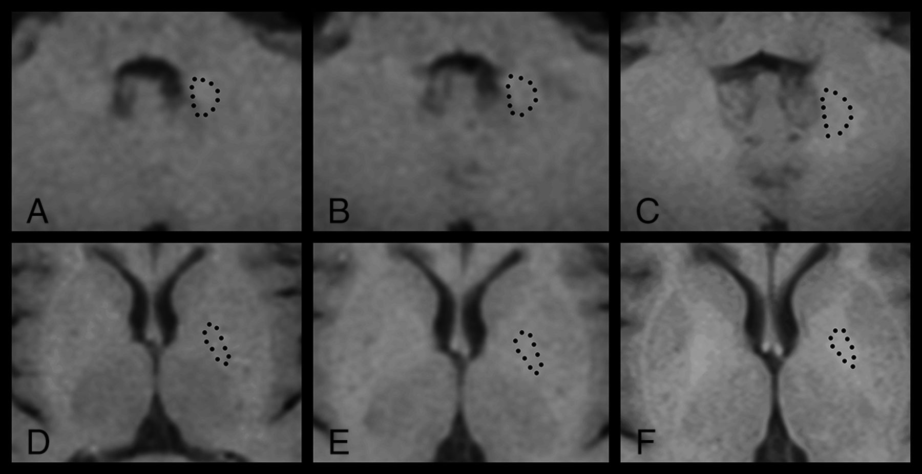
As detailed in the Table, the SII in the DN was higher in patients with MS than in healthy controls (P < .001, by the unpaired t test), while there was no significant difference for the GP (P = .19). Within the MS group, an increased number of GBCA administrations was associated with increased SIIs in both the DN (β = 0.45, P < .001) and GP (β = 0.60, P < .001), illustrated in Fig 1. The associations between high SIIs in the DN (β = 0.45, P < .001) and GP (β = 0.58, P < .001) remained significant when using only the number of linear GBCAs as an independent variable. When adding corrections for MS disease duration and EDSS, a higher number of GBCA administrations was still associated with higher SIIs in the DN (β = 0.43, P < .001) and GP (β = 0.58, P < .001). When we analyzed the later time points when lesion volume and brain parenchymal fraction were available (as a further characterization of disease severity) and by adding correction for those factors in the regression model, a higher number of GBCA administrations remained associated with higher SIIs in the DN (β = 0.39, P = .007) and GP (β = 0.64, P < .001). Figure 2 illustrates the longitudinal increase in DN signal intensity in relation to more GBCA administrations in 1 patient with MS.

Fig 1.
  • Download figure
  • Open in new tab
  • Download powerpoint
Fig 1.

Scatterplots with linear regression lines showing that the number of GBCA administrations is associated with a higher SII in the DN and GP (β = 0.45, P < .001 and β = 0.60, P < .001, respectively) with corrections for age, scanner, and sequence type.

Fig 2.
  • Download figure
  • Open in new tab
  • Download powerpoint
Fig 2.

Longitudinal T1-weighted spin-echo signal-intensity changes in the DN (A–C) and GP (D–F) in a female patient with MS after receiving 0 (A and D), 1 (B and E), and 8 (C and F) GBCA administrations. This patient had an increasingly higher SII in both regions: DN 1.03 (A), 1.05(B), 1.09 (C), GP 1.00 (D), 1.12 (E), 1.16 (F). Dotted lines illustrate the outlining of the ROIs.

From a longitudinal analysis perspective, the average SII change between baseline and the 18-year follow-up in patients was 0.06 in the DN and 0.07 in the GP, equaling a change of 0.009 in both the DN and GP per additional GBCA administration. Repeated-measures ANOVA (with the Greenhouse-Geisser correction) showed that the SIIs were significantly different with respect to the number of administrations for the DN [F(2) = 22.98, P < .001] and the GP [F(1.07, 10.67= 6.29), P = .028]. Pair-wise comparisons were significant between 0 and 5–12 administrations for both the DN (1.01 ± 0.24 and 1.08 ± 0.032, P < .001) and the GP (0.96 ± 0.128 and 1.09 ± 0.53, P = .013). Similar trends were seen in the comparison between 0 and 1–4 administrations (DN, P = .049; GP, P = .064).

Associations with Cognition in Patients with MS

A high SII in the GP (β = −0.45, P < .001) was associated with low verbal fluency performance, and a similar trend was seen for the DN (β = −0.25, P = .03). After we corrected for disease duration and the EDSS, the associations were significant in both the DN (β = −0.28, P = .012) and GP (β = −0.49, P < .001). A higher SII in the GP (β = −0.35, P = .003), but not in the DN, was associated with low auditory verbal learning encoding (β = −0.35, P = .003) and low auditory verbal learning retention (β = −0.29, P = .01). These associations remained significant when correcting for disease duration and EDSS (encoding, β = −0.33, P = .006; retention, β = −0.34, P = .004). There were no significant associations between the SII in the DN or GP with either the Rey–Osterrieth Complex Figure Test or the Symbol Digit Modalities Test. After we corrected for disease duration and EDSS, a higher SII in the GP tended to be associated with a lower Symbol Digit Modalities Test score (β = −0.24, P = .043). When we analyzed the later time points and added correction for lesion volume and brain parenchymal fraction, only the association between a higher SII and low verbal fluency remained significant in the DN (β = −0.40, P = .013), with a similar trend in the GP (β = −0.36, P = .034).

Discussion

In the current study, we show that an increasing number of GBCA administrations is longitudinally associated with a higher SII in both the DN and GP in patients with MS, adding to the growing body of evidence of gadolinium retention in the brain. We further show some initial tentative results regarding associations of the SII in these structures with cognitive performance, which remained significant for verbal fluency when correcting for MS disease severity.

Investigations on gadolinium retention in the brain have studied different types of GBCAs. Multiple studies have demonstrated associations between the SII in the DN and the number of linear GBCA administrations.23 More recently, a similar initial association was reported for macrocyclic GBCAs,24 and small amounts of retained gadolinium were recently found in human brain tissue after macrocyclic GBCA.13 However, more recent imaging studies regarding gadolinium retention support the use of macrocyclic GBCA as a safer choice even for patients with normal renal function.23,25,26 In our study, all patients received linear GBCAs, and 6 of 23 patients also received 1 administration of a macrocyclic GBCA. However, when we studied the patients with MS who received a linear GBCA (gadodiamide and gadopentetic acid) solely, the results were relatively unchanged.

Some studies have used the pons as a reference region for the DN, but here we chose the left middle cerebellar peduncle instead because it was easier to find nonlesioned areas for reference that were not affected by phase-dependent artifacts on T1-weighted spin-echo scans.9,11,25 In line with previous studies, the thalamus was used as a reference point for the GP with special care to avoid lesions, though we note that diffuse MS disease–related changes in the thalamus may be a possible confounder.27

We have tried to carefully mitigate the confounding effects of MS pathology or progression in our analysis. However, MS typically causes hypointensities in both white and gray matter on T1-weighted imaging,28 which would likely cause an underestimation of the SII rather than an overestimation in the ROI. On the other hand, we note that a hyperintense DN on native T1-weighted MR imaging has previously been reported in secondary-progressive MS.29 Whether these previous results have been confounded by GBCA administrations (before current knowledge) or actually reflect disease-related signal changes remains unclear. The different scanners and sequences used are a limitation but also unavoidable when performing a long-term study with such long-term follow-up because there is a clinical need for scanner upgrades with time. Because these patients were part of a prospective MS study, they had been scanned with the follow-up similar to that of MR imaging protocols for T1 spin-echo and MPRAGE, respectively, but on different scanners during the years. A subgroup analysis in a previous study did not show any significant difference when using different sequences.23 However, a later study showed that this significantly influenced the results, highlighting the importance of using the same sequences and scanners if possible.30,31 On the basis of these previous results, we therefore corrected the measurements for these factors to mitigate any scanner-related effects.

In the current study, we showed that a higher SII in the DN in patients with MS is associated with lower word fluency scores, which was the only cognitive measurement that remained associated with the SII after correction for several aspects of MS severity. Most interesting, executive function, including verbal fluency, is less frequently affected compared with episodic memory and information-processing speed in MS.32 Verbal fluency also declines after pallidotomy and cerebellar damage; this finding highlights the importance of these structures in verbal fluency.33,34 Furthermore, a recent study in MS showed that higher functional connectivity between the DN and prefrontal, motor, and parietal cortices is associated with lower structural parenchymal damage and less clinical impairment.35 However, many other areas of the brain are affected by MS, which may be more closely related to the decline in our neuropsychological test results.32 Naturally, it is of great clinical interest to explore any possible negative neurologic or cognitive adverse effects of gadolinium retention. It is, however, important to stress that our retrospective results must be interpreted with caution and do not necessarily imply causation.

It is not clear to what degree factors such as renal failure, liver dysfunction, and genetic variations might influence the risk of developing a higher SII in the brain after repeated GBCA administrations. MS is one of the largest patient groups receiving repeat GBCA administrations and may, therefore, be at special risk of accumulation in the brain.36 MS also has a negative impact on cognitive function.32 Consequently, it is difficult to separate the effects of MS progression and a hypothetic effect on cognition attributed to GBCA, though we statistically corrected for the potential impact of disease severity. Furthermore, disease duration, lesion volume, and brain parenchymal fraction might not fully adjust for an MS-dependent decline of brain function because MS is a heterogeneous disease with individualized treatment regimens, different progression rates, and cognitive reserve,37 which are not completely represented by these biomarkers. Last, another limitation was the lack of a matched MS group not exposed to GBCAs or solely to macrocyclic GBCAs, because it was not possible to find a large-enough group of such patients with a disease duration similar to that of the studied cohort.

The strengths of the study are the long follow-up with available clinical, laboratory, and radiologic information and a study population representative of the MS panorama with different subtypes, disease duration, and EDSS scores.

Conclusions

Our data add support to the existing framework that gadolinium may have lasting effects on the T1-weighted intensities in the DN and GP. Although we show that a higher SII is associated with worse verbal fluency scores, these results are only exploratory and have to be interpreted with caution. Future studies are important to further investigate these cognitive aspects and other clinical outcome variables in other cohorts with a repeat number of GBCA administrations to understand their clinical implications.

Acknowledgments

We thank the study participants for making this study possible.

Footnotes

  • Disclosures: Yngve Forslin—RELATED: Grant: ALF, Comments: This research was funded by Stockholm County Council and Karolinska Institutet through the regional agreement on medical training and clinical research (ALF grant)*. Farouk Hashim—RELATED: Grant: ALF*. Gösta Bergendal—RELATED: Grant: ALF*. Sten Fredrikson—UNRELATED: Board Membership: Chairman of the Educational and Examination Committee of the Swedish Neurological Society and Swedish delegate of the EFNS, European Board of Neurology and MSIF International Medical and Scientific Board; OTHER RELATIONSHIPS: received honorarium for lectures, educational activities, and/or consultancy from Genzyme/Sanofi, Merck, Novartis, and Teva Pharmaceutical Industries. Tobias Granberg—RELATED: Grant: Stockholm City Council*. *Money paid to the institution.

  • This research was funded by the Stockholm County Council and Karolinska Institutet through the regional agreement on medical training and clinical research.

Indicates open access to non-subscribers at www.ajnr.org

References

  1. 1.↵
    1. Lohrke J,
    2. Frenzel T,
    3. Endrikat J, et al
    . 25 years of contrast-enhanced MRI: developments, current challenges and future perspectives. Adv Ther 2016;33:1–28 doi:10.1007/s12325-015-0275-4 pmid:26809251
    CrossRefPubMed
  2. 2.↵
    1. Idée JM,
    2. Fretellier N,
    3. Robic C, et al
    . The role of gadolinium chelates in the mechanism of nephrogenic systemic fibrosis: a critical update. Crit Rev Toxicol 2014;44:895–913 doi:10.3109/10408444.2014.955568 pmid:25257840
    CrossRefPubMed
  3. 3.↵
    1. Frenzel T,
    2. Lengsfeld P,
    3. Schirmer H, et al
    . Stability of gadolinium-based magnetic resonance imaging contrast agents in human serum at 37 degrees C. Invest Radiol 2008;43:817–28 doi:10.1097/RLI.0b013e3181852171 pmid:19002053
    CrossRefPubMed
  4. 4.↵
    1. Thomsen HS,
    2. Morcos SK,
    3. Almén T, et al
    ; ESUR Contrast Medium Safety Committee. Nephrogenic systemic fibrosis and gadolinium-based contrast media: updated ESUR Contrast Medium Safety Committee guidelines. Eur Radiol 2013;23:307–18 doi:10.1007/s00330-012-2597-9 pmid:22865271
    CrossRefPubMed
  5. 5.↵
    1. White GW,
    2. Gibby WA,
    3. Tweedle MF
    . Comparison of Gd(DTPA-BMA) (Omniscan) versus Gd(HP-DO3A) (ProHance) relative to gadolinium retention in human bone tissue by inductively coupled plasma mass spectroscopy. Invest Radiol 2006;41:272–78 doi:10.1097/01.rli.0000186569.32408.95 pmid:16481910
    CrossRefPubMed
  6. 6.↵
    1. Idée JM,
    2. Port M,
    3. Dencausse A, et al
    . Involvement of gadolinium chelates in the mechanism of nephrogenic systemic fibrosis: an update. Radiol Clin North Am 2009;47:855–69, vii doi:10.1016/j.rcl.2009.06.006 pmid:19744600
    CrossRefPubMed
  7. 7.↵
    1. Roberts DR,
    2. Lindhorst SM,
    3. Welsh CT, et al
    . High levels of gadolinium deposition in the skin of a patient with normal renal function. Invest Radiol 2016;51:280–89 doi:10.1097/RLI.0000000000000266 pmid:26953564
    CrossRefPubMed
  8. 8.↵
    1. Kanda T,
    2. Ishii K,
    3. Kawaguchi H, et al
    . High signal intensity in the dentate nucleus and globus pallidus on unenhanced T1-weighted MR images: relationship with increasing cumulative dose of a gadolinium-based contrast material. Radiology 2014;270:834–41 doi:10.1148/radiol.13131669 pmid:24475844
    CrossRefPubMed
  9. 9.↵
    1. Errante Y,
    2. Cirimele V,
    3. Mallio CA, et al
    . Progressive increase of T1 signal intensity of the dentate nucleus on unenhanced magnetic resonance images is associated with cumulative doses of intravenously administered gadodiamide in patients with normal renal function, suggesting dechelation. Invest Radiol 2014;49:685–90 doi:10.1097/RLI.0000000000000072 pmid:24872007
    CrossRefPubMed
  10. 10.↵
    1. McDonald RJ,
    2. McDonald JS,
    3. Kallmes DF, et al
    . Intracranial gadolinium deposition after contrast-enhanced MR imaging. Radiology 2015;275:772–82 doi:10.1148/radiol.15150025 pmid:25742194
    CrossRefPubMed
  11. 11.↵
    1. Quattrocchi CC,
    2. Mallio CA,
    3. Errante Y, et al
    . Gadodiamide and dentate nucleus T1 hyperintensity in patients with meningioma evaluated by multiple follow-up contrast-enhanced magnetic resonance examinations with no systemic interval therapy. Invest Radiol 2015;50:470–72 doi:10.1097/RLI.0000000000000154 pmid:25756685
    CrossRefPubMed
  12. 12.↵
    1. Kanda T,
    2. Fukusato T,
    3. Matsuda M, et al
    . Gadolinium-based contrast agent accumulates in the brain even in subjects without severe renal dysfunction: evaluation of autopsy brain specimens with inductively coupled plasma mass spectroscopy. Radiology 2015;276:228–32 doi:10.1148/radiol.2015142690 pmid:25942417
    CrossRefPubMed
  13. 13.↵
    1. Murata N,
    2. Gonzalez-Cuyar LF,
    3. Murata K, et al
    . Macrocyclic and other non-group 1 gadolinium contrast agents deposit low levels of gadolinium in brain and bone tissue: preliminary results from 9 patients with normal renal function. Invest Radiol 2016;51:447–53 doi:10.1097/RLI.0000000000000252 pmid:26863577
    CrossRefPubMed
  14. 14.↵
    1. Robert P,
    2. Lehericy S,
    3. Grand S, et al
    . T1-weighted hypersignal in the deep cerebellar nuclei after repeated administrations of gadolinium-based contrast agents in healthy rats: difference between linear and macrocyclic agents. Invest Radiol 2015;50:473–80 doi:10.1097/RLI.0000000000000181 pmid:26107651
    CrossRefPubMed
  15. 15.↵
    1. Goischke HK
    . MRI with gadolinium-based contrast agents: practical help to ensure patient safety. J Am Coll Radiol 2016;13:890 doi:10.1016/j.jacr.2016.05.007 pmid:27325471
    CrossRefPubMed
  16. 16.↵
    1. Welk B,
    2. McArthur E,
    3. Morrow SA, et al
    . Association between gadolinium contrast exposure and the risk of parkinsonism. JAMA 2016;316:96–98 doi:10.1001/jama.2016.8096 pmid:27380348
    CrossRefPubMed
  17. 17.↵
    1. Poser CM,
    2. Paty DW,
    3. Scheinberg L, et al
    . New diagnostic criteria for multiple sclerosis: guidelines for research protocols. Ann Neurol 1983;13:227–31 doi:10.1002/ana.410130302 pmid:6847134
    CrossRefPubMed
  18. 18.↵
    1. Reuter M,
    2. Schmansky NJ,
    3. Rosas HD, et al
    . Within-subject template estimation for unbiased longitudinal image analysis. Neuroimage 2012;61:1402–18 doi:10.1016/j.neuroimage.2012.02.084 pmid:22430496
    CrossRefPubMed
  19. 19.↵
    1. Schmidt P,
    2. Gaser C,
    3. Arsic M, et al
    . An automated tool for detection of FLAIR-hyperintense white-matter lesions in multiple sclerosis. Neuroimage 2012;59:3774–83 doi:10.1016/j.neuroimage.2011.11.032 pmid:22119648
    CrossRefPubMed
  20. 20.↵
    1. Kurtzke JF
    . Rating neurologic impairment in multiple sclerosis: an expanded disability status scale (EDSS). Neurology 1983;33:1444–52 doi:10.1212/WNL.33.11.1444 pmid:6685237
    Abstract/FREE Full Text
  21. 21.↵
    1. Lezak MD
    , ed. Neuropsychological Assessment. 5th ed. Oxford: Oxford University Press; 2012
  22. 22.↵
    1. Benjamini Y,
    2. Krieger AM,
    3. Yekutieli D
    . Adaptive linear step-up procedures that control the false discovery rate. Biometrika 2006;93:491–507 doi:10.1093/biomet/93.3.491
    Abstract/FREE Full Text
  23. 23.↵
    1. Radbruch A,
    2. Weberling LD,
    3. Kieslich PJ, et al
    . Gadolinium retention in the dentate nucleus and globus pallidus is dependent on the class of contrast agent. Radiology 2015;275:783–91 doi:10.1148/radiol.2015150337 pmid:25848905
    CrossRefPubMed
  24. 24.↵
    1. Stojanov DA,
    2. Aracki-Trenkic A,
    3. Vojinovic S, et al
    . Increasing signal intensity within the dentate nucleus and globus pallidus on unenhanced T1W magnetic resonance images in patients with relapsing-remitting multiple sclerosis: correlation with cumulative dose of a macrocyclic gadolinium-based contrast agent, gadobutrol. Eur Radiol 2016;26:807–15 doi:10.1007/s00330-015-3879-9 pmid:26105022
    CrossRefPubMed
  25. 25.↵
    1. Kanda T,
    2. Osawa M,
    3. Oba H, et al
    . High signal intensity in dentate nucleus on unenhanced T1-weighted MR images: association with linear versus macrocyclic gadolinium chelate administration. Radiology 2015;275:803–09 doi:10.1148/radiol.14140364 pmid:25633504
    CrossRefPubMed
  26. 26.↵
    1. Thomsen HS
    . T1 hyperintensity in the brain after multiple intravenous injections of gadolinium-based contrast agents. Acta Radiol 2016;57:389–91 doi:10.1177/0284185115626479 pmid:26792824
    CrossRefPubMed
  27. 27.↵
    1. Minagar A,
    2. Barnett MH,
    3. Benedict RH, et al
    . The thalamus and multiple sclerosis: modern views on pathologic, imaging, and clinical aspects. Neurology 2013;80:210–19 doi:10.1212/WNL.0b013e31827b910b pmid:23296131
    Abstract/FREE Full Text
  28. 28.↵
    1. Filippi M,
    2. Rocca MA,
    3. Barkhof F, et al
    ; Attendees of the Correlation between Pathological MRI Findings in MS Workshop. Association between pathological and MRI findings in multiple sclerosis. Lancet Neurol 2012;11:349–60 doi:10.1016/S1474-4422(12)70003-0 pmid:22441196
    CrossRefPubMed
  29. 29.↵
    1. Roccatagliata L,
    2. Vuolo L,
    3. Bonzano L, et al
    . Multiple sclerosis: hyperintense dentate nucleus on unenhanced T1-weighted MR images is associated with the secondary progressive subtype. Radiology 2009;251:503–10 doi:10.1148/radiol.2511081269 pmid:19401576
    CrossRefPubMed
  30. 30.↵
    1. Ramalho J,
    2. Ramalho M,
    3. AlObaidy M, et al
    . T1 signal-intensity increase in the dentate nucleus after multiple exposures to gadodiamide: intraindividual comparison between 2 commonly used sequences. AJNR Am J Neuroradiol 2016;37:1427–31 doi:10.3174/ajnr.A4757 pmid:27032972
    Abstract/FREE Full Text
  31. 31.↵
    1. Ramalho J,
    2. Ramalho M,
    3. AlObaidy M, et al
    . Technical aspects of MRI signal change quantification after gadolinium-based contrast agents' administration. Magn Reson Imaging 2016;34:1355–58 doi:10.1016/j.mri.2016.09.004 pmid:27693606
    CrossRefPubMed
  32. 32.↵
    1. Rocca MA,
    2. Amato MP,
    3. Enzinger C, et al
    ; MAGNIMS Study Group. Clinical and imaging assessment of cognitive dysfunction in multiple sclerosis. Lancet Neurol 2015;14:302–17 doi:10.1016/S1474-4422(14)70250-9 pmid:25662900
    CrossRefPubMed
  33. 33.↵
    1. Tröster AI,
    2. Woods SP,
    3. Fields JA
    . Verbal fluency declines after pallidotomy: an interaction between task and lesion laterality. Appl Neuropsychol 2003;10:69–75 doi:10.1207/S15324826AN1002_02 pmid:12788681
    CrossRefPubMed
  34. 34.↵
    1. Mariën P,
    2. Ackermann H,
    3. Adamaszek M, et al
    . Consensus paper: language and the cerebellum—an ongoing enigma. Cerebellum 2014;13:386–410 doi:10.1007/s12311-013-0540-5 pmid:24318484
    CrossRefPubMed
  35. 35.↵
    1. Sbardella E,
    2. Upadhyay N,
    3. Tona F, et al
    . Dentate nucleus connectivity in adult patients with multiple sclerosis: functional changes at rest and correlation with clinical features. Mult Scler 2017;23:546–55 doi:10.1177/1352458516657438 pmid:27411700
    CrossRefPubMed
  36. 36.↵
    1. Filippi M,
    2. Rocca MA,
    3. De Stefano N, et al
    . Magnetic resonance techniques in multiple sclerosis: the present and the future. Arch Neurol 2011;68:1514–20 doi:10.1001/archneurol.2011.914 pmid:22159052
    CrossRefPubMed
  37. 37.↵
    1. Sandroff BM,
    2. Schwartz CE,
    3. DeLuca J
    . Measurement and maintenance of reserve in multiple sclerosis. J Neurol 2016;263:2158–69 doi:10.1007/s00415-016-8104-05 pmid:27072141
    CrossRefPubMed
  • Received October 22, 2016.
  • Accepted after revision March 3, 2017.
  • © 2017 by American Journal of Neuroradiology
View Abstract
PreviousNext
Back to top

In this issue

American Journal of Neuroradiology: 38 (7)
American Journal of Neuroradiology
Vol. 38, Issue 7
1 Jul 2017
  • Table of Contents
  • Index by author
  • Complete Issue (PDF)
Advertisement
Print
Download PDF
Email Article

Thank you for your interest in spreading the word on American Journal of Neuroradiology.

NOTE: We only request your email address so that the person you are recommending the page to knows that you wanted them to see it, and that it is not junk mail. We do not capture any email address.

Enter multiple addresses on separate lines or separate them with commas.
Retention of Gadolinium-Based Contrast Agents in Multiple Sclerosis: Retrospective Analysis of an 18-Year Longitudinal Study
(Your Name) has sent you a message from American Journal of Neuroradiology
(Your Name) thought you would like to see the American Journal of Neuroradiology web site.
CAPTCHA
This question is for testing whether or not you are a human visitor and to prevent automated spam submissions.
Cite this article
Y. Forslin, S. Shams, F. Hashim, P. Aspelin, G. Bergendal, J. Martola, S. Fredrikson, M. Kristoffersen-Wiberg, T. Granberg
Retention of Gadolinium-Based Contrast Agents in Multiple Sclerosis: Retrospective Analysis of an 18-Year Longitudinal Study
American Journal of Neuroradiology Jul 2017, 38 (7) 1311-1316; DOI: 10.3174/ajnr.A5211

Citation Manager Formats

  • BibTeX
  • Bookends
  • EasyBib
  • EndNote (tagged)
  • EndNote 8 (xml)
  • Medlars
  • Mendeley
  • Papers
  • RefWorks Tagged
  • Ref Manager
  • RIS
  • Zotero
0 Responses
Respond to this article
Share
Bookmark this article
Retention of Gadolinium-Based Contrast Agents in Multiple Sclerosis: Retrospective Analysis of an 18-Year Longitudinal Study
Y. Forslin, S. Shams, F. Hashim, P. Aspelin, G. Bergendal, J. Martola, S. Fredrikson, M. Kristoffersen-Wiberg, T. Granberg
American Journal of Neuroradiology Jul 2017, 38 (7) 1311-1316; DOI: 10.3174/ajnr.A5211
del.icio.us logo Twitter logo Facebook logo Mendeley logo
  • Tweet Widget
  • Facebook Like
  • Google Plus One
Purchase

Jump to section

  • Article
    • Abstract
    • ABBREVIATIONS:
    • Materials and Methods
    • Results
    • Discussion
    • Conclusions
    • Acknowledgments
    • Footnotes
    • References
  • Figures & Data
  • Supplemental
  • Info & Metrics
  • Responses
  • References
  • PDF

Related Articles

  • PubMed
  • Google Scholar

Cited By...

  • MR Imaging Signs of Gadolinium Retention Are Not Associated with Long-Term Motor and Cognitive Outcomes in Multiple Sclerosis
  • Does Gadolinium Deposition Lead to Metabolite Alteration in the Dentate Nucleus? An MRS Study in Patients with MS
  • Safety of Gadolinium-Based Contrast Agents in Patients with Stage 4 and 5 Chronic Kidney Disease: a Radiologists Perspective
  • Cumulative gadodiamide administration leads to brain gadolinium deposition in early MS
  • Gadolinium Retention in the Brain: An MRI Relaxometry Study of Linear and Macrocyclic Gadolinium-Based Contrast Agents in Multiple Sclerosis
  • Signal Hyperintensity on Unenhanced T1-Weighted Brain and Cervical Spinal Cord MR Images after Multiple Doses of Linear Gadolinium-Based Contrast Agent
  • Crossref (48)
  • Google Scholar

This article has been cited by the following articles in journals that are participating in Crossref Cited-by Linking.

  • Gadolinium Retention: A Research Roadmap from the 2018 NIH/ACR/RSNA Workshop on Gadolinium Chelates
    Robert J. McDonald, Deborah Levine, Jeffrey Weinreb, Emanuel Kanal, Matthew S. Davenport, James H. Ellis, Paula M. Jacobs, Robert E. Lenkinski, Kenneth R. Maravilla, Martin R. Prince, Howard A. Rowley, Michael F. Tweedle, Herbert Y. Kressel
    Radiology 2018 289 2
  • Gadolinium-based Contrast Media, Cerebrospinal Fluid and the Glymphatic System: Possible Mechanisms for the Deposition of Gadolinium in the Brain
    Toshiaki Taoka, Shinji Naganawa
    Magnetic Resonance in Medical Sciences 2018 17 2
  • Gadolinium retention after administration of contrast agents based on linear chelators and the recommendations of the European Medicines Agency
    Ilona A. Dekkers, Rick Roos, Aart J. van der Molen
    European Radiology 2018 28 4
  • Gadolinium-Based MRI Contrast Agents Induce Mitochondrial Toxicity and Cell Death in Human Neurons, and Toxicity Increases With Reduced Kinetic Stability of the Agent
    Danielle V. Bower, Johannes K. Richter, Hendrik von Tengg-Kobligk, Johannes T. Heverhagen, Val M. Runge
    Investigative Radiology 2019 54 8
  • Gadolinium: pharmacokinetics and toxicity in humans and laboratory animals following contrast agent administration
    Julie Davies, Petra Siebenhandl-Wolff, Francois Tranquart, Paul Jones, Paul Evans
    Archives of Toxicology 2022 96 2
  • Gadolinium-based contrast agents — review of recent literature on magnetic resonance imaging signal intensity changes and tissue deposits, with emphasis on pediatric patients
    Einat Blumfield, David W. Swenson, Ramesh S. Iyer, A. Luana Stanescu
    Pediatric Radiology 2019 49 4
  • Gadolinium-based contrast agents in children
    Michael N. Rozenfeld, Daniel J. Podberesky
    Pediatric Radiology 2018 48 9
  • A Review of the Current Evidence on Gadolinium Deposition in the Brain
    Richard Pullicino, Mark Radon, Shubhabrata Biswas, Maneesh Bhojak, Kumar Das
    Clinical Neuroradiology 2018 28 2
  • Exposure to gadolinium and neurotoxicity: current status of preclinical and clinical studies
    Carlo A. Mallio, Àlex Rovira, Paul M. Parizel, Carlo C. Quattrocchi
    Neuroradiology 2020 62 8
  • Current and Future MR Contrast Agents
    Eric Lancelot, Jean-Sébastien Raynaud, Pierre Desché
    Investigative Radiology 2020 55 9

More in this TOC Section

ADULT BRAIN

  • Diagnostic Neuroradiology of Monoclonal Antibodies
  • Clinical Outcomes After Chiari I Decompression
  • Segmentation of Brain Metastases with BLAST
Show more ADULT BRAIN

PATIENT SAFETY

  • Safety of Intrathecal Gadobutrol in Various Doses
  • Impact of Kidney Function on CNS Gadolinium Deposition in Patients Receiving Repeated Doses of Gadobutrol
  • Contrast-Induced Acute Kidney Injury in Radiologic Management of Acute Ischemic Stroke in the Emergency Setting
Show more PATIENT SAFETY

Similar Articles

Advertisement

Indexed Content

  • Current Issue
  • Accepted Manuscripts
  • Article Preview
  • Past Issues
  • Editorials
  • Editor's Choice
  • Fellows' Journal Club
  • Letters to the Editor
  • Video Articles

Cases

  • Case Collection
  • Archive - Case of the Week
  • Archive - Case of the Month
  • Archive - Classic Case

More from AJNR

  • Trainee Corner
  • Imaging Protocols
  • MRI Safety Corner
  • Book Reviews

Multimedia

  • AJNR Podcasts
  • AJNR Scantastics

Resources

  • Turnaround Time
  • Submit a Manuscript
  • Submit a Video Article
  • Submit an eLetter to the Editor/Response
  • Manuscript Submission Guidelines
  • Statistical Tips
  • Fast Publishing of Accepted Manuscripts
  • Graphical Abstract Preparation
  • Imaging Protocol Submission
  • Evidence-Based Medicine Level Guide
  • Publishing Checklists
  • Author Policies
  • Become a Reviewer/Academy of Reviewers
  • News and Updates

About Us

  • About AJNR
  • Editorial Board
  • Editorial Board Alumni
  • Alerts
  • Permissions
  • Not an AJNR Subscriber? Join Now
  • Advertise with Us
  • Librarian Resources
  • Feedback
  • Terms and Conditions
  • AJNR Editorial Board Alumni

American Society of Neuroradiology

  • Not an ASNR Member? Join Now

© 2025 by the American Society of Neuroradiology All rights, including for text and data mining, AI training, and similar technologies, are reserved.
Print ISSN: 0195-6108 Online ISSN: 1936-959X

Powered by HighWire