
Article Figures & Data
Figures
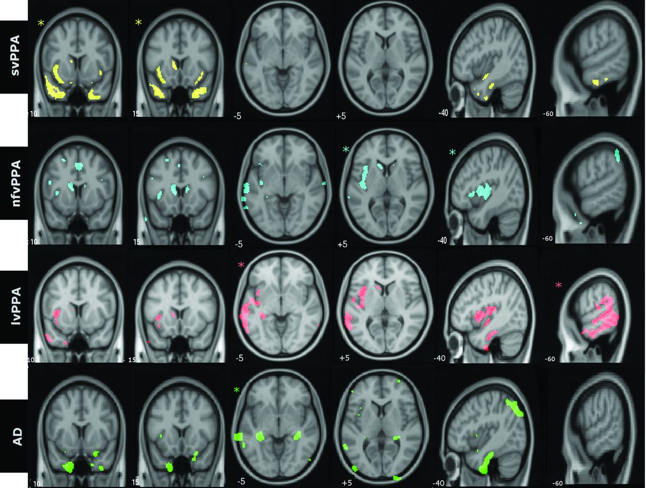
- Fig 1.
Group-level patterns of atrophy in identical axial, sagittal, and coronal sections of the brain. Images are displayed in neurologic orientation. Asterisks demonstrate the section most representative for the particular groups. All comparisons were made at false discovery rate–corrected P < .01.
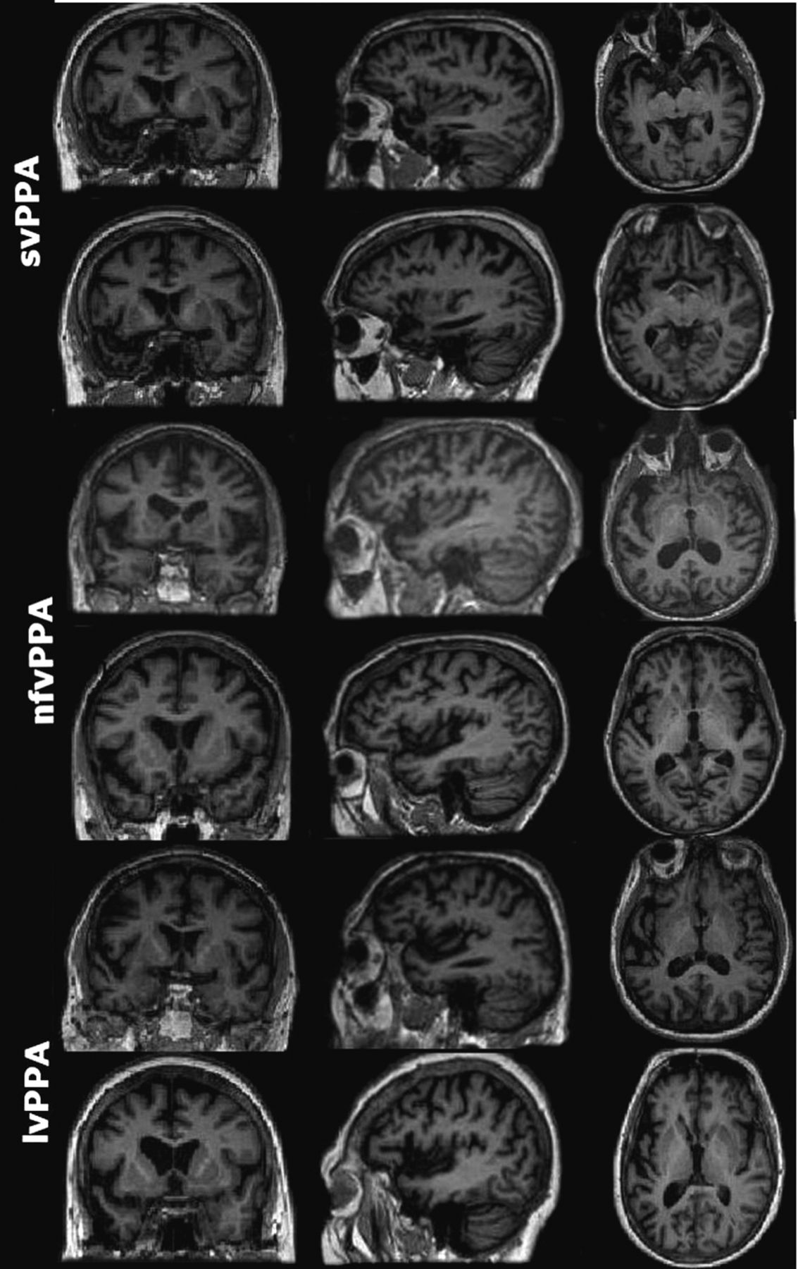
- Fig 2.
Representative sections of MR images of 6 patients with PPA (2 per subtype) with comparable Mini-Mental State Examination scores. Both patients with svPPA were reported by all neuroradiologists as having left anterior temporal atrophy. None of the patients with nfvPPA or lvPPA in the study had unanimous reports of the prescribed atrophy patterns (left posterior frontoinsular and posterior peri-Sylvian/inferior parietal, respectively). Images are displayed in neurologic orientation.


Tables
Demographics svPPA Mean (Range) nfvPPA Mean (Range) lvPPA Mean (Range) AD Mean (Range) Control Mean (Range) Omnibus Sig P Value Age at test (yr) 67 (60–79) 68.9 (53–79) 70.8 (60–83) 68 (60–79) 67.5 (51–80) NS Disease duration (mo)a 56 (24–108) 38.57 (18–60) 48.7 (24–108) 58 (24–96) NA NS Education (yr)a 13.6 (10–18) 12.6 (10–20) 11.6 (9–16) 12.5 (10–19) 12.8 (10–20) NS Sex 11 M, 10 F 5 M, 9 F 6 M, 10 F 12 M, 13 F 11 M, 15 F – ADL-Q 1 (0–5) 0.53 (0–4) 0.68 (0–4) 2.3 (0–6) NA NS Note:—ADL-Q indicates Activities of Daily Living Questionnaire; Sig, significant; NA, not applicable; NS, not significant.
↵a Nonparametric test.
- Table 2:
Sensitivity of the proposed imaging markers for the diagnosis of PPA variants and typical AD based on the lobar distribution of the atrophy and specific consensus recommendations
svPPA nfvPPA lvPPA AD Sensitivity based on lobar distribution Rater 1 100% 50% 50% 36% Rater 2 95% 8% 57% 64% Rater 3 100% 29% 64% 60% Mean (SD) 98% (2.9%) 29% (21%) 57% (7%) 53% (15%) Sensitivity based on recommendations Rater 1 90% 14% 50% 60% Rater 2 92% 20% 56% 46% Rater 3 95% 28% 42% 24% Mean (SD) 92% (2.5%) 21% (7%) 49% (7%) 43% (18%) - Table 3:
Specificity of the proposed imaging markers for the diagnosis of PPA variants and typical AD based on the specific consensus recommendations
Specificity Based on Recommendations svPPA nfvPPA lvPPA AD Rater 1 93% 92% 95% 93% Rater 2 95% 89% 93% 92% Rater 3 93% 92% 97% 91% Mean (SD) 93% (0.01) 91% (0.02) 95% (0.02) 92% (0.01) - Table 4:
Intraobserver agreement for the reported lobar distribution of abnormalities and recommendations
Rater 1 (κ) (SE) Rater 2 (κ) (SE) Rater 3 (κ) (SE) Mean (SD) Lobar distribution 0.61 (0.1) 0.95 (0.04) 0.68 (0.1) 0.75 (0.18) Recommendations 0.5 (0.09) 0.81 (0.07) 0.57 (0.1) 0.63 (0.16) - Table 5:
Interobserver agreement for the reported lobar distribution of abnormalities and recommendations
Raters 1 and 2 (κ) (SE) Raters 1 and 3 (κ) (SE) Raters 2 and 3 (κ) (SE) Mean (SD) Lobar distribution 0.56 (0.06) 0.48 (0.06) 0.64 (0.06) 0.56 (0.08) Recommendations 0.31 (0.07) 0.47 (0.07) 0.44 (0.06) 0.41 (0.09)


