Advertisement

Main menu

  • Home
  • Content
    • Current Issue
    • Accepted Manuscripts
    • Article Preview
    • Past Issue Archive
    • Video Articles
    • AJNR Case Collection
    • Case of the Week Archive
    • Case of the Month Archive
    • Classic Case Archive
  • Special Collections
    • AJNR Awards
    • Low-Field MRI
    • Alzheimer Disease
    • ASNR Foundation Special Collection
    • Photon-Counting CT
    • View All
  • Multimedia
    • AJNR Podcasts
    • AJNR SCANtastic
    • Trainee Corner
    • MRI Safety Corner
    • Imaging Protocols
  • For Authors
    • Submit a Manuscript
    • Submit a Video Article
    • Submit an eLetter to the Editor/Response
    • Manuscript Submission Guidelines
    • Statistical Tips
    • Fast Publishing of Accepted Manuscripts
    • Graphical Abstract Preparation
    • Imaging Protocol Submission
    • Author Policies
  • About Us
    • About AJNR
    • Editorial Board
    • Editorial Board Alumni
  • More
    • Become a Reviewer/Academy of Reviewers
    • Subscribers
    • Permissions
    • Alerts
    • Feedback
    • Advertisers
    • ASNR Home

User menu

  • Alerts
  • Log in

Search

  • Advanced search
American Journal of Neuroradiology

American Journal of Neuroradiology

ASHNR American Society of Functional Neuroradiology ASHNR American Society of Pediatric Neuroradiology ASSR
  • Alerts
  • Log in

Advanced Search

  • Home
  • Content
    • Current Issue
    • Accepted Manuscripts
    • Article Preview
    • Past Issue Archive
    • Video Articles
    • AJNR Case Collection
    • Case of the Week Archive
    • Case of the Month Archive
    • Classic Case Archive
  • Special Collections
    • AJNR Awards
    • Low-Field MRI
    • Alzheimer Disease
    • ASNR Foundation Special Collection
    • Photon-Counting CT
    • View All
  • Multimedia
    • AJNR Podcasts
    • AJNR SCANtastic
    • Trainee Corner
    • MRI Safety Corner
    • Imaging Protocols
  • For Authors
    • Submit a Manuscript
    • Submit a Video Article
    • Submit an eLetter to the Editor/Response
    • Manuscript Submission Guidelines
    • Statistical Tips
    • Fast Publishing of Accepted Manuscripts
    • Graphical Abstract Preparation
    • Imaging Protocol Submission
    • Author Policies
  • About Us
    • About AJNR
    • Editorial Board
    • Editorial Board Alumni
  • More
    • Become a Reviewer/Academy of Reviewers
    • Subscribers
    • Permissions
    • Alerts
    • Feedback
    • Advertisers
    • ASNR Home
  • Follow AJNR on Twitter
  • Visit AJNR on Facebook
  • Follow AJNR on Instagram
  • Join AJNR on LinkedIn
  • RSS Feeds

AJNR Awards, New Junior Editors, and more. Read the latest AJNR updates

Research ArticleADULT BRAIN

A Multiparametric Model for Mapping Cellularity in Glioblastoma Using Radiographically Localized Biopsies

P.D. Chang, H.R. Malone, S.G. Bowden, D.S. Chow, B.J.A. Gill, T.H. Ung, J. Samanamud, Z.K. Englander, A.M. Sonabend, S.A. Sheth, G.M. McKhann, M.B. Sisti, L.H. Schwartz, A. Lignelli, J. Grinband, J.N. Bruce and P. Canoll
American Journal of Neuroradiology May 2017, 38 (5) 890-898; DOI: https://doi.org/10.3174/ajnr.A5112
P.D. Chang
aFrom the Departments of Radiology (P.D.C., L.H.S., A.L., J.G.)
  • Find this author on Google Scholar
  • Find this author on PubMed
  • Search for this author on this site
  • ORCID record for P.D. Chang
H.R. Malone
bNeurological Surgery (H.R.M., S.G.B., B.J.A.G., T.H.U., Z.K.E., A.M.S., S.A.S., G.M.M., M.B.S., J.N.B.)
dGabriele Bartoli Brain Tumor Laboratory and the Irving Cancer Research Center (H.R.M., S.G.B., B.J.A.G., T.H.U., J.S., Z.K.E., A.M.S., J.N.B., P.C.), New York, New York
  • Find this author on Google Scholar
  • Find this author on PubMed
  • Search for this author on this site
  • ORCID record for H.R. Malone
S.G. Bowden
bNeurological Surgery (H.R.M., S.G.B., B.J.A.G., T.H.U., Z.K.E., A.M.S., S.A.S., G.M.M., M.B.S., J.N.B.)
dGabriele Bartoli Brain Tumor Laboratory and the Irving Cancer Research Center (H.R.M., S.G.B., B.J.A.G., T.H.U., J.S., Z.K.E., A.M.S., J.N.B., P.C.), New York, New York
  • Find this author on Google Scholar
  • Find this author on PubMed
  • Search for this author on this site
  • ORCID record for S.G. Bowden
D.S. Chow
eDepartment of Radiology (D.S.C.), University of San Francisco School of Medicine, San Francisco, California.
  • Find this author on Google Scholar
  • Find this author on PubMed
  • Search for this author on this site
  • ORCID record for D.S. Chow
B.J.A. Gill
bNeurological Surgery (H.R.M., S.G.B., B.J.A.G., T.H.U., Z.K.E., A.M.S., S.A.S., G.M.M., M.B.S., J.N.B.)
dGabriele Bartoli Brain Tumor Laboratory and the Irving Cancer Research Center (H.R.M., S.G.B., B.J.A.G., T.H.U., J.S., Z.K.E., A.M.S., J.N.B., P.C.), New York, New York
  • Find this author on Google Scholar
  • Find this author on PubMed
  • Search for this author on this site
  • ORCID record for B.J.A. Gill
T.H. Ung
bNeurological Surgery (H.R.M., S.G.B., B.J.A.G., T.H.U., Z.K.E., A.M.S., S.A.S., G.M.M., M.B.S., J.N.B.)
dGabriele Bartoli Brain Tumor Laboratory and the Irving Cancer Research Center (H.R.M., S.G.B., B.J.A.G., T.H.U., J.S., Z.K.E., A.M.S., J.N.B., P.C.), New York, New York
  • Find this author on Google Scholar
  • Find this author on PubMed
  • Search for this author on this site
  • ORCID record for T.H. Ung
J. Samanamud
dGabriele Bartoli Brain Tumor Laboratory and the Irving Cancer Research Center (H.R.M., S.G.B., B.J.A.G., T.H.U., J.S., Z.K.E., A.M.S., J.N.B., P.C.), New York, New York
  • Find this author on Google Scholar
  • Find this author on PubMed
  • Search for this author on this site
  • ORCID record for J. Samanamud
Z.K. Englander
bNeurological Surgery (H.R.M., S.G.B., B.J.A.G., T.H.U., Z.K.E., A.M.S., S.A.S., G.M.M., M.B.S., J.N.B.)
dGabriele Bartoli Brain Tumor Laboratory and the Irving Cancer Research Center (H.R.M., S.G.B., B.J.A.G., T.H.U., J.S., Z.K.E., A.M.S., J.N.B., P.C.), New York, New York
  • Find this author on Google Scholar
  • Find this author on PubMed
  • Search for this author on this site
  • ORCID record for Z.K. Englander
A.M. Sonabend
bNeurological Surgery (H.R.M., S.G.B., B.J.A.G., T.H.U., Z.K.E., A.M.S., S.A.S., G.M.M., M.B.S., J.N.B.)
dGabriele Bartoli Brain Tumor Laboratory and the Irving Cancer Research Center (H.R.M., S.G.B., B.J.A.G., T.H.U., J.S., Z.K.E., A.M.S., J.N.B., P.C.), New York, New York
  • Find this author on Google Scholar
  • Find this author on PubMed
  • Search for this author on this site
  • ORCID record for A.M. Sonabend
S.A. Sheth
bNeurological Surgery (H.R.M., S.G.B., B.J.A.G., T.H.U., Z.K.E., A.M.S., S.A.S., G.M.M., M.B.S., J.N.B.)
  • Find this author on Google Scholar
  • Find this author on PubMed
  • Search for this author on this site
  • ORCID record for S.A. Sheth
G.M. McKhann II
bNeurological Surgery (H.R.M., S.G.B., B.J.A.G., T.H.U., Z.K.E., A.M.S., S.A.S., G.M.M., M.B.S., J.N.B.)
  • Find this author on Google Scholar
  • Find this author on PubMed
  • Search for this author on this site
  • ORCID record for G.M. McKhann II
M.B. Sisti
bNeurological Surgery (H.R.M., S.G.B., B.J.A.G., T.H.U., Z.K.E., A.M.S., S.A.S., G.M.M., M.B.S., J.N.B.)
  • Find this author on Google Scholar
  • Find this author on PubMed
  • Search for this author on this site
  • ORCID record for M.B. Sisti
L.H. Schwartz
aFrom the Departments of Radiology (P.D.C., L.H.S., A.L., J.G.)
  • Find this author on Google Scholar
  • Find this author on PubMed
  • Search for this author on this site
  • ORCID record for L.H. Schwartz
A. Lignelli
aFrom the Departments of Radiology (P.D.C., L.H.S., A.L., J.G.)
  • Find this author on Google Scholar
  • Find this author on PubMed
  • Search for this author on this site
  • ORCID record for A. Lignelli
J. Grinband
aFrom the Departments of Radiology (P.D.C., L.H.S., A.L., J.G.)
  • Find this author on Google Scholar
  • Find this author on PubMed
  • Search for this author on this site
  • ORCID record for J. Grinband
J.N. Bruce
bNeurological Surgery (H.R.M., S.G.B., B.J.A.G., T.H.U., Z.K.E., A.M.S., S.A.S., G.M.M., M.B.S., J.N.B.)
dGabriele Bartoli Brain Tumor Laboratory and the Irving Cancer Research Center (H.R.M., S.G.B., B.J.A.G., T.H.U., J.S., Z.K.E., A.M.S., J.N.B., P.C.), New York, New York
  • Find this author on Google Scholar
  • Find this author on PubMed
  • Search for this author on this site
  • ORCID record for J.N. Bruce
P. Canoll
cPathology and Cell Biology (P.C.), College of Physicians and Surgeons at Columbia University, New York, New York
dGabriele Bartoli Brain Tumor Laboratory and the Irving Cancer Research Center (H.R.M., S.G.B., B.J.A.G., T.H.U., J.S., Z.K.E., A.M.S., J.N.B., P.C.), New York, New York
  • Find this author on Google Scholar
  • Find this author on PubMed
  • Search for this author on this site
  • ORCID record for P. Canoll
  • Article
  • Figures & Data
  • Supplemental
  • Info & Metrics
  • Responses
  • References
  • PDF
Loading

Article Figures & Data

Figures

  • Tables
  • Fig 1.
    • Download figure
    • Open in new tab
    • Download powerpoint
    Fig 1.

    Whole-cell counting. A, Digitized, low-power magnification view of a single H&E-stained slide. Two representative 400× fields from this single tissue specimen of relatively lower (B) and higher (C) cell density illustrate the tissue heterogeneity present at a microscopic level. Stained cellular nuclei identified by the automated counting algorithm are outlined in green. D, The “heat map” demonstrates distribution of cell density at the level of a HPF throughout the tissue sample.

  • Fig 2.
    • Download figure
    • Open in new tab
    • Download powerpoint
    Fig 2.

    Cell-counting statistics. A, Comparison between manual and automated cell counts for 25 high-power fields of various cellular densities. Correlation is high (r = 0.984), suggesting that the automated algorithm accurately reflects manual counts. B, For each biopsy sample, the median cell density of all HPFs is compared with that of the 98th percentile. A relatively strong linear correlation is preserved (r = 0.901), suggesting that the 98th percentile cell density simply represents a linear translation of the median cell density. C, Correlation analysis is repeated for all percentiles (0–100). With the exception of extreme values, most percentiles retain a strong linear correlation (r > 90%) with the median cell density.
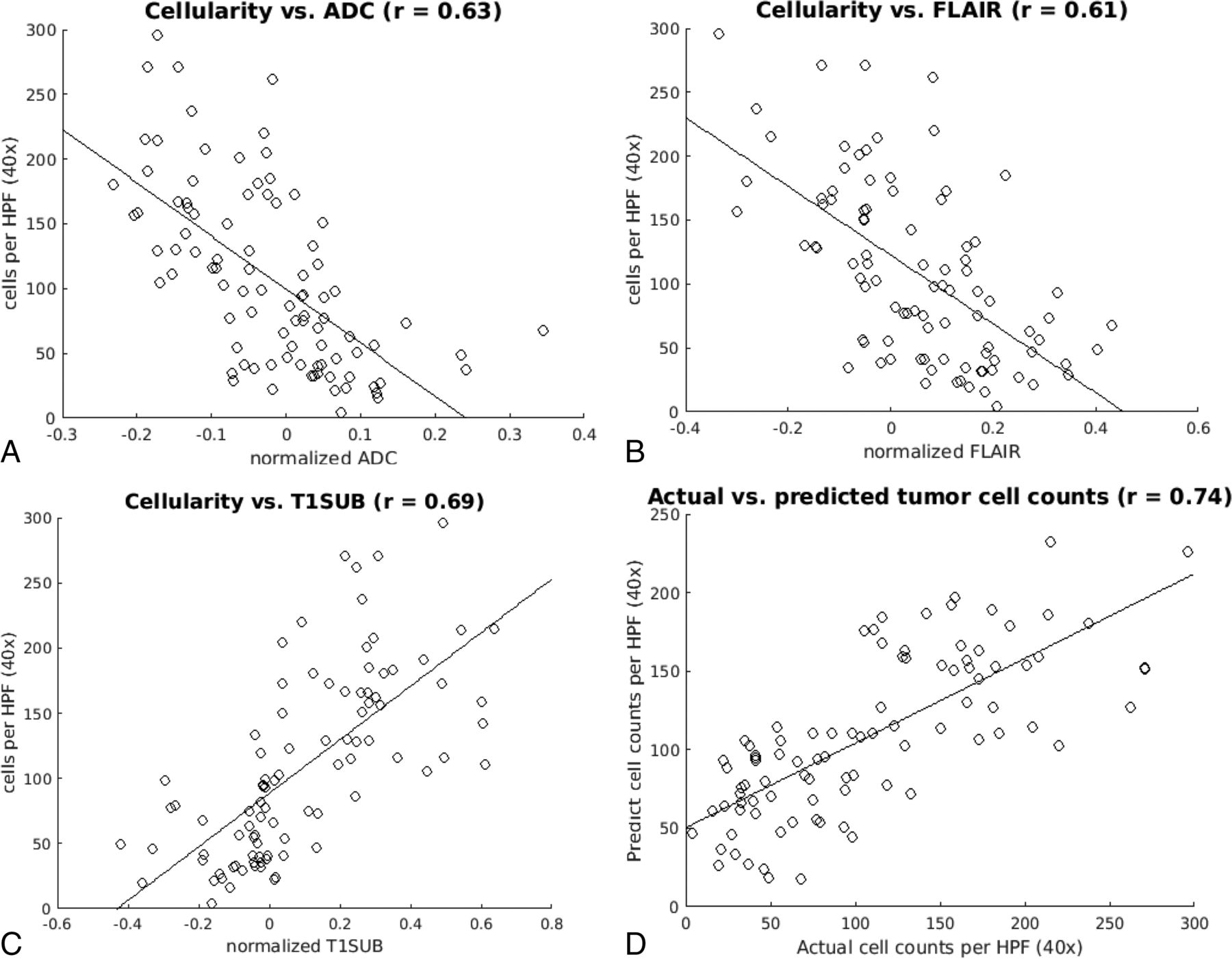
  • Fig 3.
    • Download figure
    • Open in new tab
    • Download powerpoint
    Fig 3.

    Cell count versus MR signal intensity. Scatterplots demonstrate median cell density as a function of signal intensity on ADC (A), T2-FLAIR (B), and T1-postcontrast subtraction sequences (C) correlated by using single-variable regression analysis. The linear regression and Pearson correlation (r) were significant (P < .05) for all 3 sequences. D, The scatterplot shows the actual and predicted cell counts as estimated by combining all 3 imaging modalities in a multiple-variable regression model.

  • Fig 4.
    • Download figure
    • Open in new tab
    • Download powerpoint
    Fig 4.

    Correlation versus distance from the biopsy. Scatterplots demonstrate the correlation between cell density and signal intensity for each MR image (ADC, T2-FLAIR, T1-postcontrast subtraction) obtained by taking the mean of concentric spheric shells of voxels at an increasing distance from the original biopsy point. Notably, the correlations drop to 0 at a radius of approximately 5 mm (∼10 voxels), providing an estimate of the spatial accuracy of the biopsy location. T1SUB indicates T1-subtraction.

  • Fig 5.
    • Download figure
    • Open in new tab
    • Download powerpoint
    Fig 5.

    Whole-tumor model overlay. Estimated cellularity by applying the multiple regression model on a voxelwise basis across the tumor. The model is derived from linear regression by using ADC, T2-FLAIR, and T1-postcontrast sequences shown in the inset on the left. In the right panels, corresponding biopsy specimens (400× magnification, H&E stained sections) are shown from 2 regions obtained on the same section, highlighting the considerable variation in cellularity in and around the region of contrast enhancement (demarcated by a white outline).

Tables

  • Figures
  • Multivariate linear regression model coefficients

    βSET-ScoreP Value
    Constant1025.9817.1<.001
    ADC−10632.0−3.30<.001
    FLAIR−56.023.5−2.38<.001
    T1-subtracted12924.65.27<.001
    • Note:—SE indicates standard error.

PreviousNext
Back to top

In this issue

American Journal of Neuroradiology: 38 (5)
American Journal of Neuroradiology
Vol. 38, Issue 5
1 May 2017
  • Table of Contents
  • Index by author
  • Complete Issue (PDF)
Advertisement
Print
Download PDF
Email Article

Thank you for your interest in spreading the word on American Journal of Neuroradiology.

NOTE: We only request your email address so that the person you are recommending the page to knows that you wanted them to see it, and that it is not junk mail. We do not capture any email address.

Enter multiple addresses on separate lines or separate them with commas.
A Multiparametric Model for Mapping Cellularity in Glioblastoma Using Radiographically Localized Biopsies
(Your Name) has sent you a message from American Journal of Neuroradiology
(Your Name) thought you would like to see the American Journal of Neuroradiology web site.
CAPTCHA
This question is for testing whether or not you are a human visitor and to prevent automated spam submissions.
Cite this article
P.D. Chang, H.R. Malone, S.G. Bowden, D.S. Chow, B.J.A. Gill, T.H. Ung, J. Samanamud, Z.K. Englander, A.M. Sonabend, S.A. Sheth, G.M. McKhann, M.B. Sisti, L.H. Schwartz, A. Lignelli, J. Grinband, J.N. Bruce, P. Canoll
A Multiparametric Model for Mapping Cellularity in Glioblastoma Using Radiographically Localized Biopsies
American Journal of Neuroradiology May 2017, 38 (5) 890-898; DOI: 10.3174/ajnr.A5112

Citation Manager Formats

  • BibTeX
  • Bookends
  • EasyBib
  • EndNote (tagged)
  • EndNote 8 (xml)
  • Medlars
  • Mendeley
  • Papers
  • RefWorks Tagged
  • Ref Manager
  • RIS
  • Zotero
0 Responses
Respond to this article
Share
Bookmark this article
A Multiparametric Model for Mapping Cellularity in Glioblastoma Using Radiographically Localized Biopsies
P.D. Chang, H.R. Malone, S.G. Bowden, D.S. Chow, B.J.A. Gill, T.H. Ung, J. Samanamud, Z.K. Englander, A.M. Sonabend, S.A. Sheth, G.M. McKhann, M.B. Sisti, L.H. Schwartz, A. Lignelli, J. Grinband, J.N. Bruce, P. Canoll
American Journal of Neuroradiology May 2017, 38 (5) 890-898; DOI: 10.3174/ajnr.A5112
del.icio.us logo Twitter logo Facebook logo Mendeley logo
  • Tweet Widget
  • Facebook Like
  • Google Plus One
Purchase

Jump to section

  • Article
    • Abstract
    • ABBREVIATIONS:
    • Materials and Methods
    • Results
    • Discussion
    • Conclusions
    • Footnotes
    • REFERENCES
  • Figures & Data
  • Supplemental
  • Info & Metrics
  • Responses
  • References
  • PDF

Related Articles

  • PubMed
  • Google Scholar

Cited By...

  • Advanced Distance-Resolved Evaluation of the Perienhancing Tumor Areas with FLAIR Hyperintensity Indicates Different ADC Profiles by MGMT Promoter Methylation Status in Glioblastoma
  • Imaging Genomics of Glioma Revisited: Analytic Methods to Understand Spatial and Temporal Heterogeneity
  • Biologically-informed deep neural networks provide quantitative assessment of intratumoral heterogeneity in post-treatment glioblastoma
  • Revealing the biology behind MRI signatures in high grade glioma
  • Interactions between ploidy and resource availability shape clonal interference at initiation and recurrence of glioblastoma
  • Cellular Density in Adult Glioma, Estimated with MR Imaging Data and a Machine Learning Algorithm, Has Prognostic Power Approaching World Health Organization Histologic Grading in a Cohort of 1181 Patients
  • Glioblastoma states are defined by cohabitating cellular populations with progression-, imaging- and sex-distinct patterns
  • Associations Between ADC Texture Analysis and Tumor Infiltrating Lymphocytes in Brain Metastasis - A Preliminary Study
  • Radio-pathomic maps of cell density identify glioma invasion beyond traditional MR imaging defined margins
  • Non-Contrast-Enhancing Tumor: A New Frontier in Glioblastoma Research
  • Accurate Patient-Specific Machine Learning Models of Glioblastoma Invasion Using Transfer Learning
  • Deep-Learning Convolutional Neural Networks Accurately Classify Genetic Mutations in Gliomas
  • Local Glioma Cells Are Associated with Vascular Dysregulation
  • Radiomics in Brain Tumor: Image Assessment, Quantitative Feature Descriptors, and Machine-Learning Approaches
  • Crossref (91)
  • Google Scholar

This article has been cited by the following articles in journals that are participating in Crossref Cited-by Linking.

  • Artificial intelligence in cancer imaging: Clinical challenges and applications
    Wenya Linda Bi, Ahmed Hosny, Matthew B. Schabath, Maryellen L. Giger, Nicolai J. Birkbak, Alireza Mehrtash, Tavis Allison, Omar Arnaout, Christopher Abbosh, Ian F. Dunn, Raymond H. Mak, Rulla M. Tamimi, Clare M. Tempany, Charles Swanton, Udo Hoffmann, Lawrence H. Schwartz, Robert J. Gillies, Raymond Y. Huang, Hugo J. W. L. Aerts
    CA: A Cancer Journal for Clinicians 2019 69 2
  • Deep-Learning Convolutional Neural Networks Accurately Classify Genetic Mutations in Gliomas
    P. Chang, J. Grinband, B.D. Weinberg, M. Bardis, M. Khy, G. Cadena, M.-Y. Su, S. Cha, C.G. Filippi, D. Bota, P. Baldi, L.M. Poisson, R. Jain, D. Chow
    American Journal of Neuroradiology 2018 39 7
  • Radiomics in Brain Tumor: Image Assessment, Quantitative Feature Descriptors, and Machine-Learning Approaches
    M. Zhou, J. Scott, B. Chaudhury, L. Hall, D. Goldgof, K.W. Yeom, M. Iv, Y. Ou, J. Kalpathy-Cramer, S. Napel, R. Gillies, O. Gevaert, R. Gatenby
    American Journal of Neuroradiology 2018 39 2
  • Emerging Applications of Artificial Intelligence in Neuro-Oncology
    Jeffrey D. Rudie, Andreas M. Rauschecker, R. Nick Bryan, Christos Davatzikos, Suyash Mohan
    Radiology 2019 290 3
  • Radiomic MRI Phenotyping of Glioblastoma: Improving Survival Prediction
    Sohi Bae, Yoon Seong Choi, Sung Soo Ahn, Jong Hee Chang, Seok-Gu Kang, Eui Hyun Kim, Se Hoon Kim, Seung-Koo Lee
    Radiology 2018 289 3
  • Fully automated hybrid approach to predict theIDHmutation status of gliomas via deep learning and radiomics
    Yoon Seong Choi, Sohi Bae, Jong Hee Chang, Seok-Gu Kang, Se Hoon Kim, Jinna Kim, Tyler Hyungtaek Rim, Seung Hong Choi, Rajan Jain, Seung-Koo Lee
    Neuro-Oncology 2021 23 2
  • Non-Contrast-Enhancing Tumor: A New Frontier in Glioblastoma Research
    A. Lasocki, F. Gaillard
    American Journal of Neuroradiology 2019 40 5
  • Imaging of intratumoral heterogeneity in high-grade glioma
    Leland S. Hu, Andrea Hawkins-Daarud, Lujia Wang, Jing Li, Kristin R. Swanson
    Cancer Letters 2020 477
  • Clinical applications of artificial intelligence and radiomics in neuro-oncology imaging
    Ahmed Abdel Khalek Abdel Razek, Ahmed Alksas, Mohamed Shehata, Amr AbdelKhalek, Khaled Abdel Baky, Ayman El-Baz, Eman Helmy
    Insights into Imaging 2021 12 1
  • Optimizing Neuro-Oncology Imaging: A Review of Deep Learning Approaches for Glioma Imaging
    Madeleine Shaver, Paul Kohanteb, Catherine Chiou, Michelle Bardis, Chanon Chantaduly, Daniela Bota, Christopher Filippi, Brent Weinberg, Jack Grinband, Daniel Chow, Peter Chang
    Cancers 2019 11 6

More in this TOC Section

  • Diagnostic Neuroradiology of Monoclonal Antibodies
  • Clinical Outcomes After Chiari I Decompression
  • Segmentation of Brain Metastases with BLAST
Show more ADULT BRAIN

Similar Articles

Advertisement

Indexed Content

  • Current Issue
  • Accepted Manuscripts
  • Article Preview
  • Past Issues
  • Editorials
  • Editor's Choice
  • Fellows' Journal Club
  • Letters to the Editor
  • Video Articles

Cases

  • Case Collection
  • Archive - Case of the Week
  • Archive - Case of the Month
  • Archive - Classic Case

More from AJNR

  • Trainee Corner
  • Imaging Protocols
  • MRI Safety Corner
  • Book Reviews

Multimedia

  • AJNR Podcasts
  • AJNR Scantastics

Resources

  • Turnaround Time
  • Submit a Manuscript
  • Submit a Video Article
  • Submit an eLetter to the Editor/Response
  • Manuscript Submission Guidelines
  • Statistical Tips
  • Fast Publishing of Accepted Manuscripts
  • Graphical Abstract Preparation
  • Imaging Protocol Submission
  • Evidence-Based Medicine Level Guide
  • Publishing Checklists
  • Author Policies
  • Become a Reviewer/Academy of Reviewers
  • News and Updates

About Us

  • About AJNR
  • Editorial Board
  • Editorial Board Alumni
  • Alerts
  • Permissions
  • Not an AJNR Subscriber? Join Now
  • Advertise with Us
  • Librarian Resources
  • Feedback
  • Terms and Conditions
  • AJNR Editorial Board Alumni

American Society of Neuroradiology

  • Not an ASNR Member? Join Now

© 2025 by the American Society of Neuroradiology All rights, including for text and data mining, AI training, and similar technologies, are reserved.
Print ISSN: 0195-6108 Online ISSN: 1936-959X

Powered by HighWire