Advertisement

Main menu

  • Home
  • Content
    • Current Issue
    • Accepted Manuscripts
    • Article Preview
    • Past Issue Archive
    • Video Articles
    • AJNR Case Collection
    • Case of the Week Archive
    • Case of the Month Archive
    • Classic Case Archive
  • Special Collections
    • Advancing NeuroMRI with High-Relaxivity Contrast Agents
    • AJNR Awards
    • Low-Field MRI
    • Alzheimer Disease
    • ASNR Foundation Special Collection
    • Photon-Counting CT
    • View All
  • Multimedia
    • AJNR Podcasts
    • AJNR SCANtastic
    • Trainee Corner
    • MRI Safety Corner
    • Imaging Protocols
  • For Authors
    • Submit a Manuscript
    • Submit a Video Article
    • Submit an eLetter to the Editor/Response
    • Manuscript Submission Guidelines
    • Statistical Tips
    • Fast Publishing of Accepted Manuscripts
    • Graphical Abstract Preparation
    • Imaging Protocol Submission
    • Author Policies
  • About Us
    • About AJNR
    • Editorial Board
    • Editorial Board Alumni
  • More
    • Become a Reviewer/Academy of Reviewers
    • Subscribers
    • Permissions
    • Alerts
    • Feedback
    • Advertisers
    • ASNR Home

User menu

  • Alerts
  • Log in

Search

  • Advanced search
American Journal of Neuroradiology

American Journal of Neuroradiology

ASHNR American Society of Functional Neuroradiology ASHNR American Society of Pediatric Neuroradiology ASSR
  • Alerts
  • Log in

Advanced Search

  • Home
  • Content
    • Current Issue
    • Accepted Manuscripts
    • Article Preview
    • Past Issue Archive
    • Video Articles
    • AJNR Case Collection
    • Case of the Week Archive
    • Case of the Month Archive
    • Classic Case Archive
  • Special Collections
    • Advancing NeuroMRI with High-Relaxivity Contrast Agents
    • AJNR Awards
    • Low-Field MRI
    • Alzheimer Disease
    • ASNR Foundation Special Collection
    • Photon-Counting CT
    • View All
  • Multimedia
    • AJNR Podcasts
    • AJNR SCANtastic
    • Trainee Corner
    • MRI Safety Corner
    • Imaging Protocols
  • For Authors
    • Submit a Manuscript
    • Submit a Video Article
    • Submit an eLetter to the Editor/Response
    • Manuscript Submission Guidelines
    • Statistical Tips
    • Fast Publishing of Accepted Manuscripts
    • Graphical Abstract Preparation
    • Imaging Protocol Submission
    • Author Policies
  • About Us
    • About AJNR
    • Editorial Board
    • Editorial Board Alumni
  • More
    • Become a Reviewer/Academy of Reviewers
    • Subscribers
    • Permissions
    • Alerts
    • Feedback
    • Advertisers
    • ASNR Home
  • Follow AJNR on Twitter
  • Visit AJNR on Facebook
  • Follow AJNR on Instagram
  • Join AJNR on LinkedIn
  • RSS Feeds

AJNR Awards, New Junior Editors, and more. Read the latest AJNR updates


Improved Turnaround Times | Median time to first decision: 12 days

Research ArticlePediatric Neuroimaging

Diagnostic Performance of Ultrafast Brain MRI for Evaluation of Abusive Head Trauma

S.F. Kralik, M. Yasrebi, N. Supakul, C. Lin, L.G. Netter, R.A. Hicks, R.A. Hibbard, L.L. Ackerman, M.L. Harris and C.Y. Ho
American Journal of Neuroradiology April 2017, 38 (4) 807-813; DOI: https://doi.org/10.3174/ajnr.A5093
S.F. Kralik
aFrom the Department of Radiology and Imaging Sciences (S.F.K., M.Y., N.S., C.L., L.G.N., C.Y.H.)
  • Find this author on Google Scholar
  • Find this author on PubMed
  • Search for this author on this site
  • ORCID record for S.F. Kralik
M. Yasrebi
aFrom the Department of Radiology and Imaging Sciences (S.F.K., M.Y., N.S., C.L., L.G.N., C.Y.H.)
  • Find this author on Google Scholar
  • Find this author on PubMed
  • Search for this author on this site
  • ORCID record for M. Yasrebi
N. Supakul
aFrom the Department of Radiology and Imaging Sciences (S.F.K., M.Y., N.S., C.L., L.G.N., C.Y.H.)
  • Find this author on Google Scholar
  • Find this author on PubMed
  • Search for this author on this site
  • ORCID record for N. Supakul
C. Lin
aFrom the Department of Radiology and Imaging Sciences (S.F.K., M.Y., N.S., C.L., L.G.N., C.Y.H.)
  • Find this author on Google Scholar
  • Find this author on PubMed
  • Search for this author on this site
  • ORCID record for C. Lin
L.G. Netter
aFrom the Department of Radiology and Imaging Sciences (S.F.K., M.Y., N.S., C.L., L.G.N., C.Y.H.)
  • Find this author on Google Scholar
  • Find this author on PubMed
  • Search for this author on this site
  • ORCID record for L.G. Netter
R.A. Hicks
bChild Protective Program (R.A. Hicks, R.A. Hibbard)
  • Find this author on Google Scholar
  • Find this author on PubMed
  • Search for this author on this site
  • ORCID record for R.A. Hicks
R.A. Hibbard
bChild Protective Program (R.A. Hicks, R.A. Hibbard)
  • Find this author on Google Scholar
  • Find this author on PubMed
  • Search for this author on this site
  • ORCID record for R.A. Hibbard
L.L. Ackerman
cDepartments of Neurological Surgery (L.L.A.)
  • Find this author on Google Scholar
  • Find this author on PubMed
  • Search for this author on this site
  • ORCID record for L.L. Ackerman
M.L. Harris
dNeurology (M.L.H.), Indiana University School of Medicine, Indianapolis, Indiana.
  • Find this author on Google Scholar
  • Find this author on PubMed
  • Search for this author on this site
  • ORCID record for M.L. Harris
C.Y. Ho
aFrom the Department of Radiology and Imaging Sciences (S.F.K., M.Y., N.S., C.L., L.G.N., C.Y.H.)
  • Find this author on Google Scholar
  • Find this author on PubMed
  • Search for this author on this site
  • ORCID record for C.Y. Ho
  • Article
  • Figures & Data
  • Info & Metrics
  • Responses
  • References
  • PDF
Loading

Article Figures & Data

Figures

  • Tables
  • Fig 1.
    • Download figure
    • Open in new tab
    • Download powerpoint
    Fig 1.

    A 4-month-old infant with suspected abusive head trauma found to have bilateral subdural collections identified on coronal T2 TSE (A); however, the right subdural collection was not prospectively identified on ultrafast coronal T2 HASTE (B).

  • Fig 2.
    • Download figure
    • Open in new tab
    • Download powerpoint
    Fig 2.

    A 31-month-old child with a suspected abusive head trauma with a subdural hematoma (not shown) found to have subarachnoid hemorrhage in the sulci of the left superior frontal and parietal lobes on axial SWI (A), which was prospectively detected by only 1 reviewer on ultrafast axial EPI T2* (B).
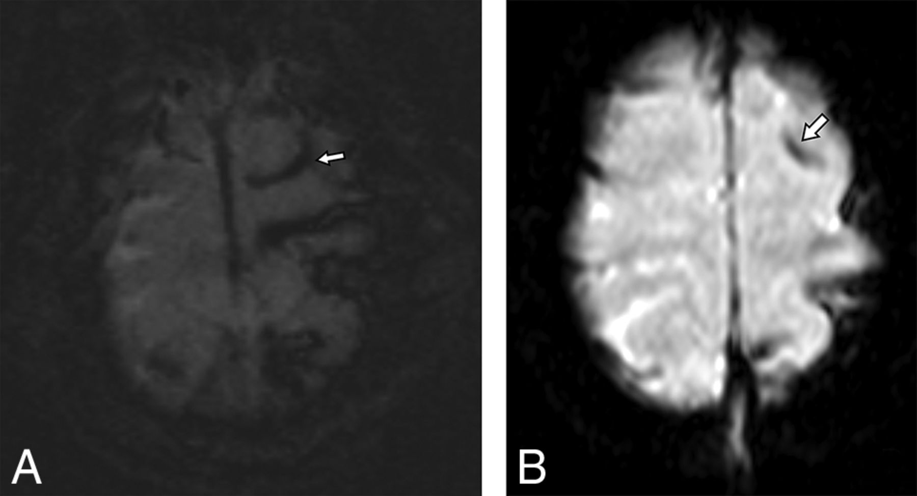
  • Fig 3.
    • Download figure
    • Open in new tab
    • Download powerpoint
    Fig 3.

    A 10-month-old child with suspected abusive head trauma found to have subtle parenchymal edema identified in the left parietal lobe on axial and coronal T2 TSE (A and B), which was not prospectively identified on ultrafast axial or coronal HASTE (C and D).

Tables

  • Figures
    • View popup
    Table 1:

    Ultrafast MRI brain protocols

    SequenceParametersTotal Time: 1.5T: 1m 43s; 3T: 1m 54s
    Magnet StrengthTE (ms)TR (ms)MatrixSection Thickness (mm)
    Axial T2 HASTE1.5T96550192 × 154423s
    3T98536192 × 154419s
    Coronal T2 HASTE96550123 × 192423s
    98536123 × 192419s
    Axial DWI774508128 × 128436s
    7812,600128 × 128446s
    Axial EPI T2*394190192 × 154421s
    393350192 × 154430s
    • Note:—m indicates minute; s, second.

    • View popup
    Table 2:

    stMRI brain protocols

    SequenceParametersTotal Time: 1.5T: 17m 15s; 3T: 14m 42s
    Magnet StrengthTE (ms)TR (ms)MatrixSection Thickness (mm)
    Sagittal 3D T11.5T2.982180192 × 2561.23m 53s
    MPRAGE3T2.181460251 × 2560.93m 16s
    Axial T2 TSE993950320 × 32021m 51s
    1163980307 × 38422m 12s
    Coronal T2 TSE1093870320 × 32022m 12s
    1163520320 × 32024m 6s
    Axial T2 FLAIR15210,000256 × 25643m 0s
    1077000180 × 32041m 24s
    Axial DWI774508128 × 128436s
    7812,600128 × 128446s
    Axial SWI4049195 × 3201.55m 43s
    4027182 × 2561.52m 58s
    • Note:—m indicates minute; s, second.

    • View popup
    Table 3:

    Prevalence of imaging findings per patient on stMRI

    FindingPrevalence
    Subdural collection11/24 (46%)
    Bilateral subdural collection10/11 (44%)
    Subarachnoid hemorrhage8/24 (33%)
    Intraparenchymal hemorrhage7/24 (29%)
    Intraventricular hemorrhage1/24 (4%)
    Epidural hemorrhage3/24 (13%)
    Cytotoxic edema4/24 (17%)
    Parenchymal laceration0/24 (0%)
    Vasogenic edema2/24 (8%)
    Herniation or midline shift0/24 (0%)
    Hydrocephalus0/24 (0%)
    Encephalomalacia2/24 (8%)
    Large subarachnoid spaces5/24 (21%)
    Total No. of patients with any abnormal finding20/24 (83%)
    • View popup
    Table 4:

    Discrepancy rates for consensus ufMRI, nHCT, and combined versus stMRI

    Ultrafast vs stMRInHCT vs stMRIUltrafast + nHCT vs stMRI
    Subdural collection0/24 (0%)0/24 (0%)0/24 (0%)
    Bilateral subdural collection3/24 (13%)1/24 (4%)1/24 (4%)
    Tentorial subdural hemorrhage3/24 (13%)3/24 (13%)3/24 (13%)
    Subdural membrane formation0/24 (0%)2/24 (8%)0/24 (0%)
    Subdural fluid-fluid level2/24 (8%)2/24 (8%)2/24 (8%)
    Subarachnoid hemorrhage4/24 (17%)4/24 (17%)4/24 (17%)
    Intraparenchymal hemorrhage0/24 (0%)6/24 (25%)a0/24 (0%)
    Intraventricular hemorrhage0/24 (0%)1/24 (4%)0/24 (0%)
    Epidural hemorrhage0/24 (0%)0/24 (0%)0/24 (0%)
    Cytotoxic edema0/24 (0%)4/24 (17%)0/24 (0%)
    Parenchymal laceration0/24 (0%)0/24 (0%)0/24 (0%)
    Vasogenic edema0/24 (0%)1/24 (4%)0/24 (0%)
    Herniation or midline shift0/24 (0%)0/24 (0%)0/24 (0%)
    Hydrocephalus0/24 (0%)0/24 (0%)0/24 (0%)
    Encephalomalacia0/24 (0%)0/24 (0%)0/24 (0%)
    Large subarachnoid spaces0/24 (0%)1/24 (4%)0/24 (0%)
    Any discrepancy10/24 (42%)a15/24 (63%)a8/24 (33%)a
    • ↵a Statistically significant McNemar test (P < .05).

    • View popup
    Table 5:

    Diagnostic performance of consensus ufMRI, nHCT, and combined ufMRI and nHCT compared with stMRIa

    SensitivitySpecificityPPVNPV
    ufMRI50%100%100%31%
    (27%–73%)(40%–100%)(69%–100%)(8%–58%)
    nHCT25%100%100%21%
    (9%–49%)(40%–100%)(48%–100%)(6%–46%)
    Combined ultrafast with nHCT60%100%100%33%
    (36%–81%)(40%–100%)(74%–100%)(10%–65%)
    • Note:—PPV indicates positive predictive value; NPV, negative predictive value.

    • ↵a Parentheses denote 95% confidence intervals.

PreviousNext
Back to top

In this issue

American Journal of Neuroradiology: 38 (4)
American Journal of Neuroradiology
Vol. 38, Issue 4
1 Apr 2017
  • Table of Contents
  • Index by author
  • Complete Issue (PDF)
Advertisement
Print
Download PDF
Email Article

Thank you for your interest in spreading the word on American Journal of Neuroradiology.

NOTE: We only request your email address so that the person you are recommending the page to knows that you wanted them to see it, and that it is not junk mail. We do not capture any email address.

Enter multiple addresses on separate lines or separate them with commas.
Diagnostic Performance of Ultrafast Brain MRI for Evaluation of Abusive Head Trauma
(Your Name) has sent you a message from American Journal of Neuroradiology
(Your Name) thought you would like to see the American Journal of Neuroradiology web site.
CAPTCHA
This question is for testing whether or not you are a human visitor and to prevent automated spam submissions.
Cite this article
S.F. Kralik, M. Yasrebi, N. Supakul, C. Lin, L.G. Netter, R.A. Hicks, R.A. Hibbard, L.L. Ackerman, M.L. Harris, C.Y. Ho
Diagnostic Performance of Ultrafast Brain MRI for Evaluation of Abusive Head Trauma
American Journal of Neuroradiology Apr 2017, 38 (4) 807-813; DOI: 10.3174/ajnr.A5093

Citation Manager Formats

  • BibTeX
  • Bookends
  • EasyBib
  • EndNote (tagged)
  • EndNote 8 (xml)
  • Medlars
  • Mendeley
  • Papers
  • RefWorks Tagged
  • Ref Manager
  • RIS
  • Zotero
0 Responses
Respond to this article
Share
Bookmark this article
Diagnostic Performance of Ultrafast Brain MRI for Evaluation of Abusive Head Trauma
S.F. Kralik, M. Yasrebi, N. Supakul, C. Lin, L.G. Netter, R.A. Hicks, R.A. Hibbard, L.L. Ackerman, M.L. Harris, C.Y. Ho
American Journal of Neuroradiology Apr 2017, 38 (4) 807-813; DOI: 10.3174/ajnr.A5093
del.icio.us logo Twitter logo Facebook logo Mendeley logo
  • Tweet Widget
  • Facebook Like
  • Google Plus One
Purchase

Jump to section

  • Article
    • Abstract
    • ABBREVIATIONS:
    • Materials and Methods
    • Results
    • Discussion
    • Conclusions
    • Footnotes
    • REFERENCES
  • Figures & Data
  • Info & Metrics
  • Responses
  • References
  • PDF

Related Articles

  • No related articles found.
  • PubMed
  • Google Scholar

Cited By...

  • Association of volume and prehospital paediatric care quality in emergency medical services: retrospective analysis of a national sample
  • Implementation of a Survey Spine MR Imaging Protocol for Cord Compression in the Emergency Department: Experience at a Level 1 Trauma Center
  • Clinical Experience of 1-Minute Brain MRI Using a Multicontrast EPI Sequence in a Different Scan Environment
  • Rapid-Sequence MRI of the Brain: A Distinct Imaging Study
  • Crossref
  • Google Scholar

This article has not yet been cited by articles in journals that are participating in Crossref Cited-by Linking.

More in this TOC Section

  • Pediatric Glioma:Imaging Clusters,Molecular Traits
  • Clinical SVR of Fetal Brain MRI
  • Comparing MRI Perfusion in Pediatric Brain Tumors
Show more PEDIATRIC NEUROIMAGING

Similar Articles

Advertisement

Indexed Content

  • Current Issue
  • Accepted Manuscripts
  • Article Preview
  • Past Issues
  • Editorials
  • Editor's Choice
  • Fellows' Journal Club
  • Letters to the Editor
  • Video Articles

Cases

  • Case Collection
  • Archive - Case of the Week
  • Archive - Case of the Month
  • Archive - Classic Case

More from AJNR

  • Trainee Corner
  • Imaging Protocols
  • MRI Safety Corner
  • Book Reviews

Multimedia

  • AJNR Podcasts
  • AJNR Scantastics

Resources

  • Turnaround Time
  • Submit a Manuscript
  • Submit a Video Article
  • Submit an eLetter to the Editor/Response
  • Manuscript Submission Guidelines
  • Statistical Tips
  • Fast Publishing of Accepted Manuscripts
  • Graphical Abstract Preparation
  • Imaging Protocol Submission
  • Evidence-Based Medicine Level Guide
  • Publishing Checklists
  • Author Policies
  • Become a Reviewer/Academy of Reviewers
  • News and Updates

About Us

  • About AJNR
  • Editorial Board
  • Editorial Board Alumni
  • Alerts
  • Permissions
  • Not an AJNR Subscriber? Join Now
  • Advertise with Us
  • Librarian Resources
  • Feedback
  • Terms and Conditions
  • AJNR Editorial Board Alumni

American Society of Neuroradiology

  • Not an ASNR Member? Join Now

© 2025 by the American Society of Neuroradiology All rights, including for text and data mining, AI training, and similar technologies, are reserved.
Print ISSN: 0195-6108 Online ISSN: 1936-959X

Powered by HighWire