
Article Figures & Data
Figures
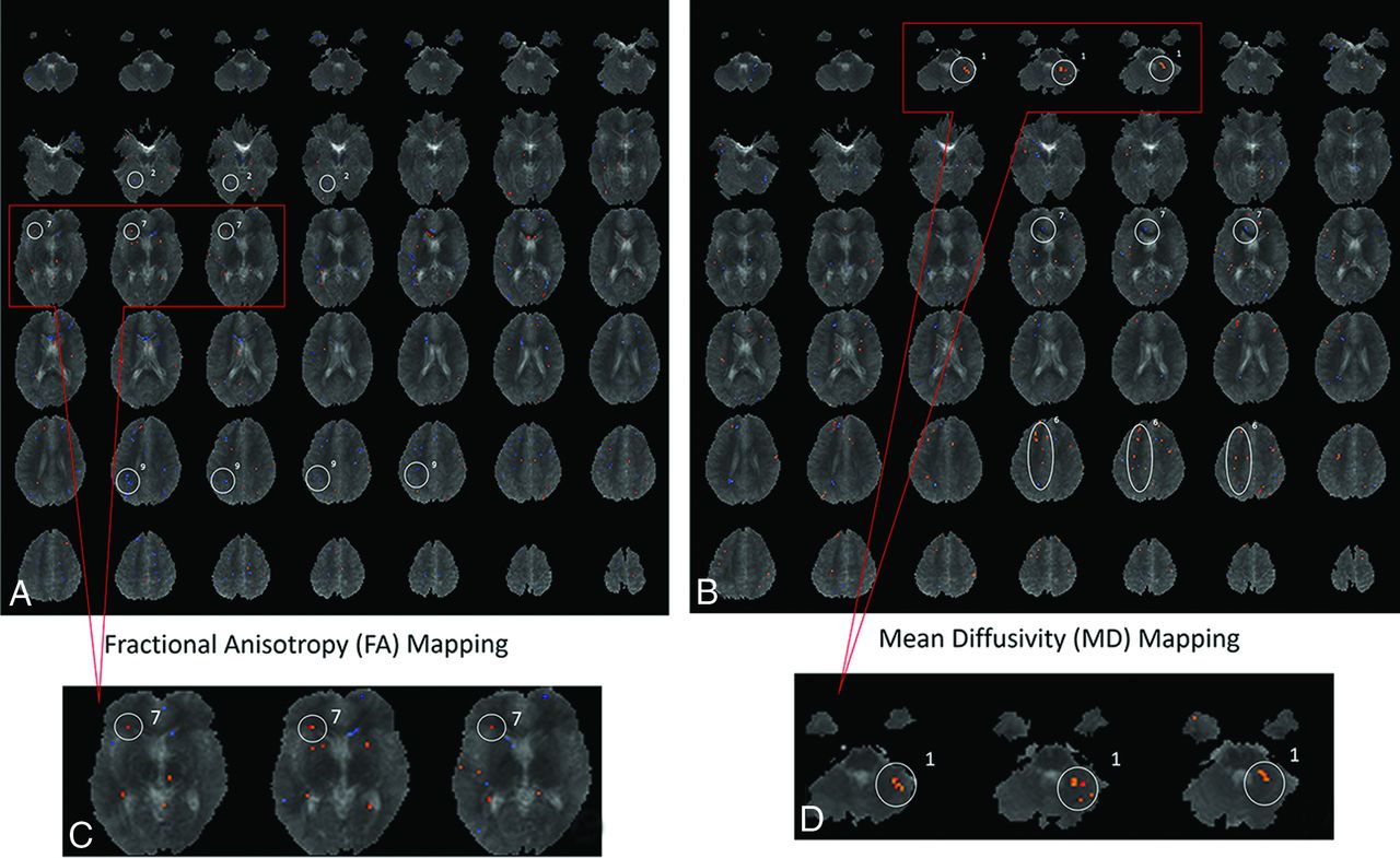
- Fig 1.
Voxel-based analysis comparing the brains of children born from preeclamptic pregnancies with the brains of children born from typical healthy pregnancies (controls). A custom template was created on the basis of the anatomic images of all 20 children enrolled in this study. Voxel-based analysis was performed to compare white matter measurements in PE-F1s and control children. The blue and red dots represent areas where there might have been differences between the fractional anisotropy values (A and C) or mean diffusivity values (B and D). From the voxel-based analysis, areas in which a cluster of voxels persisted through at least ≥3 sections were identified and used to define the specific ROIs to be used as seeds for tractography. Magnification of an area (C and D) is used as an example of how ROIs were defined. The 3 most dot-clustered regions in voxel-based analysis for FA and MD were selected, yielding 6 ROIs for more detailed analysis. The ROIs identified on FA were the following: 2, middle occipital gyrus; 7, caudate nucleus; and 9, precuneus. The ROIs identified on MD were the following: 1, cerebellum; 6, superior longitudinal fasciculus; and 7, cingulate gyrus.

Tables
Comparison of DTI, MRI, and MRA findings by brain region for PE-F1
Brain Anatomic Regions DTI Findings in PE-F1 Brains (Present Study) MRI/MRA, Previous Findings in PE-F1 Brains14 ROIs Comparisons with Significant Differences between PE-F1s and Controls Morphologic Findings in PE-F1 Vascular Findings in PE-F1 Temporal lobe Superior longitudinal fasciculus Volume of tract: PE-F1s > controls (P = .03) Larger volume in temporal lobe NS Limbic area Caudate nucleus FA: PE-F1s > controls (P = .008); volume of tract: PE-F1s > controls (P = .05) Larger volume in right and left amygdalae NS Cingulate gyrus Parallel diffusion: PE-F1s > controls (P = .0.04) NS NS Parietal lobe Precuneus NS NS Smaller radii globally Occipital lobe Middle occipital gyrus NS NS Smaller radii globally Cerebellum Cerebellum NS Larger volume in cerebellum NS Note:—NS indicates that no significant correlation or comparison was found.


