Advertisement

Main menu

  • Home
  • Content
    • Current Issue
    • Accepted Manuscripts
    • Article Preview
    • Past Issue Archive
    • Video Articles
    • AJNR Case Collection
    • Case of the Week Archive
    • Case of the Month Archive
    • Classic Case Archive
  • Special Collections
    • AJNR Awards
    • Low-Field MRI
    • Alzheimer Disease
    • ASNR Foundation Special Collection
    • Photon-Counting CT
    • View All
  • Multimedia
    • AJNR Podcasts
    • AJNR SCANtastic
    • Trainee Corner
    • MRI Safety Corner
    • Imaging Protocols
  • For Authors
    • Submit a Manuscript
    • Submit a Video Article
    • Submit an eLetter to the Editor/Response
    • Manuscript Submission Guidelines
    • Statistical Tips
    • Fast Publishing of Accepted Manuscripts
    • Graphical Abstract Preparation
    • Imaging Protocol Submission
    • Author Policies
  • About Us
    • About AJNR
    • Editorial Board
    • Editorial Board Alumni
  • More
    • Become a Reviewer/Academy of Reviewers
    • Subscribers
    • Permissions
    • Alerts
    • Feedback
    • Advertisers
    • ASNR Home

User menu

  • Alerts
  • Log in

Search

  • Advanced search
American Journal of Neuroradiology

American Journal of Neuroradiology

ASHNR American Society of Functional Neuroradiology ASHNR American Society of Pediatric Neuroradiology ASSR
  • Alerts
  • Log in

Advanced Search

  • Home
  • Content
    • Current Issue
    • Accepted Manuscripts
    • Article Preview
    • Past Issue Archive
    • Video Articles
    • AJNR Case Collection
    • Case of the Week Archive
    • Case of the Month Archive
    • Classic Case Archive
  • Special Collections
    • AJNR Awards
    • Low-Field MRI
    • Alzheimer Disease
    • ASNR Foundation Special Collection
    • Photon-Counting CT
    • View All
  • Multimedia
    • AJNR Podcasts
    • AJNR SCANtastic
    • Trainee Corner
    • MRI Safety Corner
    • Imaging Protocols
  • For Authors
    • Submit a Manuscript
    • Submit a Video Article
    • Submit an eLetter to the Editor/Response
    • Manuscript Submission Guidelines
    • Statistical Tips
    • Fast Publishing of Accepted Manuscripts
    • Graphical Abstract Preparation
    • Imaging Protocol Submission
    • Author Policies
  • About Us
    • About AJNR
    • Editorial Board
    • Editorial Board Alumni
  • More
    • Become a Reviewer/Academy of Reviewers
    • Subscribers
    • Permissions
    • Alerts
    • Feedback
    • Advertisers
    • ASNR Home
  • Follow AJNR on Twitter
  • Visit AJNR on Facebook
  • Follow AJNR on Instagram
  • Join AJNR on LinkedIn
  • RSS Feeds

AJNR Awards, New Junior Editors, and more. Read the latest AJNR updates

Research ArticleADULT BRAIN
Open Access

Cerebral Temperature Dysregulation: MR Thermographic Monitoring in a Nonhuman Primate Study of Acute Ischemic Stroke

S. Dehkharghani, C.C. Fleischer, D. Qiu, M. Yepes and F. Tong
American Journal of Neuroradiology April 2017, 38 (4) 712-720; DOI: https://doi.org/10.3174/ajnr.A5059
S. Dehkharghani
aFrom the Departments of Radiology and Imaging Sciences (S.D., D.Q., F.T.)
bNeurology (S.D., M.Y.), Emory University Hospital, Atlanta, Georgia
  • Find this author on Google Scholar
  • Find this author on PubMed
  • Search for this author on this site
  • ORCID record for S. Dehkharghani
C.C. Fleischer
cDepartment of Biomedical Engineering (C.C.F.), Emory University and Georgia Institute of Technology, Atlanta, Georgia.
  • Find this author on Google Scholar
  • Find this author on PubMed
  • Search for this author on this site
  • ORCID record for C.C. Fleischer
D. Qiu
aFrom the Departments of Radiology and Imaging Sciences (S.D., D.Q., F.T.)
  • Find this author on Google Scholar
  • Find this author on PubMed
  • Search for this author on this site
  • ORCID record for D. Qiu
M. Yepes
bNeurology (S.D., M.Y.), Emory University Hospital, Atlanta, Georgia
  • Find this author on Google Scholar
  • Find this author on PubMed
  • Search for this author on this site
  • ORCID record for M. Yepes
F. Tong
aFrom the Departments of Radiology and Imaging Sciences (S.D., D.Q., F.T.)
  • Find this author on Google Scholar
  • Find this author on PubMed
  • Search for this author on this site
  • ORCID record for F. Tong
  • Article
  • Figures & Data
  • Info & Metrics
  • Responses
  • References
  • PDF
Loading

Article Figures & Data

Figures

  • Tables
  • Fig 1.
    • Download figure
    • Open in new tab
    • Download powerpoint
    Fig 1.

    MR thermographs in subject 3 obtained during t−7 (A) and t0 (B) sessions, overlaid on T1-MPRAGE images. Terminal DWI (C) obtained at the conclusion of the t0 session following endovascular stroke induction demonstrates a large MCA territory infarction. Thermographs in A and B are presented in equivalent color scales, depicting voxelwise brain versus systemic temperature gradients. Disturbance to the geographic distribution of brain temperature gradients present during physiologic t0 conditions are present, with notable, diffuse cerebral hyperthermia affecting both hemispheres in B. Generalized cerebral heating following infarction corresponds to significant elevation above decoupled systemic temperatures (not shown) as a function of time and infarction volume (see text).

  • Fig 2.
    • Download figure
    • Open in new tab
    • Download powerpoint
    Fig 2.

    Subject-specific absolute temperature versus time for paired prestroke and stroke sessions. For each subject, the vertical columns represent t0 above and t−7 below. The y-axis in t0 plots represents MR imaging–derived temperatures for the hemisphere ipsilateral and contralateral to the infarction, as well as systemic temperatures. All plots are presented in the same vertical scale, with errors bars (SD) as indicated. The final time point in all t0 plots represents values from the poststroke session. Progressive heating of both cerebral hemispheres is present in all 3 subjects, outpacing the progressive systemic febrile temperatures in stroke aftermath. Brain hyperthermia is noted to resolve in the t1 session for all subjects. By comparison, all baseline t−7 scans exhibit closely coupled brain systemic temperatures, despite fluctuations related to early postanesthetic hypothermia during subject preparation.
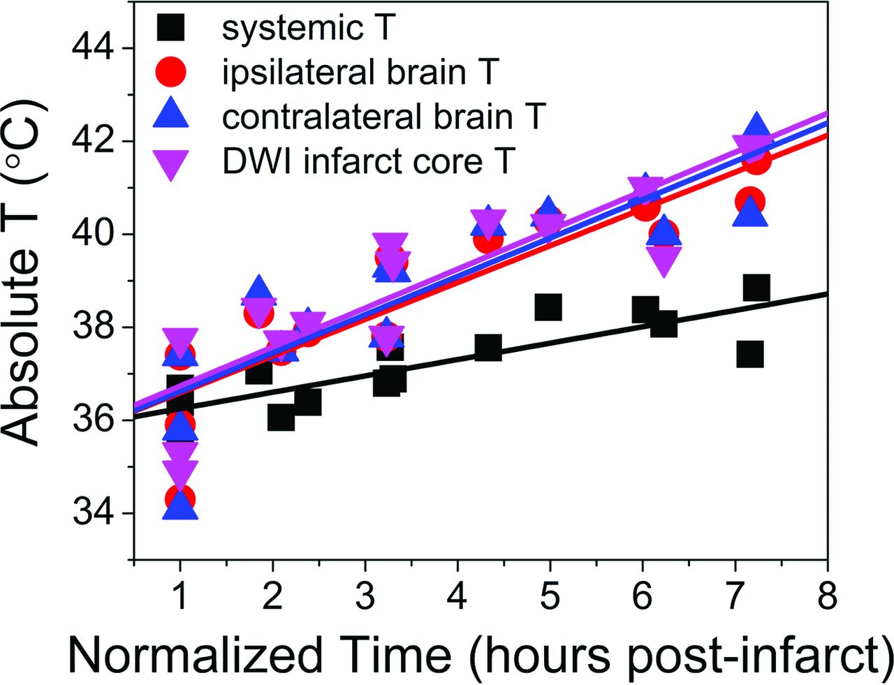
  • Fig 3.
    • Download figure
    • Open in new tab
    • Download powerpoint
    Fig 3.

    Aggregated fit from individual regressions from each subject, depicted for systemic temperatures, average hemispheric temperature ipsilateral and contralateral to infarction, and those voxels defined within the infarction territory. Individual regressions derived from a linear fixed-effects model demonstrate highly significant associations among all variables relative to time, as well as significant differences between brain temperatures and systemic temperatures following multiple-comparison correction (Table 2).
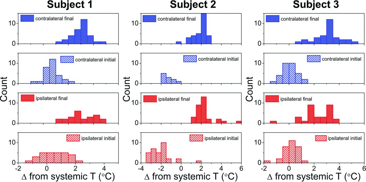
  • Fig 4.
    • Download figure
    • Open in new tab
    • Download powerpoint
    Fig 4.

    Subject-specific histograms of hemispheric cerebral temperature versus time reflecting divergence of systemic and brain temperatures. The x-axis represents the brain-systemic temperature offset, represented respectively for the initial and final time points, both for ipsilateral and contralateral brain temperatures. The rightward shift of all histograms reflects progressive cerebral hyperthermia decoupled from systemic temperatures present for both hemispheres of all 3 subjects.

Tables

  • Figures
    • View popup
    Table 1:

    Subject-specific temperature and infarction volumesa

    ParameterSubject 1Subject 2Subject 3
    T−7 scan session
        Systemic temperature range36.3–37.236.8–37.936.0–38.2
        Mean brain temperature range36.6 ± 1.0–37.1 ± 1.236.7 ± 0.6–37.8 ± 1.136.3 ± 0.7–38.8 ± 0.7
        Temperature difference: brain, systemicb−0.1 ± 1.2–0.2 ± 1.0−0.1 ± 0.6–0.4 ± 1.10.3 ± 0.7–1.2 ± 0.7
    T0 scan session
        Infarct volume 1 hr postsurgery (cm3)0.420.286.70
        Systemic temperature range36.7–37.636.4–38.435.7–38.8
        Mean ipsilateral brain temperature range37.4 ± 1.1–40.7 ± 1.434.3 ± 1.1–40.3 ± 1.235.9 ± 0.6–41.6 ± 2.9
        Mean contralateral brain temperature range37.4 ± 0.8–40.4 ± 0.734.1 ± 1.0–40.4 ± 0.735.8 ± 0.5–42.2 ± 1.8
        Temperature difference: ipsilateral brain, systemic0.7 ± 1.1–3.2 ± 1.4−2.1 ± 1.1–2.5 ± 1.30.2 ± 0.6–2.8 ± 2.9
        Temperature difference: contralateral brain, systemic0.7 ± 0.8–3.0 ± 0.7−2.3 ± 1.0–2.3 ± 1.20.1 ± 0.5–3.4 ± 1.8
    T1 scan session
        Infarct volume 24 hr postsurgery (cm3)23.821.2925.58
        Systemic temperature35.635.134.6
        Mean brain temperature37.9 ± 1.036.9 ± 1.236.3 ± 0.9
        Temperature difference: brain, systemic2.3 ± 1.01.7 ± 1.21.7 ± 0.9
    • ↵a All temperatures are reported in degrees Celsius. Reported temperature ranges represent the full dynamic range from the indicated imaging session beginning from matched initial systemic and imaging temperature time points.

    • ↵b Indicates a range of systemic-versus-brain temperature offsets.

    • View popup
    Table 2:

    Absolute systemic and cerebral temperatures versus timea

    Temperature ParameterSlope (°C/hr)Intercept (°C)F-Stat of Regression (df)P Value (F-Stat)Difference in Slope versus Systemic TemperatureP Value of Difference versus Systemic
    Systemic0.352 (0.062)35.9 (0.3)32.2 (1,13)<.0005bNANA
    Ipsilateral brain0.791 (0.116)35.8 (0.5)47.0 (1,13)<.0005b0.439 (0.156).007b
    Contralateral brain0.824 (0.129)35.8 (0.5)40.7 (1,13)<.0005b0.471 (0.156).004b
    DWI infarct core0.838 (0.122)35.9 (0.5)47.5 (1,13)<.0005b0.486 (0.156).003b
    • Note:—F-Stat indicates F-statistic; NA, not applicable.

    • ↵a Parameter estimates (standard error when applicable) from a linear fixed-effects model reported relative to normalized time since stroke onset (defined at initiation of scanning 1 hour following stroke induction).

    • ↵b Indicates statistical significance at P < .05, results of 2-tailed t test of F-statistics.

PreviousNext
Back to top

In this issue

American Journal of Neuroradiology: 38 (4)
American Journal of Neuroradiology
Vol. 38, Issue 4
1 Apr 2017
  • Table of Contents
  • Index by author
  • Complete Issue (PDF)
Advertisement
Print
Download PDF
Email Article

Thank you for your interest in spreading the word on American Journal of Neuroradiology.

NOTE: We only request your email address so that the person you are recommending the page to knows that you wanted them to see it, and that it is not junk mail. We do not capture any email address.

Enter multiple addresses on separate lines or separate them with commas.
Cerebral Temperature Dysregulation: MR Thermographic Monitoring in a Nonhuman Primate Study of Acute Ischemic Stroke
(Your Name) has sent you a message from American Journal of Neuroradiology
(Your Name) thought you would like to see the American Journal of Neuroradiology web site.
CAPTCHA
This question is for testing whether or not you are a human visitor and to prevent automated spam submissions.
Cite this article
S. Dehkharghani, C.C. Fleischer, D. Qiu, M. Yepes, F. Tong
Cerebral Temperature Dysregulation: MR Thermographic Monitoring in a Nonhuman Primate Study of Acute Ischemic Stroke
American Journal of Neuroradiology Apr 2017, 38 (4) 712-720; DOI: 10.3174/ajnr.A5059

Citation Manager Formats

  • BibTeX
  • Bookends
  • EasyBib
  • EndNote (tagged)
  • EndNote 8 (xml)
  • Medlars
  • Mendeley
  • Papers
  • RefWorks Tagged
  • Ref Manager
  • RIS
  • Zotero
0 Responses
Respond to this article
Share
Bookmark this article
Cerebral Temperature Dysregulation: MR Thermographic Monitoring in a Nonhuman Primate Study of Acute Ischemic Stroke
S. Dehkharghani, C.C. Fleischer, D. Qiu, M. Yepes, F. Tong
American Journal of Neuroradiology Apr 2017, 38 (4) 712-720; DOI: 10.3174/ajnr.A5059
del.icio.us logo Twitter logo Facebook logo Mendeley logo
  • Tweet Widget
  • Facebook Like
  • Google Plus One
Purchase

Jump to section

  • Article
    • Abstract
    • ABBREVIATIONS:
    • Materials and Methods
    • Results
    • Discussion
    • Conclusions
    • Footnotes
    • REFERENCES
  • Figures & Data
  • Info & Metrics
  • Responses
  • References
  • PDF

Related Articles

  • No related articles found.
  • PubMed
  • Google Scholar

Cited By...

  • COmbination of Targeted temperature management and Thrombectomy after acute Ischemic Stroke (COTTIS): a pilot study
  • Diurnal brain temperature rhythms and mortality after brain injury: a prospective and retrospective cohort study
  • MR Thermometry in Cerebrovascular Disease: Physiologic Basis, Hemodynamic Dependence, and a New Frontier in Stroke Imaging
  • The Brain Thermal Response as a Potential Neuroimaging Biomarker of Cerebrovascular Impairment
  • Crossref (29)
  • Google Scholar

This article has been cited by the following articles in journals that are participating in Crossref Cited-by Linking.

  • A daily temperature rhythm in the human brain predicts survival after brain injury
    Nina M Rzechorzek, Michael J Thrippleton, Francesca M Chappell, Grant Mair, Ari Ercole, Manuel Cabeleira, Jonathan Rhodes, Ian Marshall, John S O’Neill
    Brain 2022 145 6
  • Risks associated with the stroke predisposition at young age: facts and hypotheses in light of individualized predictive and preventive approach
    Jiri Polivka, Jiri Polivka, Martin Pesta, Vladimir Rohan, Libuse Celedova, Smit Mahajani, Ondrej Topolcan, Olga Golubnitschaja
    EPMA Journal 2019 10 1
  • Reproducibility of whole‐brain temperature mapping and metabolite quantification using proton magnetic resonance spectroscopy
    Yue Zhang, Edward Taub, Christina Mueller, Jarred Younger, Gitendra Uswatte, Thomas Patrick DeRamus, David C. Knight
    NMR in Biomedicine 2020 33 7
  • Personalized predictions and non-invasive imaging of human brain temperature
    Dongsuk Sung, Peter A. Kottke, Benjamin B. Risk, Jason W. Allen, Fadi Nahab, Andrei G. Fedorov, Candace C. Fleischer
    Communications Physics 2021 4 1
  • An Ambiguous Role for Fever in Worsening Outcome After Intracerebral Hemorrhage
    Lane J. Liddle, Christine A. Dirks, Mohammed Almekhlafi, Frederick Colbourne
    Translational Stroke Research 2023 14 2
  • The Brain Thermal Response as a Potential Neuroimaging Biomarker of Cerebrovascular Impairment
    C.C. Fleischer, J. Wu, D. Qiu, S.-E. Park, F. Nahab, S. Dehkharghani
    American Journal of Neuroradiology 2017 38 11
  • MR Thermometry in Cerebrovascular Disease: Physiologic Basis, Hemodynamic Dependence, and a New Frontier in Stroke Imaging
    S. Dehkharghani, D. Qiu
    American Journal of Neuroradiology 2020 41 4
  • Resting‐State Brain Temperature: Dynamic Fluctuations in Brain Temperature and the Brain–Body Temperature Gradient
    Dongsuk Sung, Benjamin B. Risk, Kelly J. Wang, Jason W. Allen, Candace C. Fleischer
    Journal of Magnetic Resonance Imaging 2023 57 4
  • Brain Temperature Measured by Magnetic Resonance Spectroscopy to Predict Clinical Outcome in Patients with Infarction
    Tomohisa Ishida, Takashi Inoue, Tomoo Inoue, Toshiki Endo, Miki Fujimura, Kuniyasu Niizuma, Hidenori Endo, Teiji Tominaga
    Sensors 2021 21 2
  • Measuring radiofrequency field‐induced temperature variations in brain MRI exams with motion compensated MR thermometry and field monitoring
    Caroline Le Ster, Franck Mauconduit, Christian Mirkes, Alexandre Vignaud, Nicolas Boulant
    Magnetic Resonance in Medicine 2022 87 3

More in this TOC Section

  • Diagnostic Neuroradiology of Monoclonal Antibodies
  • Clinical Outcomes After Chiari I Decompression
  • Segmentation of Brain Metastases with BLAST
Show more ADULT BRAIN

Similar Articles

Advertisement

Indexed Content

  • Current Issue
  • Accepted Manuscripts
  • Article Preview
  • Past Issues
  • Editorials
  • Editor's Choice
  • Fellows' Journal Club
  • Letters to the Editor
  • Video Articles

Cases

  • Case Collection
  • Archive - Case of the Week
  • Archive - Case of the Month
  • Archive - Classic Case

More from AJNR

  • Trainee Corner
  • Imaging Protocols
  • MRI Safety Corner
  • Book Reviews

Multimedia

  • AJNR Podcasts
  • AJNR Scantastics

Resources

  • Turnaround Time
  • Submit a Manuscript
  • Submit a Video Article
  • Submit an eLetter to the Editor/Response
  • Manuscript Submission Guidelines
  • Statistical Tips
  • Fast Publishing of Accepted Manuscripts
  • Graphical Abstract Preparation
  • Imaging Protocol Submission
  • Evidence-Based Medicine Level Guide
  • Publishing Checklists
  • Author Policies
  • Become a Reviewer/Academy of Reviewers
  • News and Updates

About Us

  • About AJNR
  • Editorial Board
  • Editorial Board Alumni
  • Alerts
  • Permissions
  • Not an AJNR Subscriber? Join Now
  • Advertise with Us
  • Librarian Resources
  • Feedback
  • Terms and Conditions
  • AJNR Editorial Board Alumni

American Society of Neuroradiology

  • Not an ASNR Member? Join Now

© 2025 by the American Society of Neuroradiology All rights, including for text and data mining, AI training, and similar technologies, are reserved.
Print ISSN: 0195-6108 Online ISSN: 1936-959X

Powered by HighWire