
Article Figures & Data
Figures
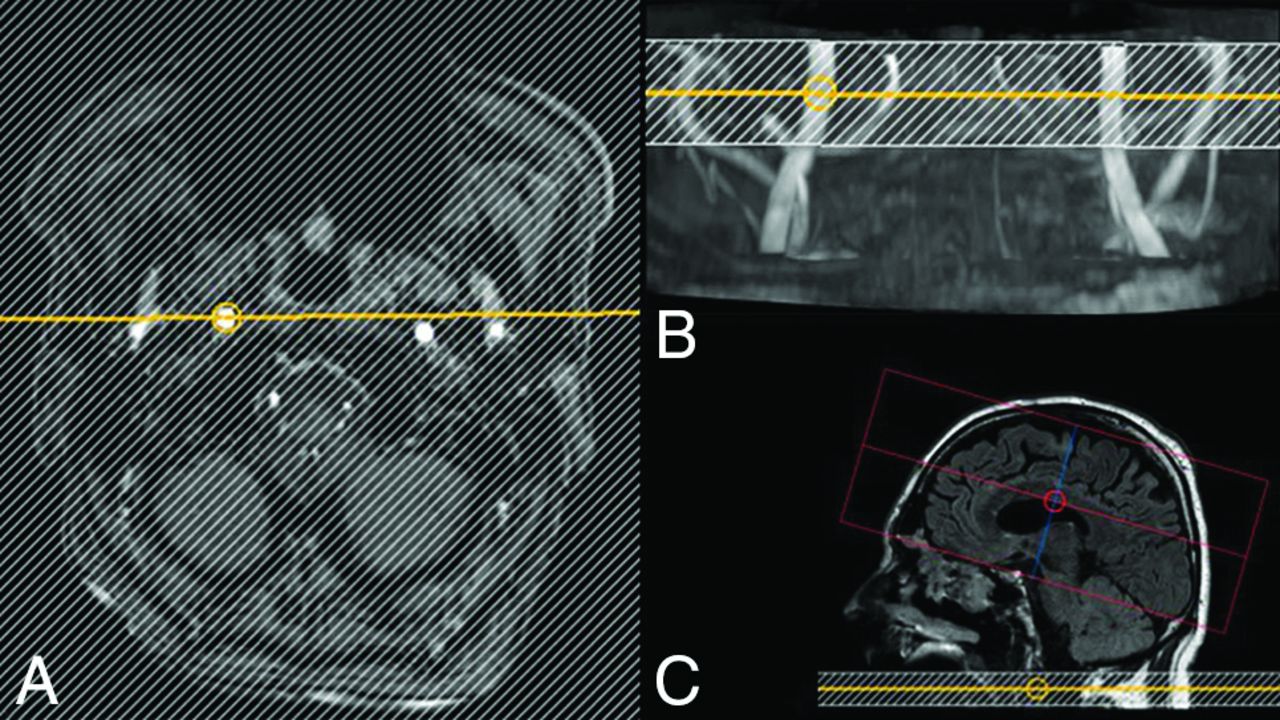
- Fig 1.
Planning steps of the superselective pCASL sequence. The labeling stack is positioned on a fast-TOF-MRA of the cervical vessels in the axial plane (A) and on the MIP projection (B). C, The readout slab is positioned covering the expected brain perfusion territory.
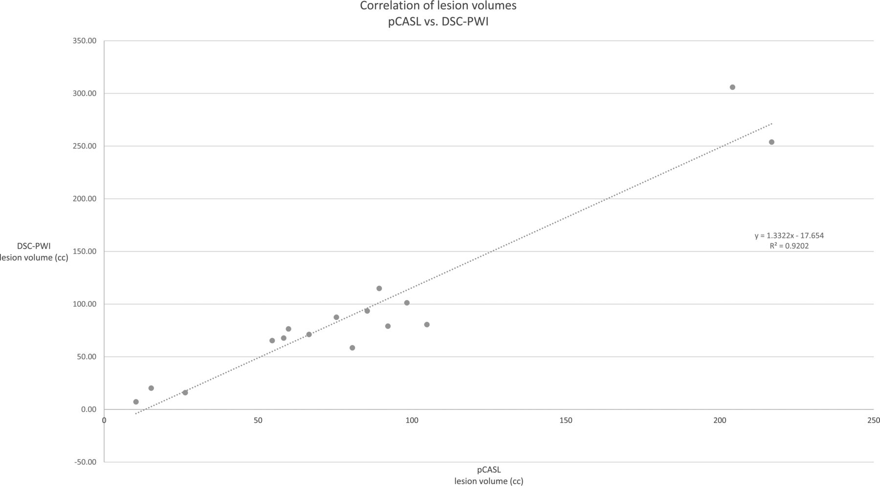
- Fig 2.
Correlation between pCASL- (global labeling) and DSC-PWI–identified infarct lesion volumes.
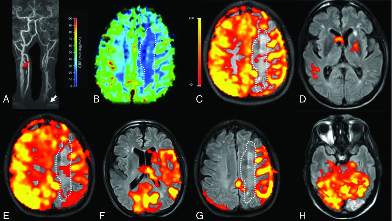
- Fig 3.
Illustration of individual perfusion patterns in 2 different patients with similar chronic high-grade (>70%) right bifurcation ICA stenosis. The red arrows indicate the stenosis in patient 1 (a) and patient 2 (A). Both patients had patent AcomAs and bilateral PcomAs. Nonselective labeling in b and B shows no significant perfusion deficits. These patients did not have any infarct lesions. Labeling was performed for the right ICA, left ICA, and right VA in both patients (the left VA was hypoplastic in both cases). In patient 1 (upper row), the stenotic right ICA (c) continues to provide perfusion to the right cerebral hemisphere; The left ICA (d) and right VA (e) do not collateralize. In patient 2 (bottom row), right ICA perfusion is diminished (C) and supplies only the MCA territory; the anterior and posterior territories are perfused by recruitment of the left ICA (D) and right VA (E). These cases illustrate situations in which the individual perfusion pattern is not predictable: Even though extracranial stenoses and circle of Willis anatomies are similar, the perfusion patterns are different.
- Fig 4.
A 78-year-old male patient with extracranial left ICA occlusion and an extracranial high-grade right ICA stenosis (red arrow) and a left VA stenosis in the V1 segment (white arrow, A). The AcomA and both PcomAs are patent. A perfusion deficit (white dotted line) is seen in the left corona radiata in both the DSC-PWI (B) and the nonselective pCASL map (C). Selective labeling was performed for the left common carotid artery (D), right ICA (E), right VA (F and G), and left VA (H). The intracranial perfusion signal is missing on labeling of the left common carotid artery (D) as a proof of left ICA occlusion. Right ICA labeling (E) shows perfusion of the left anterior cerebral artery and MCA territory. Right VA labeling (F and G) shows recruitment of the posterior circulation for the perfusion of the left MCA territory. The new watershed region with a perfusion deficit and chronic infarcts is seen between the posterior circulation and the right ICA (E and G). A subacute infarct lesion in the left occipital lobe is localized within the perfusion territory of the left VA (H) and can be categorized as a territorial infarct.
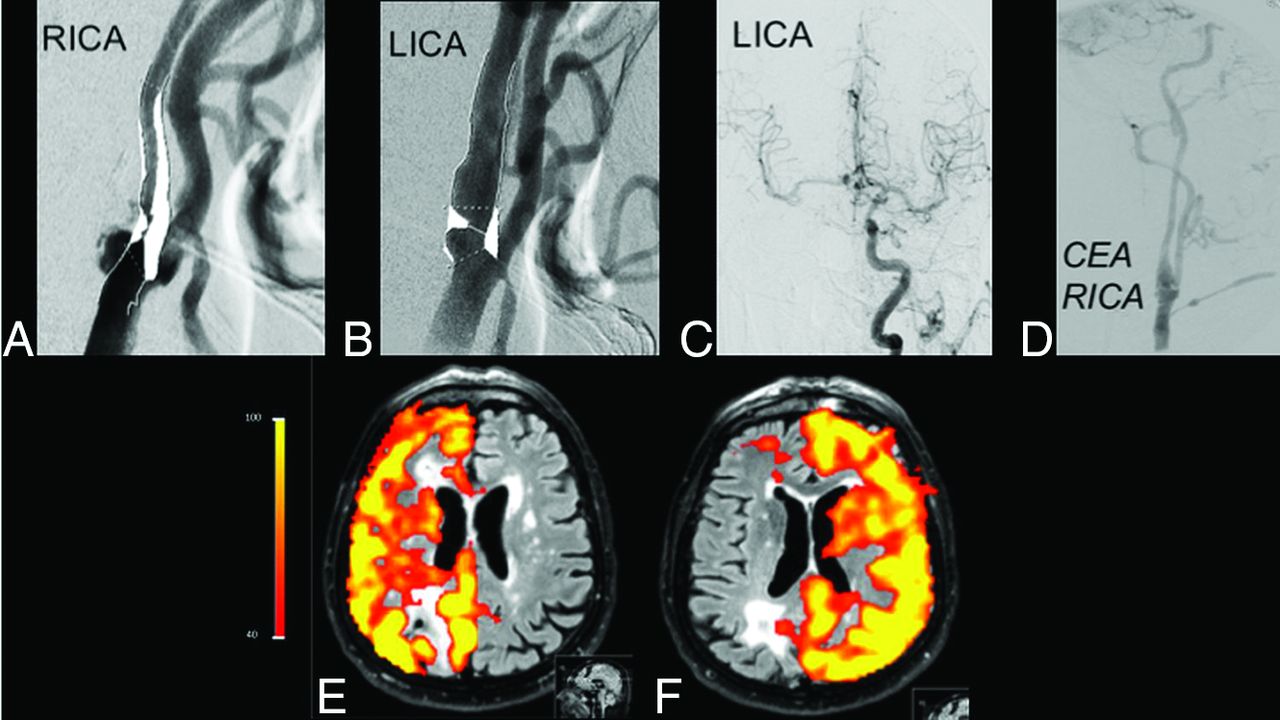
- Fig 5.
Initial cDSA shows high-grade (>70%) stenoses of both ICAs (A and B), with flow on the right MCA and anterior cerebral artery from the left ICA (C). After carotid endarterectomy (D), perfusion patterns of the right and left ICAs are normalized (E and F), demonstrating the utility of this noninvasive method for the follow-up of therapy success. RICA indicates right ICA; LICA, left ICA; CEA, carotid endarterectomy.


Tables
No. of subacute or chronic infarcts in the patient subgroups (defined on FLAIR imaging), their categorization as watershed or territorial infarcts, and the number of cases in which an infarct lesion was categorized differently after ss-pCASL imaging than with the standardized perfusion atlas
Vessel Status No. of Infarct Lesions/No. of Patients in Subgroup No. of Watershed Infarcts/No. of Infarct Lesions in Subgroup No. of Territorial Infarcts/No. of Infarct Lesions in Subgroup No. of Infarct Lesions Differently Categorized by Using the Standardized Perfusion Atlas vs ss-pCASL Unilateral ICA stenosis 11/21 (52%) 5/11 (45%) 6/11 (55%) 4/11 (36%) Bilateral ICA stenosis 4/9 (44%) 2/4 (50%) 2/4 (50%) 1/4 (25%) Unilateral ICA occlusion and contralateral ICA stenosis 15/15 (100%) 8/15 (54%) 7/15 (46%) 6/15 (40%) Bilateral ICA occlusion 3/3 (100%) 3/3 (100%) – 0/3 (0%) Sum 33/48 (69%) 18/33 (55%) 15/33 (45%) 11/33 (33%)


