
Article Figures & Data
Figures
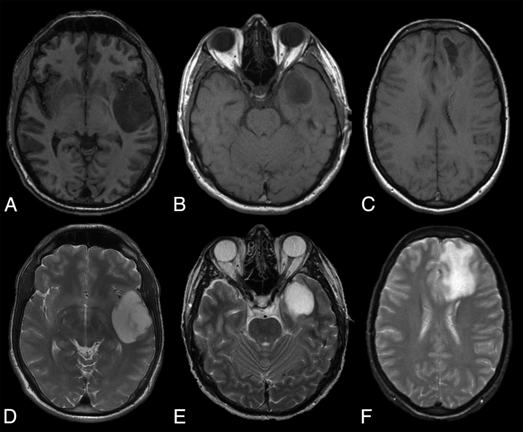
- Fig 1.
Examples of circumscribed (A and D), partially circumscribed (B and E), and noncircumscribed (C and F) tumor borders on axial T1-weighted (upper row) and T2-weighted (lower row) MR images.
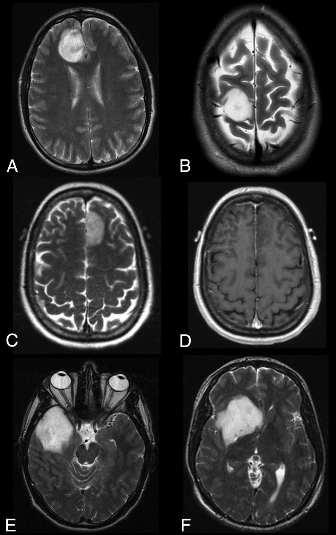
- Fig 2.
Examples of tumor classifications are as follows: circumscribed, with sharp smooth borders (A); circumscribed, with sharp borders, but not smooth due to the extent of concave inward borders (B); circumscribed with sharp borders on T2-weighted image (C) but not on T1-weighted image (D); partially circumscribed, with indistinct borders, but not smooth due to the extent of concave inward borders (E); and circumscribed, with predominantly sharp borders, but not smooth due to the extent of concave inward borders (F). All images are T2-weighted, except D as indicated.
- Fig 3.
MR images of a prototypical genetically defined (1p/19q codeleted) oligodendroglioma (upper row) and of an astrocytoma with microscopic oligodendroglial features but no 1p/19q codeletion (lower row). Axial T2-weighted (A and D), T1-weighted postcontrast (B and E), and ADC (C and F) images. Genetic oligodendrogliomas tend to be located in the frontal or parietal lobe and lack circumscription, are heterogeneous, and have lower ADC values. The mean ADC value of the tumor in C is 1.26 mm2/s. Astrocytomas in this cohort with microscopic oligodendroglial features tend to be located in the temporal or insular lobes, are frequently well circumscribed, and have higher ADC values. The mean ADC value of the tumor shown in F is 1.92 mm2/s.
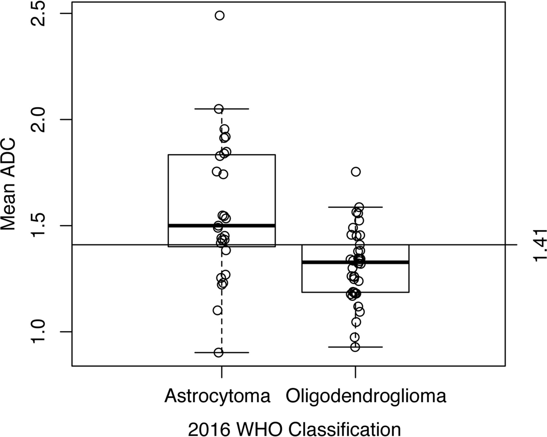
- Fig 4.
Boxplot demonstrating the distribution of mean ADC values by the WHO 2016 classification as oligodendroglioma or astrocytoma.


Tables
- Table 2:
Cross-tabulation of tumor histopathology by WHO 2007 criteria and grade with WHO 2016 criteria for oligodendroglioma (1p/19q codeletion)
Total (N = 148) WHO 2016 Oligodendroglioma (1p/19q codeleted) (n = 90) Astrocytoma (non-1p/19q codeleted) (n = 58) WHO 2007 classification Oligodendroglioma 42 (28.4%) 36 (40.0%) 6 (10.3%) Oligoastrocytoma 106 (71.6%) 54 (60.0%) 52 (89.7%) Tumor grade WHO II 98 (66.2%) 58 (64.4%) 40 (69.0%) WHO III 50 (33.8%) 32 (35.6%) 18 (31.0%) IDH mutation status No. 60 39 21 IDH-mutant 58 (96.7%) 39 (100%) 19 (90.5%) IDH-wild type 2 (3.3%) 0 (0%) 2 (9.5%) - Table 3:
Multivariate model including all standard anatomic imaging elements significantly associated with tumor classification as molecular oligodendroglioma in univariate analysisa
Odds Ratio 95% CI P Value Overall P Value Location Frontal Reference Insular 0.36 0.07–1.69 .20 .06 Parietal 0.71 0.21–2.65 .59 Temporal 0.13 0.02–0.60 .01 Circumscribed border Partial/yes Reference No 16.35 6.08–50.63 <.0001 Age, 1-yr increase 1.06 1.02–1.10 .002 ↵a C statistic = 0.84.
- Table 4:
Multivariable models of molecular oligodendroglioma status based on tumor circumscription and ADC valuea
Odds Ratio 95% CI P Value Model A Circumscribed Partial/yes Reference No 5.24 1.27–24.42 .02 Overall mean ADC, 0.5 decrease 5.66 1.53–27.49 .02 Model B Circumscribed Partial/yes Reference No 5.55 1.40–24.99 .02 Mean min ADC, 0.5 decrease 2.75 0.78–11.36 .13 Model C Circumscribed Partial/yes Reference No 7.09 1.81–33.83 .007 Mean max ADC, 0.5 decrease 3.89 1.45–12.58 .01 ↵a Model A indicates mean ADC (C statistic = 0.80); Model B, minimum ADC (C statistic = 0.74); and Model C, maximum ADC (C statistic = 0.81).


Jump to section
Related Articles
Cited By...
- Radiogenomics Provides Insights into Gliomas Demonstrating Single-Arm 1p or 19q Deletion
- Regional and Volumetric Parameters for Diffusion-Weighted WHO Grade II and III Glioma Genotyping: A Method Comparison
- Noninvasive Determination of IDH and 1p19q Status of Lower-grade Gliomas Using MRI Radiomics: A Systematic Review
- Polymorphous Low-Grade Neuroepithelial Tumor of the Young as a Partially Calcified Intra-Axial Mass in an Adult
- Neuroimaging-Based Classification Algorithm for Predicting 1p/19q-Codeletion Status in IDH-Mutant Lower Grade Gliomas
- Predicting Genotype and Survival in Glioma Using Standard Clinical MR Imaging Apparent Diffusion Coefficient Images: A Pilot Study from The Cancer Genome Atlas
- Deep-Learning Convolutional Neural Networks Accurately Classify Genetic Mutations in Gliomas
- MRI Features Can Predict 1p/19q Status in Intracranial Gliomas
- T2-FLAIR Mismatch, an Imaging Biomarker for IDH and 1p/19q Status in Lower-grade Gliomas: A TCGA/TCIA Project