
Article Figures & Data
Figures
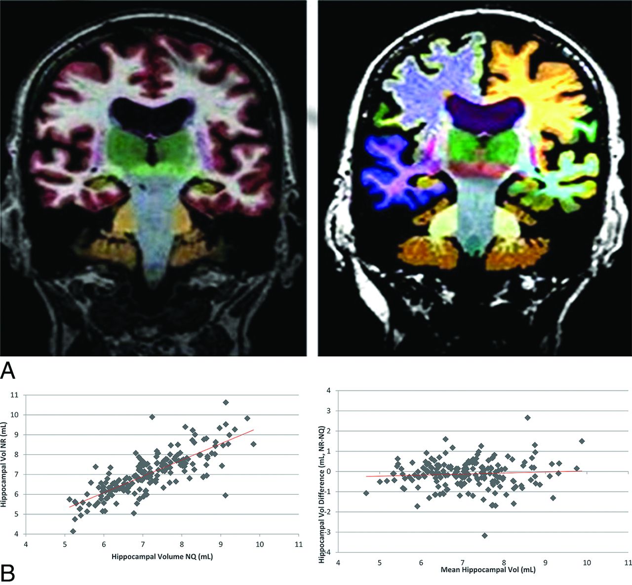
- Fig 1.
Example of NeuroQuant (A, left) and Neuroreader (A, right) color segmentations in the same subject and hippocampal scatter (B, left) and Bland-Altman plots (B, right). Note the high correlation (r = 0.79, P < .05) of hippocampal volumetrics between software packages across all subjects and the small underestimation bias (−0.12 mL, P < .05) of NR with respect to NQ.

Tables
Characteristic All Patients MCI (Nonconverter) MCI to AD (Converter) P Value No. 192 107 85 Age (yr) 74.8 ± 7.3 74.7 ± 7.6 75.0 ± 6.9 .7604 Sex .3262 Female 75 (39) 38 (36) 37 (44) Male 117 (61) 69 (64) 48 (56) Education (yr) 15.7 ± 2.9 15.7 ± 3.0 15.6 ± 2.8 .7637 ADAS-13 (baseline) 17.7 ± 6.4 15.2 ± 6.1 20.9 ± 5.3 <.0001 MMSE (baseline) 27.1 ± 1.7 27.6 ± 1.7 26.5 ± 1.6 <.0001 Note:—MMSE indicates Mini-Mental State Examination.
↵a MCI or AD status was determined at 3-year follow-up by the ADNI criteria. Data are means. Percentages listed in parentheses are rounded to the nearest percentage.
- Table 2:
AUC values for different brain regions separated by software package (NQ, NR) and method of analysis (univariable-multivariable)
Feature AUC NQ (95% CI) AUC NR (95% CI) Univariable analysis Hippocampus 0.69 (0.61–0.76) 0.68 (0.60–0.76) Amygdala 0.67 (0.59–0.74) 0.65 (0.57–0.73) Cerebellum 0.58 (0.49–0.66) 0.57 (0.49–0.66) Putamen 0.58 (0.50–0.66) 0.62 (0.54–0.70) Thalamus 0.56 (0.47–0.64) 0.56 (0.48–0.64) Lateral ventricle 0.54 (0.46–0.62) 0.54 (0.46–0.62) Pallidum 0.52 (0.43–0.60) NA Caudate 0.51 (0.42–0.59) NA Cortical gray matter 0.64 (0.56–0.72) NA Forebrain parenchyma 0.62 (0.54–0.70) NA Inferior lateral ventricle 0.60 (0.52–0.68) NA Temporal lobe NA 0.63 (0.55–0.71) Parietal lobe NA 0.61 (0.53–0.69) Frontal lobe NA 0.60 (0.52–0.68) Occipital lobe NA 0.59 (0.51–0.68) Ventral diencephalon NA 0.51 (0.43–0.60) Multivariable analysis Logistic regression 0.63 (0.55–0.71) 0.65 (0.58–0.73) Random forest 0.60 (0.52–0.68) 0.62 (0.54–0.71) Note:—NA indicates not applicable.
- Table 3:
Pearson correlation coefficients for NQ and NR volumetrics in regions with the same name
Feature Pearson Coefficient (r) 95% CI P Value Thalamus 0.60 0.51–0.69 <.001 Putamen 0.61 0.51–0.69 <.001 Lateral ventricles 0.99 0.99–0.99 <.001 Hippocampus 0.79 0.72–0.83 <.001 Cerebellum 0.87 0.83–0.90 <.001 Amygdala 0.71 0.64–0.78 <.001


Jump to section
Related Articles
Cited By...
- Brain Parcellation Repeatability and Reproducibility Using Conventional and Quantitative 3D MR Imaging
- Associations of Stages of Objective Memory Impairment With Amyloid PET and Structural MRI: The A4 Study
- Complete Evaluation of Dementia: PET and MRI Correlation and Diagnosis for the Neuroradiologist
- Predicting the Progression of Mild Cognitive Impairment Using Machine Learning: A Systematic, Quantitative and Critical Review
- Comparison of feature representations in MRI-based MCI-to-AD conversion prediction
- Regularized Bagged Canonical Component Analysis for Multiclass Learning in Brain Imaging
- Biomarker Localization, Analysis, Visualization, Extraction, and Registration (BLAzER) Workflow for Research and Clinical Brain PET Applications
- Is Hippocampal Volumetry Really All That Matters?