Advertisement

Main menu

  • Home
  • Content
    • Current Issue
    • Accepted Manuscripts
    • Article Preview
    • Past Issue Archive
    • Video Articles
    • AJNR Case Collection
    • Case of the Week Archive
    • Case of the Month Archive
    • Classic Case Archive
  • Special Collections
    • AJNR Awards
    • Low-Field MRI
    • Alzheimer Disease
    • ASNR Foundation Special Collection
    • Photon-Counting CT
    • View All
  • Multimedia
    • AJNR Podcasts
    • AJNR SCANtastic
    • Trainee Corner
    • MRI Safety Corner
    • Imaging Protocols
  • For Authors
    • Submit a Manuscript
    • Submit a Video Article
    • Submit an eLetter to the Editor/Response
    • Manuscript Submission Guidelines
    • Statistical Tips
    • Fast Publishing of Accepted Manuscripts
    • Graphical Abstract Preparation
    • Imaging Protocol Submission
    • Author Policies
  • About Us
    • About AJNR
    • Editorial Board
    • Editorial Board Alumni
  • More
    • Become a Reviewer/Academy of Reviewers
    • Subscribers
    • Permissions
    • Alerts
    • Feedback
    • Advertisers
    • ASNR Home

User menu

  • Alerts
  • Log in

Search

  • Advanced search
American Journal of Neuroradiology

American Journal of Neuroradiology

ASHNR American Society of Functional Neuroradiology ASHNR American Society of Pediatric Neuroradiology ASSR
  • Alerts
  • Log in

Advanced Search

  • Home
  • Content
    • Current Issue
    • Accepted Manuscripts
    • Article Preview
    • Past Issue Archive
    • Video Articles
    • AJNR Case Collection
    • Case of the Week Archive
    • Case of the Month Archive
    • Classic Case Archive
  • Special Collections
    • AJNR Awards
    • Low-Field MRI
    • Alzheimer Disease
    • ASNR Foundation Special Collection
    • Photon-Counting CT
    • View All
  • Multimedia
    • AJNR Podcasts
    • AJNR SCANtastic
    • Trainee Corner
    • MRI Safety Corner
    • Imaging Protocols
  • For Authors
    • Submit a Manuscript
    • Submit a Video Article
    • Submit an eLetter to the Editor/Response
    • Manuscript Submission Guidelines
    • Statistical Tips
    • Fast Publishing of Accepted Manuscripts
    • Graphical Abstract Preparation
    • Imaging Protocol Submission
    • Author Policies
  • About Us
    • About AJNR
    • Editorial Board
    • Editorial Board Alumni
  • More
    • Become a Reviewer/Academy of Reviewers
    • Subscribers
    • Permissions
    • Alerts
    • Feedback
    • Advertisers
    • ASNR Home
  • Follow AJNR on Twitter
  • Visit AJNR on Facebook
  • Follow AJNR on Instagram
  • Join AJNR on LinkedIn
  • RSS Feeds

AJNR Awards, New Junior Editors, and more. Read the latest AJNR updates

Research ArticleADULT BRAIN

A Fully Automated, Atlas-Based Approach for Superior Cerebellar Peduncle Evaluation in Progressive Supranuclear Palsy Phenotypes

G. Nicoletti, M.E. Caligiuri, A. Cherubini, M. Morelli, F. Novellino, G. Arabia, M. Salsone and A. Quattrone
American Journal of Neuroradiology March 2017, 38 (3) 523-530; DOI: https://doi.org/10.3174/ajnr.A5048
G. Nicoletti
aFrom the Institute of Bioimaging and Molecular Physiology (G.N., M.E.C., A.C., F.N., M.S., A.Q.), National Research Council, Catanzaro, Italy
  • Find this author on Google Scholar
  • Find this author on PubMed
  • Search for this author on this site
  • ORCID record for G. Nicoletti
M.E. Caligiuri
aFrom the Institute of Bioimaging and Molecular Physiology (G.N., M.E.C., A.C., F.N., M.S., A.Q.), National Research Council, Catanzaro, Italy
  • Find this author on Google Scholar
  • Find this author on PubMed
  • Search for this author on this site
  • ORCID record for M.E. Caligiuri
A. Cherubini
aFrom the Institute of Bioimaging and Molecular Physiology (G.N., M.E.C., A.C., F.N., M.S., A.Q.), National Research Council, Catanzaro, Italy
  • Find this author on Google Scholar
  • Find this author on PubMed
  • Search for this author on this site
  • ORCID record for A. Cherubini
M. Morelli
bInstitute of Neurology (M.M., G.A., A.Q.), University “Magna Graecia”, Catanzaro, Italy.
  • Find this author on Google Scholar
  • Find this author on PubMed
  • Search for this author on this site
  • ORCID record for M. Morelli
F. Novellino
aFrom the Institute of Bioimaging and Molecular Physiology (G.N., M.E.C., A.C., F.N., M.S., A.Q.), National Research Council, Catanzaro, Italy
  • Find this author on Google Scholar
  • Find this author on PubMed
  • Search for this author on this site
  • ORCID record for F. Novellino
G. Arabia
bInstitute of Neurology (M.M., G.A., A.Q.), University “Magna Graecia”, Catanzaro, Italy.
  • Find this author on Google Scholar
  • Find this author on PubMed
  • Search for this author on this site
  • ORCID record for G. Arabia
M. Salsone
aFrom the Institute of Bioimaging and Molecular Physiology (G.N., M.E.C., A.C., F.N., M.S., A.Q.), National Research Council, Catanzaro, Italy
  • Find this author on Google Scholar
  • Find this author on PubMed
  • Search for this author on this site
  • ORCID record for M. Salsone
A. Quattrone
aFrom the Institute of Bioimaging and Molecular Physiology (G.N., M.E.C., A.C., F.N., M.S., A.Q.), National Research Council, Catanzaro, Italy
bInstitute of Neurology (M.M., G.A., A.Q.), University “Magna Graecia”, Catanzaro, Italy.
  • Find this author on Google Scholar
  • Find this author on PubMed
  • Search for this author on this site
  • ORCID record for A. Quattrone
  • Article
  • Figures & Data
  • Supplemental
  • Info & Metrics
  • Responses
  • References
  • PDF
Loading

Article Figures & Data

Figures

  • Tables
  • Fig 1.
    • Download figure
    • Open in new tab
    • Download powerpoint
    Fig 1.

    Image-processing workflow. The coregistration of different sequences allows the extraction of multimodal parameters from an ROI.
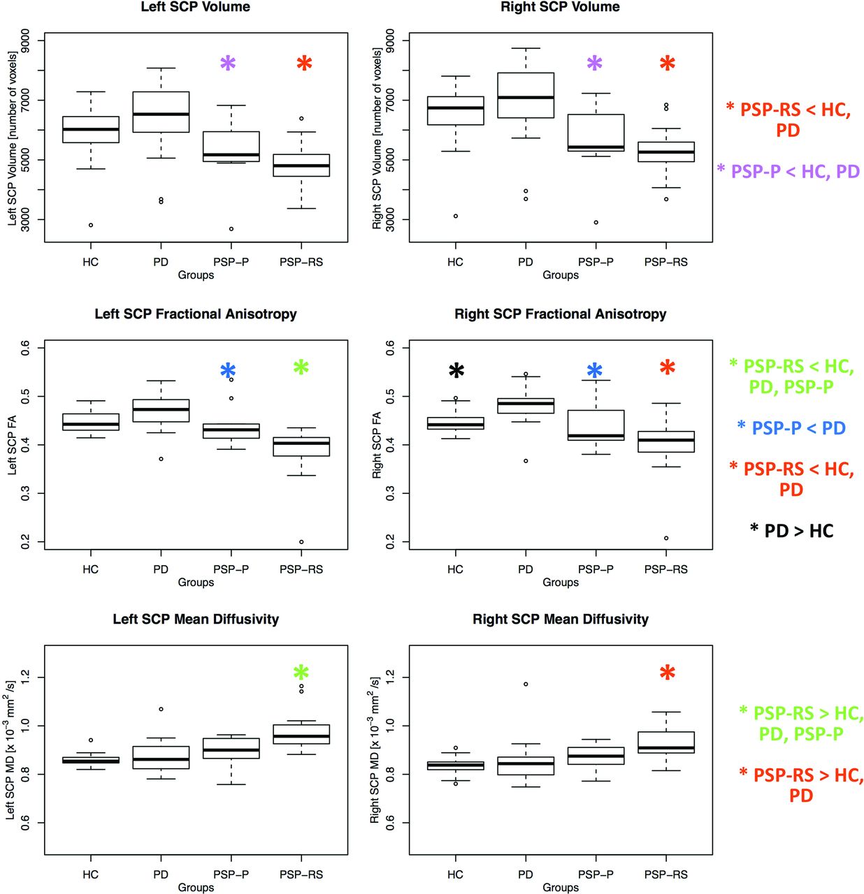
  • Fig 2.
    • Download figure
    • Open in new tab
    • Download powerpoint
    Fig 2.

    Box-and-whisker plots of volumes, FA, and MD of the right and left SCPs in patients and controls.

Tables

  • Figures
    • View popup
    Table 1:

    Demographic, clinical, and neuroimaging features in patients with PSP-P, PSP-RS, PD, and healthy controls

    PSP-P (n = 9)PSP-RS (n = 21)PD (n = 20)HC (n = 30)
    Age (mean) (yr)70.1 ± 4.871.9 ± 5.966.2 ± 3.067.2 ± 7.2
    Sex, % male100%57%50%47%
    Disease duration (mean) (yr)6.3 ± 3.73.1 ± 1.47.5 ± 3.7–
    MMSE (mean)22.1 ± 3.619.0 ± 5.423.2 ± 3.3–
    UPDRS-III (mean)22.8 ± 17.739.3 ± 7.727.1 ± 9.1–
    HY (mean)2.9 ± 0.73.4 ± 0.82.2 ± 0.5–
    SCP right
        Volume (No. of voxels) (mean)5652 ± 11725282 ± 7536907 ± 13066569 ± 944
        FA (mean)0.44 ± 0.050.40 ± 0.050.48 ± 0.040.45 ± 0.02
        MD (×10−3 mm2/s) (mean)0.873 ± 0.060.949 ± 0.1700.851 ± 0.0890.833 ± 0.031
    SCP left
        Volume (No. of voxels) (mean)5315 ± 11214812 ± 7016369 ± 12055961 ± 894
        FA (mean)0.44 ± 0.040.39 ± 0.050.47 ± 0.040.45 ± 0.02
        MD (×10−3 mm2/s (mean)0.895 ± 0.0610.974 ± 0.0740.874 ± 0.0660.857 ± 0.023
    • Note:—HC indicates healthy controls; MMSE, Mini-Mental State Examination; UPDRS-III, Unified Parkinson's Disease Rating Scale–Motor Examination; HY, Hohen and Yahr score.

    • View popup
    Table 2:

    P values from the statistical tests among different groups after correction for multiple comparisons (Tukey test)a

    PD vs HCPSP-P vs HCPSP-RS vs HCPSP-P vs PDPSP-RS vs PDPSP-P vs PSP-RS
    Age11.02b0.1.01b1
    Sex.1.0001b.1.008b1.01b
    Disease duration–––1.0001b.02b
    MMSE–––1.01b1
    UPDRS-III–––1.0022b.002b
    HY–––.0004b.94.001b
    SCP right
        Volume.66.008b.00005b.001b.000004b.95
        FA.01b.66.0005b.004b1.45 × 10−8b.16
        MD.89.38.0003b.77.01b.31
    SCP left
        Volume.44.03b.00008b.002b.000002b.81
        FA.15.58.0000005b.03b9.33 × 10−10b.007b
        MD.70.169.30 × 10−10b.67.000001b.003b
    • Note:— HC indicates healthy controls; MMSE, Mini-Mental State Examination; UPDRS-III, Unified Parkinson's Disease Rating Scale-Motor Examination; HY, Hohen and Yahr score.

    • ↵a Sex differences were assessed with χ2 tests. Age differences were assessed with ANOVA. Disease duration differences were assessed using ANCOVA, with age and sex as covariates. Other clinical and imaging differences were assessed with ANCOVA, with age, sex, and disease duration, as covariates. Correction for multiple comparisons was performed with the Tukey honest significant difference test.

    • ↵b Significant.

PreviousNext
Back to top

In this issue

American Journal of Neuroradiology: 38 (3)
American Journal of Neuroradiology
Vol. 38, Issue 3
1 Mar 2017
  • Table of Contents
  • Index by author
  • Complete Issue (PDF)
Advertisement
Print
Download PDF
Email Article

Thank you for your interest in spreading the word on American Journal of Neuroradiology.

NOTE: We only request your email address so that the person you are recommending the page to knows that you wanted them to see it, and that it is not junk mail. We do not capture any email address.

Enter multiple addresses on separate lines or separate them with commas.
A Fully Automated, Atlas-Based Approach for Superior Cerebellar Peduncle Evaluation in Progressive Supranuclear Palsy Phenotypes
(Your Name) has sent you a message from American Journal of Neuroradiology
(Your Name) thought you would like to see the American Journal of Neuroradiology web site.
CAPTCHA
This question is for testing whether or not you are a human visitor and to prevent automated spam submissions.
Cite this article
G. Nicoletti, M.E. Caligiuri, A. Cherubini, M. Morelli, F. Novellino, G. Arabia, M. Salsone, A. Quattrone
A Fully Automated, Atlas-Based Approach for Superior Cerebellar Peduncle Evaluation in Progressive Supranuclear Palsy Phenotypes
American Journal of Neuroradiology Mar 2017, 38 (3) 523-530; DOI: 10.3174/ajnr.A5048

Citation Manager Formats

  • BibTeX
  • Bookends
  • EasyBib
  • EndNote (tagged)
  • EndNote 8 (xml)
  • Medlars
  • Mendeley
  • Papers
  • RefWorks Tagged
  • Ref Manager
  • RIS
  • Zotero
0 Responses
Respond to this article
Share
Bookmark this article
A Fully Automated, Atlas-Based Approach for Superior Cerebellar Peduncle Evaluation in Progressive Supranuclear Palsy Phenotypes
G. Nicoletti, M.E. Caligiuri, A. Cherubini, M. Morelli, F. Novellino, G. Arabia, M. Salsone, A. Quattrone
American Journal of Neuroradiology Mar 2017, 38 (3) 523-530; DOI: 10.3174/ajnr.A5048
del.icio.us logo Twitter logo Facebook logo Mendeley logo
  • Tweet Widget
  • Facebook Like
  • Google Plus One
Purchase

Jump to section

  • Article
    • Abstract
    • ABBREVIATIONS:
    • Materials and Methods
    • Results
    • Discussion
    • Conclusions
    • References
  • Figures & Data
  • Supplemental
  • Info & Metrics
  • Responses
  • References
  • PDF

Related Articles

  • PubMed
  • Google Scholar

Cited By...

  • Midbrain MRI assessments in progressive supranuclear palsy subtypes
  • Crossref (20)
  • Google Scholar

This article has been cited by the following articles in journals that are participating in Crossref Cited-by Linking.

  • A new MR imaging index for differentiation of progressive supranuclear palsy-parkinsonism from Parkinson's disease
    Aldo Quattrone, Maurizio Morelli, Salvatore Nigro, Andrea Quattrone, Basilio Vescio, Gennarina Arabia, Giuseppe Nicoletti, Rita Nisticò, Maria Salsone, Fabiana Novellino, Gaetano Barbagallo, Emilio Le Piane, Pierfrancesco Pugliese, Domenico Bosco, Maria Grazia Vaccaro, Carmelina Chiriaco, Umberto Sabatini, Virginia Vescio, Carlo Stanà, Federico Rocca, Domenico Gullà, Manuela Caracciolo
    Parkinsonism & Related Disorders 2018 54
  • Neuroimaging Advances in Parkinson's Disease and Atypical Parkinsonian Syndromes
    Usman Saeed, Anthony E. Lang, Mario Masellis
    Frontiers in Neurology 2020 11
  • Cerebellum and neurodegenerative diseases: Beyond conventional magnetic resonance imaging
    Enricomaria Mormina, Maria Petracca, Giulia Bommarito, Niccolò Piaggio, Sirio Cocozza, Matilde Inglese
    World Journal of Radiology 2017 9 10
  • Midbrain MRI assessments in progressive supranuclear palsy subtypes
    Marina Picillo, Maria Francesca Tepedino, Filomena Abate, Roberto Erro, Sara Ponticorvo, Salvatore Tartaglione, Giampiero Volpe, Daniela Frosini, Paolo Cecchi, Mirco Cosottini, Roberto Ceravolo, Fabrizio Esposito, Maria Teresa Pellecchia, Paolo Barone, Renzo Manara
    Journal of Neurology, Neurosurgery & Psychiatry 2020 91 1
  • Automated MRI Classification in Progressive Supranuclear Palsy: A Large International Cohort Study
    Salvatore Nigro, Angelo Antonini, David E. Vaillancourt, Klaus Seppi, Roberto Ceravolo, Antonio P. Strafella, Antonio Augimeri, Andrea Quattrone, Maurizio Morelli, Luca Weis, Eleonora Fiorenzato, Roberta Biundo, Roxana G. Burciu, Florian Krismer, Nikolaus R. McFarland, Christoph Mueller, Elke R. Gizewski, Mirco Cosottini, Eleonora Del Prete, Sonia Mazzucchi, Aldo Quattrone
    Movement Disorders 2020 35 6
  • Diagnostic potential of dentatorubrothalamic tract analysis in progressive supranuclear palsy
    Morinobu Seki, Klaus Seppi, Christoph Mueller, Thomas Potrusil, Georg Goebel, Eva Reiter, Michael Nocker, Ruth Steiger, Matthias Wildauer, Elke R. Gizewski, Gregor K. Wenning, Werner Poewe, Christoph Scherfler
    Parkinsonism & Related Disorders 2018 49
  • Imaging counterpart of postural instability and vertical ocular dysfunction in patients with PSP: A multimodal MRI study
    Andrea Quattrone, Maria Eugenia Caligiuri, Maurizio Morelli, Salvatore Nigro, Basilio Vescio, Gennarina Arabia, Giuseppe Nicoletti, Rita Nisticò, Maria Salsone, Fabiana Novellino, Gaetano Barbagallo, Maria Grazia Vaccaro, Umberto Sabatini, Virginia Vescio, Carlo Stanà, Federico Rocca, Manuela Caracciolo, Aldo Quattrone
    Parkinsonism & Related Disorders 2019 63
  • Clinical Spectrum of Tauopathies
    Nahid Olfati, Ali Shoeibi, Irene Litvan
    Frontiers in Neurology 2022 13
  • “Parkinson’s disease” on the way to progressive supranuclear palsy: a review on PSP-parkinsonism
    Ján Necpál, Miroslav Borsek, Bibiána Jeleňová
    Neurological Sciences 2021 42 12
  • Diagnostic potential of automated tractography in progressive supranuclear palsy variants
    Thomas Potrusil, Florian Krismer, Vincent Beliveau, Klaus Seppi, Christoph Müller, Felix Troger, Georg Göbel, Ruth Steiger, Elke R. Gizewski, Werner Poewe, Christoph Scherfler
    Parkinsonism & Related Disorders 2020 72

More in this TOC Section

  • Diagnostic Neuroradiology of Monoclonal Antibodies
  • Clinical Outcomes After Chiari I Decompression
  • Segmentation of Brain Metastases with BLAST
Show more ADULT BRAIN

Similar Articles

Advertisement

Indexed Content

  • Current Issue
  • Accepted Manuscripts
  • Article Preview
  • Past Issues
  • Editorials
  • Editor's Choice
  • Fellows' Journal Club
  • Letters to the Editor
  • Video Articles

Cases

  • Case Collection
  • Archive - Case of the Week
  • Archive - Case of the Month
  • Archive - Classic Case

More from AJNR

  • Trainee Corner
  • Imaging Protocols
  • MRI Safety Corner

Multimedia

  • AJNR Podcasts
  • AJNR Scantastics

Resources

  • Turnaround Time
  • Submit a Manuscript
  • Submit a Video Article
  • Submit an eLetter to the Editor/Response
  • Manuscript Submission Guidelines
  • Statistical Tips
  • Fast Publishing of Accepted Manuscripts
  • Graphical Abstract Preparation
  • Imaging Protocol Submission
  • Evidence-Based Medicine Level Guide
  • Publishing Checklists
  • Author Policies
  • Become a Reviewer/Academy of Reviewers
  • News and Updates

About Us

  • About AJNR
  • Editorial Board
  • Editorial Board Alumni
  • Alerts
  • Permissions
  • Not an AJNR Subscriber? Join Now
  • Advertise with Us
  • Librarian Resources
  • Feedback
  • Terms and Conditions
  • AJNR Editorial Board Alumni

American Society of Neuroradiology

  • Not an ASNR Member? Join Now

© 2025 by the American Society of Neuroradiology All rights, including for text and data mining, AI training, and similar technologies, are reserved.
Print ISSN: 0195-6108 Online ISSN: 1936-959X

Powered by HighWire