
Article Figures & Data
Figures
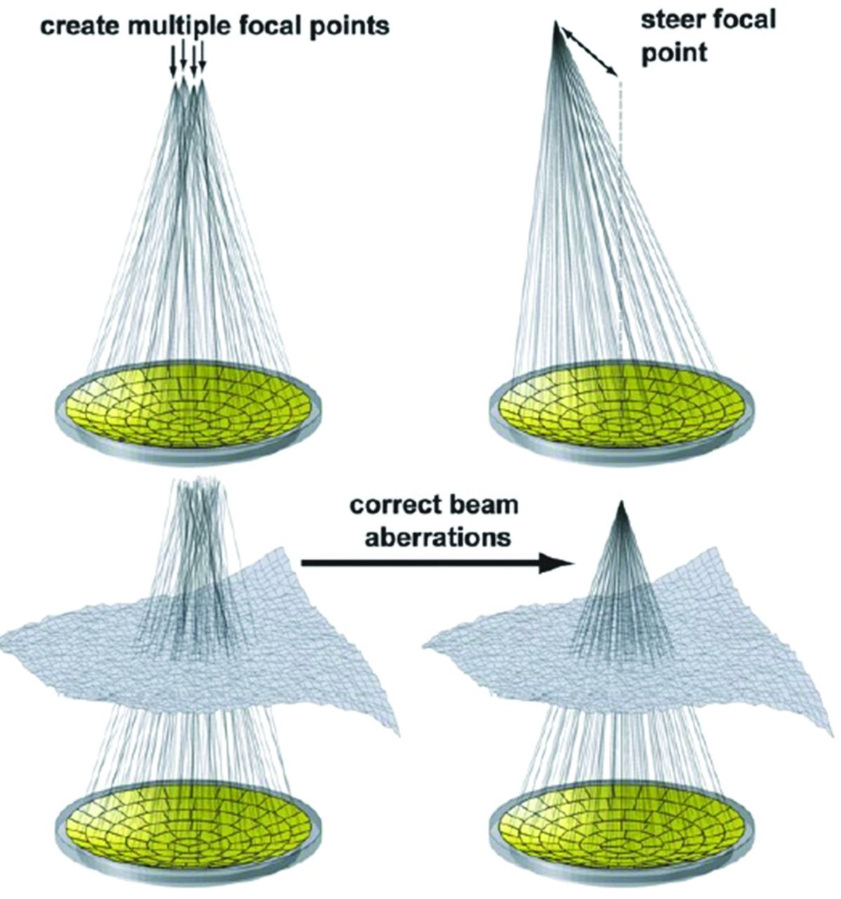
- Fig 1.
With a multielement, hemispheric phased array transducer, a single focus can be electronically steered (upper right), multiple focal points can be generated (upper left), and corrections can be achieved for aberrations in the beam path. Reprinted with permission from Tempany et al.37
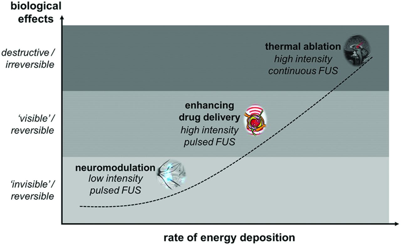
- Fig 2.
Unique biologic effects can be achieved over a range of energy-deposition rates by manipulating the intensity and duty cycle of the ultrasound application. These include neuromodulation, localized reversible enhancement of blood-brain barrier permeability, and thermal ablation.
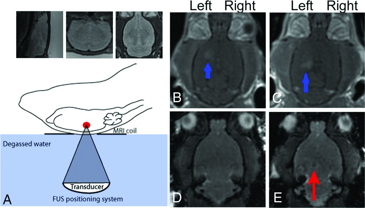
- Fig 3.
Application of MRgFUS for the delivery of iron-labeled neural stem cells. Schematic of the FUS apparatus (lower left). Sagittal, coronal, and axial T2-weighted images (A) are used to identify the intended targets of neural stem cell delivery in the left hippocampus and left striatum. T1-weighted, postcontrast images after local FUS sonication (B and C) demonstrate enhancement in the striatum and hippocampus (blue arrows) compatible with enhanced BBB permeability. Fast gradient-echo sequences are obtained before (D) and after (E) sonication, localizing a focus of hypointense signal (red arrow) confirming delivery of iron-labeled neural stem cells. Reprinted with permission from Burgess et al.40
- Fig 4.
Comparison of TMZ versus TMZ + FUS. At day 10, tumors in all groups are similar in size, designating the start of treatments. At day 17, the FUS + TMZ group demonstrate the slowest rate of tumor growth as evidenced by the degree of T2-weighted signal. The FUS + TMZ group also shows the longest survival of any of the TMZ-only groups (not shown). Reprinted with permission from Wei et al.18
- Fig 5.
Neuromodulation of the rabbit motor cortex. An fMRI activation map shows increased blood oxygen level–dependent–weighted signal in the right motor cortex (A and B). The blue crosshairs on the fMRI images correspond to the sonication focus. Cartoon schematic illustrates the experimental setup and spatial orientation. The graph (C) demonstrates the percentage of blood oxygen level–dependent signal change as a function of the time/acquisition number at 2 different FUS intensities: 6 W/cm2 (red curve) and 3 W/cm2 (blue curve). The green dataset represents the control group. The gray bars indicate the sonication time. Reprinted with permission from Yoo et al.28


Tables
Clinical applications of focused ultrasound and the proposed mechanisms of action
FUS Exposures and Effects on Biologic Tissues Mechanism of Action Applications High-intensity continuous application Thermal → coagulative necrosis Thalamotomy: essential tremor,11 chronic neuropathic pain,13 obsessive- compulsive disorder36 Pallidotomy: Parkinson disease12 Solid tumor ablation14 High-intensity pulsed application Oscillation of ultrasound “microbubble” contrast agents Enhanced agent CNS delivery22 Thermal → regional hyperthermia Enhanced chemotherapy/radiotherapy37 Mechanical → radiation force–induced displacements Induction of microglial activation27 Sonothrombolysis in ischemic stroke38 Intracranial hematoma evacuation39 Low-intensity pulsed application Mechanical → activation/inhibition of Na+ and Ca2+-gated ion channels Noninvasive neuromodulation33,34


