
Article Figures & Data
Figures
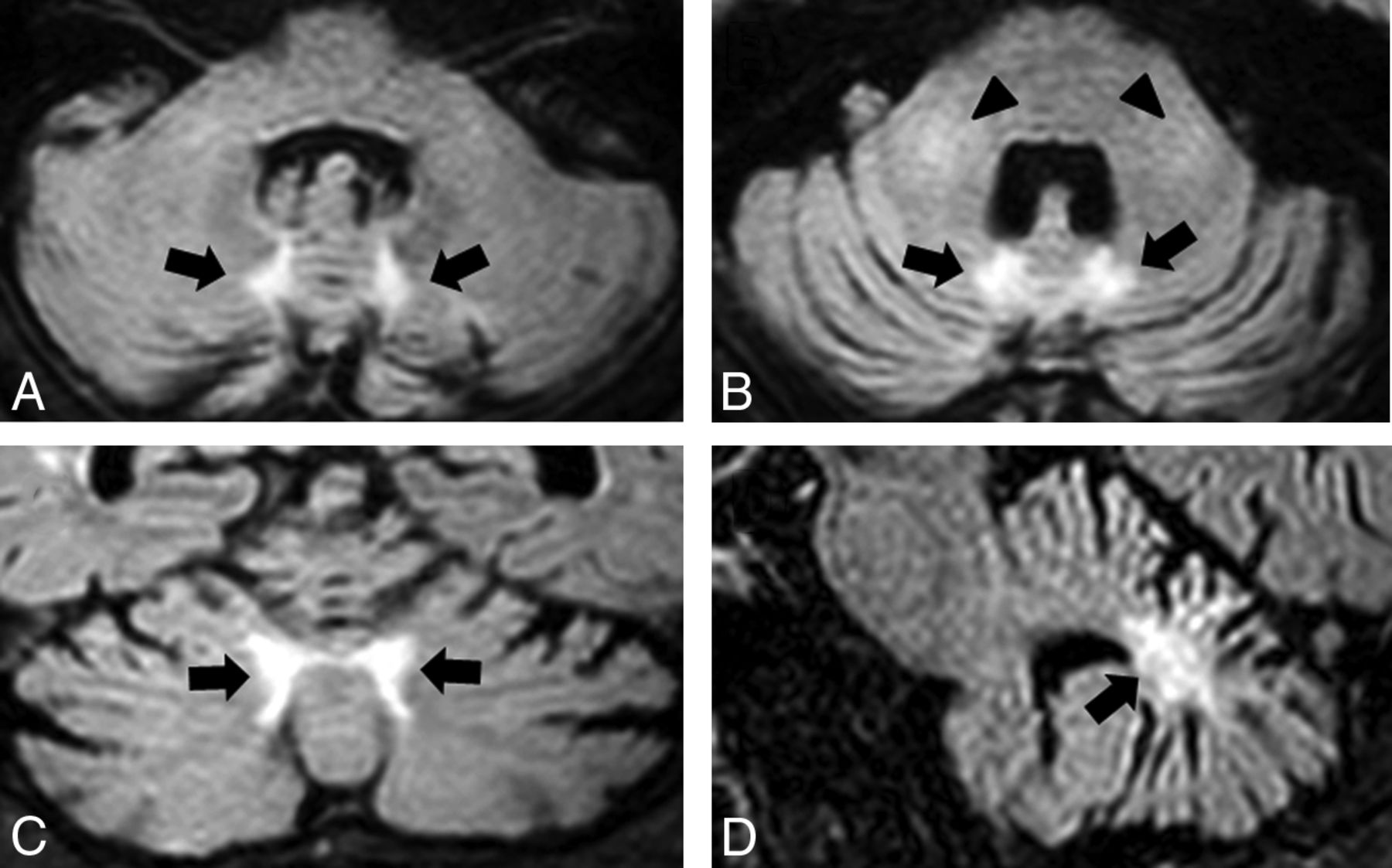
- Fig 1.
A representative case (patient 1) showing the abnormal signals at the paravermal area and the middle cerebellar peduncle. FLAIR axial images (A and B), a coronal image (C), and a sagittal image (D) show atrophy of the cerebellum and bilateral high signal intensity in the medial part of the cerebellar hemisphere immediately beside the vermis (the paravermal area) (black arrows) and in the middle cerebellar peduncle (black arrowheads).
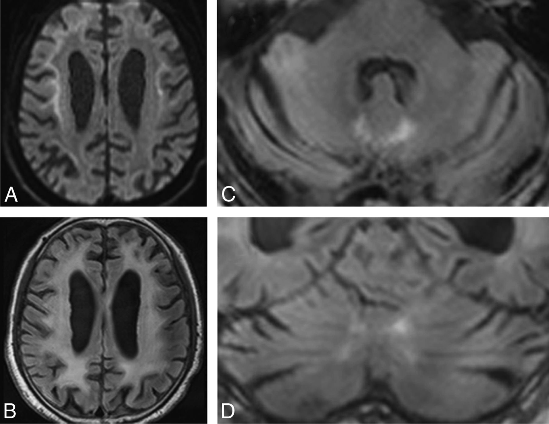
- Fig 2.
Patient 4. DWI (A) shows high-intensity signal along the corticomedullary junction. A FLAIR axial image (B) shows diffuse high intensity in the bilateral cerebral hemispheres. FLAIR axial (C) and coronal (D) images show atrophy of the cerebellum and high-intensity signal in the medial part of the cerebellar hemisphere right beside the vermis (the paravermal area).
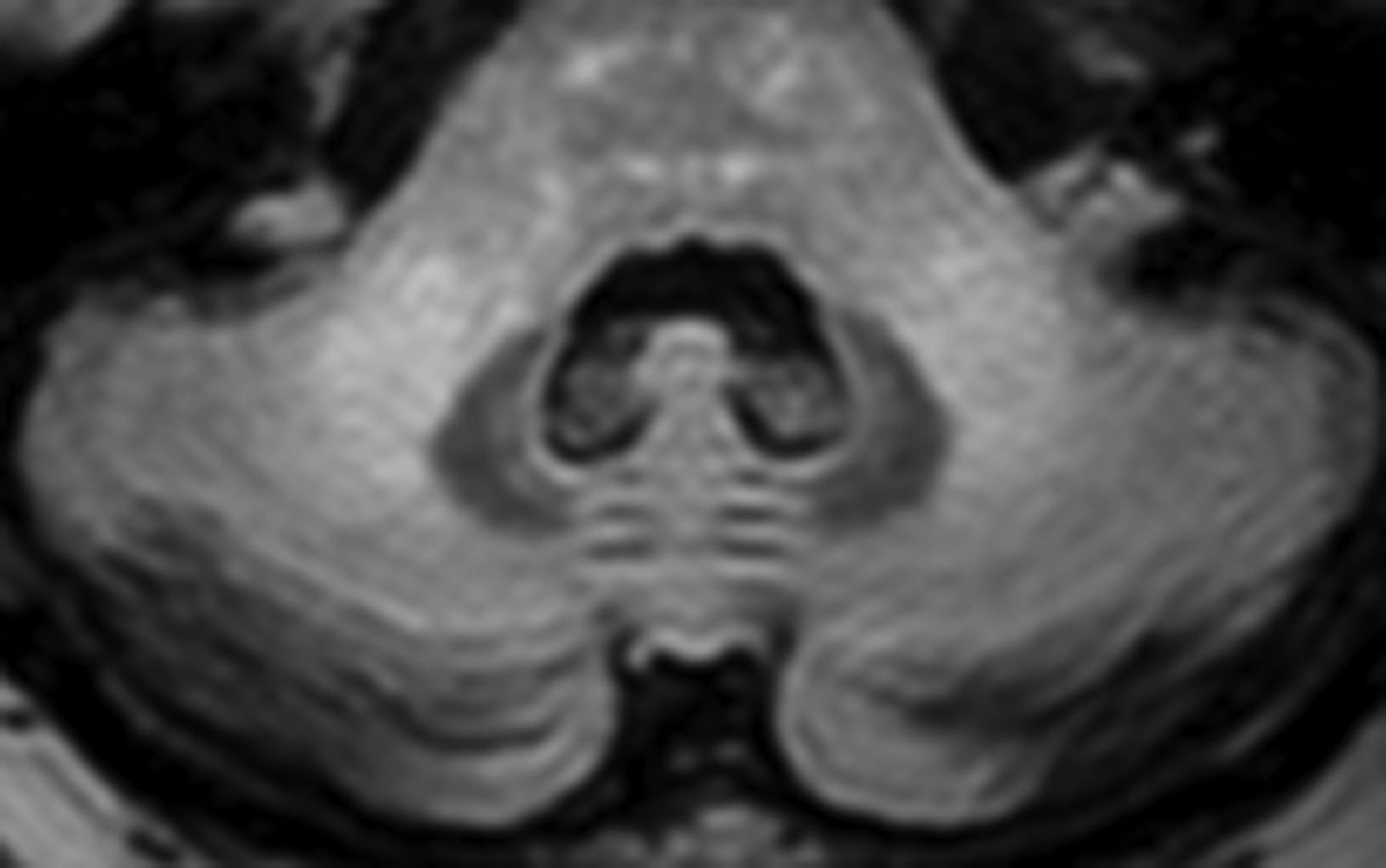
- Fig 3.
Patient 2. A FLAIR axial image shows bilateral high signal intensities in the middle cerebellar peduncle.


Tables
Patient No. 1 2 3 4 5 6 7 8 Age at onset (yr) 64 52 62 62 67 67 67 64 Sex M F F M F M F M Disease duration at MRI (yr) 2 9 8 3 5 6 1 5 Clinical manifestations Dementia – – – + + – + – Delusion – – + – – – – – Muscle weakness – – – – – – – – Tremor – – Postural – Postural Postural – Resting, postural Rigidity – – – – – – – – Ataxia – – – + + – – + Hyporeflexia or areflexia in DTR + + + + + + + + Sensory disturbance – – – – – – – – Urinary incontinence – – – + + – – – Transient visual field abnormality + + – – – – – + Transient hemiparesis – + – – – – – – Transient abnormal behavior – + – + – + – – Generalized convulsion – – – – – – – – Disturbance of consciousness – – – – – + + + Cognitive screening test MMSE 30 29 30 14a 30 28 24 29 Frontal lobe function test FAB 13b 16 14b 11b 12b 16 8b NE CSF Cell (mm3) 3 2 4 1 1 6 NE NE Protein (mg/dL) 66 55 100 47 52 48 NE NE Patient No. 1 2 3 4 5 6 7 8 Cerebrum High-intensity signal along the corticomedullary junction on DWI + + + + + + + + Diffuse high-intensity signal of cerebral white matter on FLAIR images + + + + + + + + Cerebellum Atrophy + + + + + + + + High-intensity signal in the medial part of cerebellar hemisphere right beside the vermis on FLAIR images + – + + + + – + High-intensity signal in the middle cerebellar peduncle on FLAIR images + + + – – + – – Note:— – indicates absence of the finding; +, presence of the finding.


Jump to section
Related Articles
Cited By...
- NOTCH2NLC expanded GGC repeats in patients with cerebral small vessel disease
- Clinical Features and Classification of Neuronal Intranuclear Inclusion Disease
- Clinical features of NOTCH2NLC-related neuronal intranuclear inclusion disease
- NOTCH2NLC expanded GGC repeats in patients with cerebral small vessel disease
- Teaching NeuroImage: Paravermal Lesions in Neuronal Intranuclear Inclusion Disease
- Isolated Paravermal Hyperintensities in Neuronal Intranuclear Inclusion Disease
- Clinical Reasoning: A 65-Year-Old Woman With Tremor
- MRI diagnosis of neuronal intranuclear inclusion disease leukoencephalopathy
- MR Imaging Features of Adult-Onset Neuronal Intranuclear Inclusion Disease May Be Indistinguishable from Fragile X-Associated Tremor/Ataxia Syndrome