
Article Figures & Data
Figures
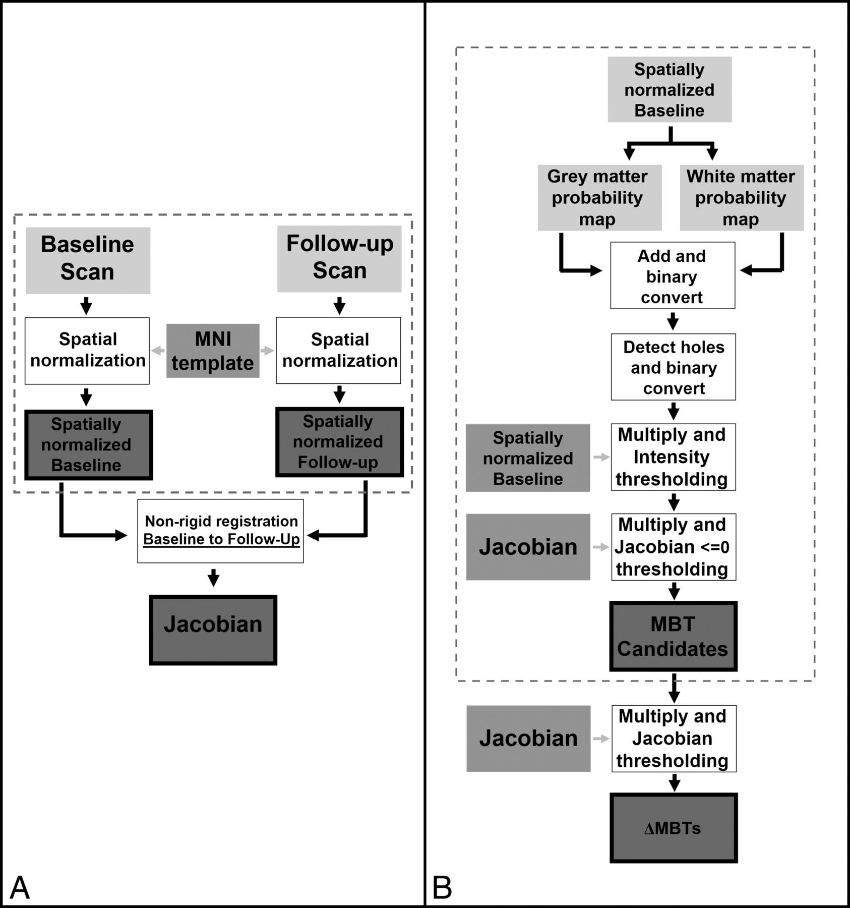
- Fig 1.
Steps summarizing the preprocessing of patient datasets (dotted box) and calculation of the Jacobian operator field in the forward direction (A) and for segmentation of metastatic brain tumor candidates (dotted box) and detection of shrinking MBTs in the forward direction (B).
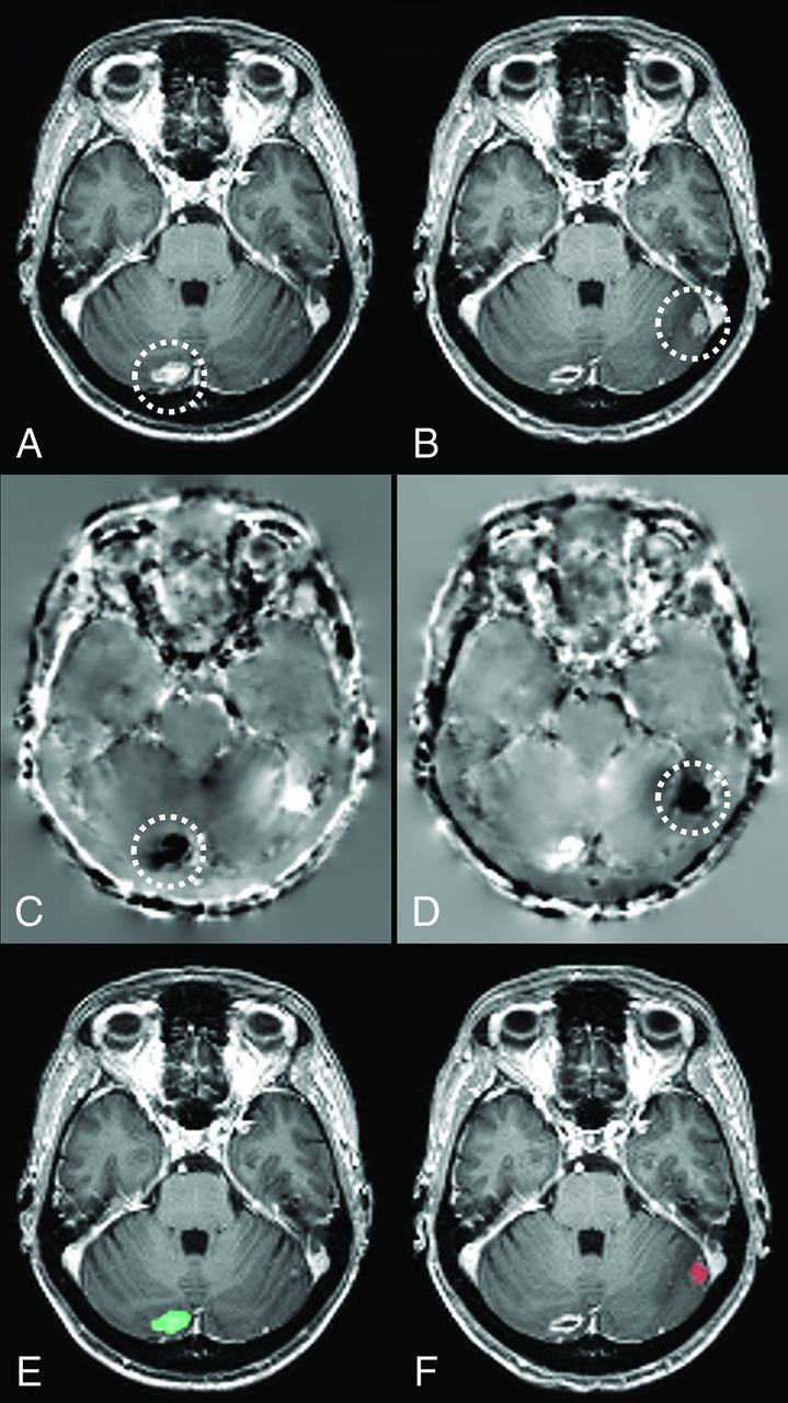
- Fig 2.
Axial sections of a patient's baseline (A) and follow-up (B) scans. The Jacobian operator field, calculated from the deformation field in the forward (C) and reverse (D) directions. The final output of our algorithm produced for baseline (E) and follow-up (F) scans, highlighting volume-changing metastatic brain tumors on each scan. Note that darker voxels on C and D correspond to negative JOF values and brighter voxels correspond to positive JOF values. The location of a metastatic brain tumor that has shrunk in size across the scans has been circled on A–D. The green on E indicates shrinkage, and the red on F indicates growth. Note that while this image is demonstrated in 2D, various operations as described here were performed in 3D.
- Fig 3.
A, Illustration of the receiver operating characteristic curve of our algorithm for detecting volume-changing metastatic brain tumors at 1.5T, constructed from 233 different thresholding values of the Jacobian operator field (from 0 to −0.232, separated by −0.001). The arrow shows the optimal point of balance. B, Illustration of the ROC curve of our algorithm for detecting ΔMBTs at 3T, constructed from 656 different thresholding values of the Jacobian operator field (from 0 to −0.655, separated by −0.001). The arrow shows the optimal point of balance between sensitivity and specificity, which happens at the Jacobian value of −0.182.


Tables
Variable 1.5T 3T Sex Male (total %) 15 (50%) 11 (55%) Female (total %) 15 (50%) 9 (45%) Age (yr) Averagea 60.3 ± 13.1 58.7 ± 15.7 Range 23.4–91.0 27.3–84.5 Time between baseline and follow-up (days) Averagea (per patient) 147 ± 155 132 ± 129 Range 26–676 17–532 Number of ΔMBTs Total 74 76 ΔMBTts 58 67 ΔMBTos 16 9 Averagea (per patient) 4.4 ± 3.1 3.8 ± 3.9 ΔMBT volume (mL) Averagea 2.4 ± 4.0 2.2 ± 3.8 Range 4.0 × 10⁻³–1.9 × 101 2.0 × 10⁻²–3.0 × 101 ΔMBT volume change (mL) Averagea 1.5 ± 2.2 2.2 ± 3.5 Range 3.4 × 10⁻³–3.5 × 101 9.1 × 10⁻²–2.0 × 101 ΔMBT VCR (%) Averagea 7.0 × 101 ± 29.5 7.6 × 101 ± 1.7 × 101 Range 7.8 × 10⁻¹–1.0 × 10² 4.2–1.0 × 10² ↵a Average ± SD.
- Table 2:
Summary of ROC analysis, for detecting all 1.5T and 3T ΔMBTs and 1.5T ΔMBTts only, and the VCR of detected and missed ΔMBTs
Variable 1.5T 3T ROC curve AUC 0.925 0.965 Optimal Jacobian threshold −0.035 −0.182 Sensitivitya (%) 85.1 92.1 Specificitya (%) 86.7 91.3 FPRa (per section) 0.208 0.227 FPRa (per scan) 25.1 27.5 Detecteda ΔMBT VCR (%) Averageb 7.1 × 101 ± 2.8 × 101 7.7 × 101 ± 1.7 × 101 Median 7.9 × 101 7.5 × 101 Range 7.8 × 10⁻¹–1.0 × 10² 4.2–1.0 × 10² Misseda ΔMBT VCR (%) Averageb 6.2 × 101 ± 3.8 × 101 7.3 × 101 ± 1.9 × 101 Median 7.5 × 101 8.2 × 10² Range 3.9–1.0 × 10² 4.3 × 101–8.6 × 101 False-Negative Categories 1.5T 3T ΔMBTos (percentage of total) 4 (36.3%) 0 (0%) ΔMBTts (percentage of total) 7 (63.7%) 6 (100%) ≤2 voxels 3 (27.3%) 1 (16.7%) Poorly segmented 4 (36.4%) 5 (83.3%) - Table 5:
Summary of ROC analysis after jackknifing datasets, for detecting all 1.5T and 3T ΔMBTs and 1.5T ΔMBTts only
Variable 1.5T 1.5T: ΔMBTts Only 3T AUC of ROC curve Averagea 0.925 ± 0.003 0.929 ± 0.003 0.965 ± 0.002 Median 0.924 0.928 0.965 Sensitivityb (%) Averagea 85.1 ± 0.8 87.9 ± 0.8 92.2 ± 0.5 Median 84.9 87.7 92.0 Specificityb (%) Averagea 86.7 ± 0.3 86.6 ± 0.3 91.3 ± 0.6 Median 86.7 86.6 91.1 FPR (per section) Averagea 0.208 ± 0.005 0.210 ± 0.005 0.227 ± 0.02 Median 0.209 0.211 0.232


