Advertisement

Main menu

  • Home
  • Content
    • Current Issue
    • Accepted Manuscripts
    • Article Preview
    • Past Issue Archive
    • Video Articles
    • AJNR Case Collection
    • Case of the Week Archive
    • Case of the Month Archive
    • Classic Case Archive
  • Special Collections
    • AJNR Awards
    • Low-Field MRI
    • Alzheimer Disease
    • ASNR Foundation Special Collection
    • Photon-Counting CT
    • View All
  • Multimedia
    • AJNR Podcasts
    • AJNR SCANtastic
    • Trainee Corner
    • MRI Safety Corner
    • Imaging Protocols
  • For Authors
    • Submit a Manuscript
    • Submit a Video Article
    • Submit an eLetter to the Editor/Response
    • Manuscript Submission Guidelines
    • Statistical Tips
    • Fast Publishing of Accepted Manuscripts
    • Graphical Abstract Preparation
    • Imaging Protocol Submission
    • Author Policies
  • About Us
    • About AJNR
    • Editorial Board
    • Editorial Board Alumni
  • More
    • Become a Reviewer/Academy of Reviewers
    • Subscribers
    • Permissions
    • Alerts
    • Feedback
    • Advertisers
    • ASNR Home

User menu

  • Alerts
  • Log in

Search

  • Advanced search
American Journal of Neuroradiology

American Journal of Neuroradiology

ASHNR American Society of Functional Neuroradiology ASHNR American Society of Pediatric Neuroradiology ASSR
  • Alerts
  • Log in

Advanced Search

  • Home
  • Content
    • Current Issue
    • Accepted Manuscripts
    • Article Preview
    • Past Issue Archive
    • Video Articles
    • AJNR Case Collection
    • Case of the Week Archive
    • Case of the Month Archive
    • Classic Case Archive
  • Special Collections
    • AJNR Awards
    • Low-Field MRI
    • Alzheimer Disease
    • ASNR Foundation Special Collection
    • Photon-Counting CT
    • View All
  • Multimedia
    • AJNR Podcasts
    • AJNR SCANtastic
    • Trainee Corner
    • MRI Safety Corner
    • Imaging Protocols
  • For Authors
    • Submit a Manuscript
    • Submit a Video Article
    • Submit an eLetter to the Editor/Response
    • Manuscript Submission Guidelines
    • Statistical Tips
    • Fast Publishing of Accepted Manuscripts
    • Graphical Abstract Preparation
    • Imaging Protocol Submission
    • Author Policies
  • About Us
    • About AJNR
    • Editorial Board
    • Editorial Board Alumni
  • More
    • Become a Reviewer/Academy of Reviewers
    • Subscribers
    • Permissions
    • Alerts
    • Feedback
    • Advertisers
    • ASNR Home
  • Follow AJNR on Twitter
  • Visit AJNR on Facebook
  • Follow AJNR on Instagram
  • Join AJNR on LinkedIn
  • RSS Feeds

AJNR Awards, New Junior Editors, and more. Read the latest AJNR updates

Research ArticleADULT BRAIN
Open Access

Diagnostic Value of Brain Calcifications in Adult-Onset Leukoencephalopathy with Axonal Spheroids and Pigmented Glia

T. Konno, D.F. Broderick, N. Mezaki, A. Isami, D. Kaneda, Y. Tashiro, T. Tokutake, B.M. Keegan, B.K. Woodruff, T. Miura, H. Nozaki, M. Nishizawa, O. Onodera, Z.K. Wszolek and T. Ikeuchi
American Journal of Neuroradiology January 2017, 38 (1) 77-83; DOI: https://doi.org/10.3174/ajnr.A4938
T. Konno
aFrom the Departments of Neurology (T.K., Z.K.W.)
cDepartments of Neurology (T.K., N.M., A.I., T.T., T.M., M.N., O.O.)
  • Find this author on Google Scholar
  • Find this author on PubMed
  • Search for this author on this site
  • ORCID record for T. Konno
D.F. Broderick
bRadiology (D.F.B.), Mayo Clinic Florida, Jacksonville, Florida
  • Find this author on Google Scholar
  • Find this author on PubMed
  • Search for this author on this site
  • ORCID record for D.F. Broderick
N. Mezaki
cDepartments of Neurology (T.K., N.M., A.I., T.T., T.M., M.N., O.O.)
  • Find this author on Google Scholar
  • Find this author on PubMed
  • Search for this author on this site
  • ORCID record for N. Mezaki
A. Isami
cDepartments of Neurology (T.K., N.M., A.I., T.T., T.M., M.N., O.O.)
  • Find this author on Google Scholar
  • Find this author on PubMed
  • Search for this author on this site
  • ORCID record for A. Isami
D. Kaneda
fDepartment of Neurology (D.K.), Tokyo Metropolitan Geriatric Hospital, Tokyo, Japan
gDepartment of Neurology (D.K.), Osaka Red Cross Hospital, Osaka, Japan
  • Find this author on Google Scholar
  • Find this author on PubMed
  • Search for this author on this site
  • ORCID record for D. Kaneda
Y. Tashiro
hDepartment of Neurology (Y.T.), National Hospital Organization Mito Medical Center, Ibaraki, Japan
  • Find this author on Google Scholar
  • Find this author on PubMed
  • Search for this author on this site
  • ORCID record for Y. Tashiro
T. Tokutake
cDepartments of Neurology (T.K., N.M., A.I., T.T., T.M., M.N., O.O.)
  • Find this author on Google Scholar
  • Find this author on PubMed
  • Search for this author on this site
  • ORCID record for T. Tokutake
B.M. Keegan
iDepartment of Neurology (B.M.K.), Mayo Clinic Rochester, Rochester, Minnesota
  • Find this author on Google Scholar
  • Find this author on PubMed
  • Search for this author on this site
  • ORCID record for B.M. Keegan
B.K. Woodruff
jDepartment of Neurology (B.K.W.), Mayo Clinic Arizona, Scottsdale, Arizona.
  • Find this author on Google Scholar
  • Find this author on PubMed
  • Search for this author on this site
  • ORCID record for B.K. Woodruff
T. Miura
cDepartments of Neurology (T.K., N.M., A.I., T.T., T.M., M.N., O.O.)
  • Find this author on Google Scholar
  • Find this author on PubMed
  • Search for this author on this site
  • ORCID record for T. Miura
H. Nozaki
eMedical Technology (H.N.), School of Health Sciences Faculty of Medicine, Niigata University, Niigata, Japan
  • Find this author on Google Scholar
  • Find this author on PubMed
  • Search for this author on this site
  • ORCID record for H. Nozaki
M. Nishizawa
cDepartments of Neurology (T.K., N.M., A.I., T.T., T.M., M.N., O.O.)
  • Find this author on Google Scholar
  • Find this author on PubMed
  • Search for this author on this site
  • ORCID record for M. Nishizawa
O. Onodera
cDepartments of Neurology (T.K., N.M., A.I., T.T., T.M., M.N., O.O.)
  • Find this author on Google Scholar
  • Find this author on PubMed
  • Search for this author on this site
  • ORCID record for O. Onodera
Z.K. Wszolek
aFrom the Departments of Neurology (T.K., Z.K.W.)
  • Find this author on Google Scholar
  • Find this author on PubMed
  • Search for this author on this site
  • ORCID record for Z.K. Wszolek
T. Ikeuchi
dMolecular Genetics (T.I.), Brain Research Institute
  • Find this author on Google Scholar
  • Find this author on PubMed
  • Search for this author on this site
  • ORCID record for T. Ikeuchi
  • Article
  • Figures & Data
  • Supplemental
  • Info & Metrics
  • Responses
  • References
  • PDF
Loading

Article Figures & Data

Figures

  • Tables
  • Fig 1.
    • Download figure
    • Open in new tab
    • Download powerpoint
    Fig 1.

    A–C, Case 1. Small bilateral calcifications in the frontal and parietal subcortical white matter on axial CT images (arrows). D–F, Sagittal CT images corresponding to the dashes in A show pericallosal calcifications. Note that these calcifications symmetrically aligned with the upper edges of the lateral ventricles have a stepping stone appearance (enlarged images are inserted); no calcifications are seen in the midline (E).
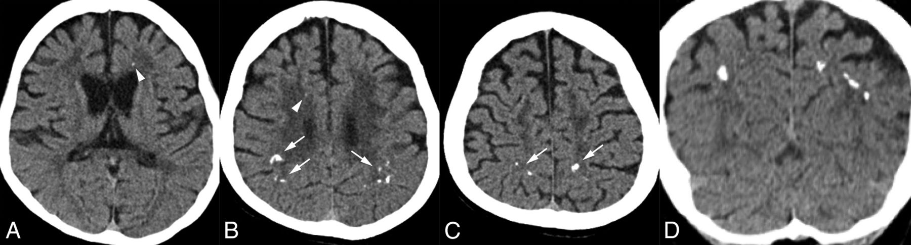
  • Fig 2.
    • Download figure
    • Open in new tab
    • Download powerpoint
    Fig 2.

    A–C, Case 2. Bilateral, scattered calcifications in the parietal subcortical white matter on axial CT images (arrows in B and C). There are also very tiny calcifications in the bilateral frontal white matter (arrowheads in A and B). D, Coronal CT imaging reveals that the parietal calcifications are located in the subcortical white matter, not in the cortex.
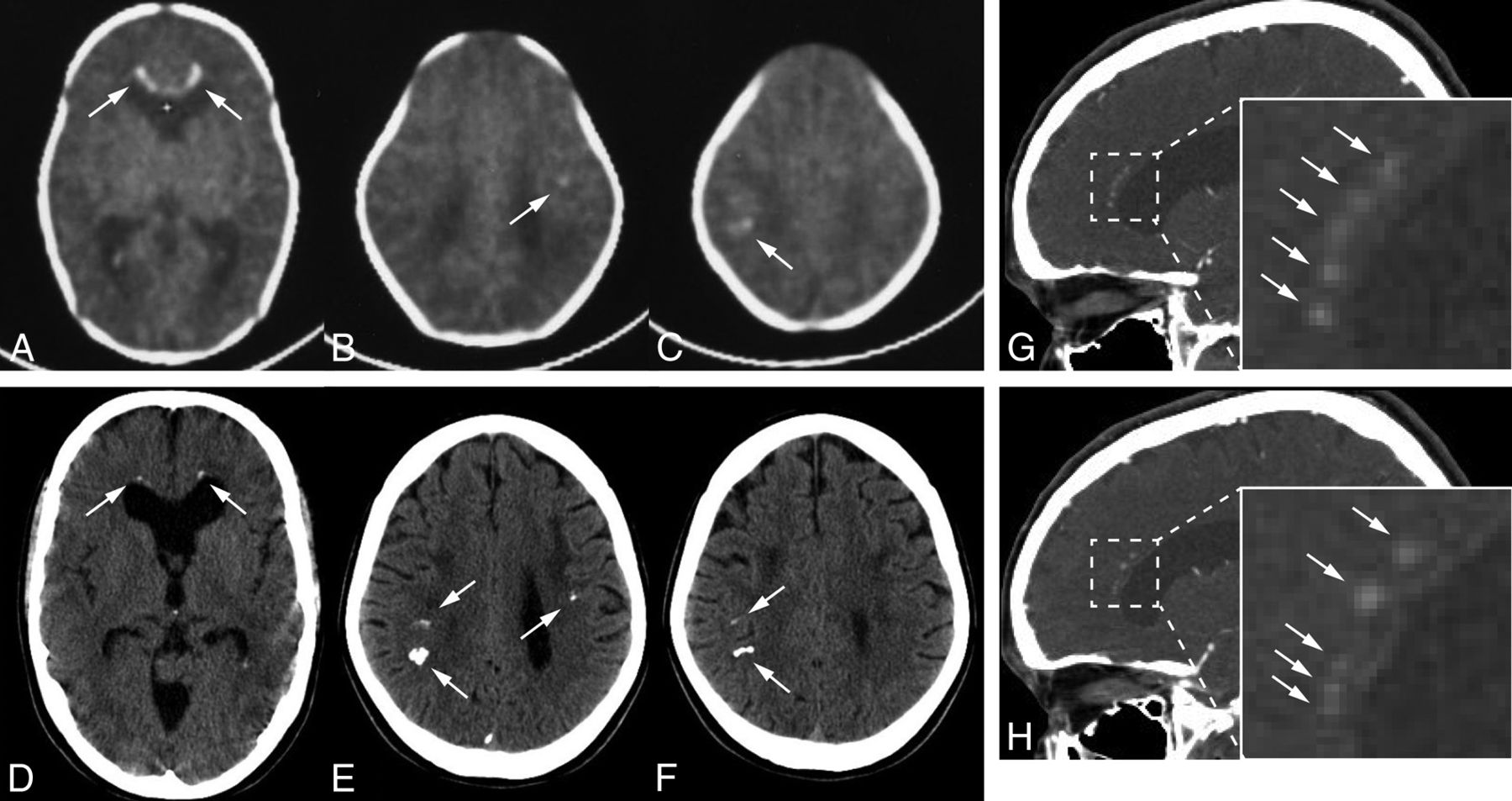
  • Fig 3.
    • Download figure
    • Open in new tab
    • Download powerpoint
    Fig 3.

    A, Case 3. Small bilateral calcifications in the frontal white matter adjacent to the anterior horns of the lateral ventricles on an axial CT image (arrows). B, Sagittal view represents the symmetric and characteristic stepping stone appearance (arrows). C, Case 4. Small bilateral calcifications in the parietal subcortical white matter on an axial CT image (arrows), but there are no calcifications in the frontal area. D, Sagittal CT image displays subtle calcifications in the anterior pericallosal region bilaterally (arrows).

  • Fig 4.
    • Download figure
    • Open in new tab
    • Download powerpoint
    Fig 4.

    A–C, Case 5. Axial CT images at 1 month after birth show bilateral frontal and parietal calcifications (arrows). D–F, These calcifications still exist when the patient is 24 years of age, but they are somewhat smaller, especially those in the frontal regions (arrows). The white cursor inside the anterior horn is included (A) because this is a captured image. G and H, Sagittal views show symmetric frontal pericallosal calcifications in the characteristic stepping stone appearance (arrows; enlarged images in the inserts: G is the right hemisphere, and H is the left hemisphere).

  • Fig 5.
    • Download figure
    • Open in new tab
    • Download powerpoint
    Fig 5.

    A–C, Case 1. Typical white matter changes involving the corpus callosum and the pyramidal tracts (A and C, arrows), dilation of the lateral ventricles and cortical atrophy seen on FLAIR axial (A and B) and T2-weighted coronal imaging (C). D–F, Case 2. Abnormal signaling in the white matter, corpus callosum, and bilateral pyramidal tracts (D, arrows) and enlargement of the lateral ventricles on FLAIR (D). Several diffusion-restricted lesions with low ADC values in the white matter on DWI (E, arrows) and ADC maps (F, arrows). G–I, Case 3. FLAIR image shows frontal-dominant white matter changes bilaterally as well as in the internal capsules (G, arrows). The genu of the corpus callosum is also involved. Several diffusion-restricted lesions in the subcortical white matter can be seen around the lateral ventricles on DWI (H, arrows). These lesions demonstrate decreased signals on ADC maps (I, arrows). J, Case 4. The severity of the white matter changes is less evident than in case 3, the daughter of case 4, on the FLAIR axial image. K, Case 6. Bifrontal white matter lesions, thinning of the corpus callosum, dilation of the lateral ventricles, and cortical atrophy on FLAIR coronal image. L, Case 7. Left-side predominant white matter changes in the frontal and parietal regions and involvement in the corpus callosum on a FLAIR axial image.

Tables

  • Figures
  • Case series of ALSP with brain calcifications

    CaseSexCSF1R MutationOriginFHAge at Onset (yr)Age at CT (yr)Section Thickness (mm)Location of CalcificationsStepping Stone Appearance on Sagittal CT
    FrontalParietal
    1Fc.1765G>A/p.G589RaJapan−37445+++
    2Fc.1954G>C/p.A652PaJapan−30312++NA
    3Fc.2442+5G>AaJapan+27281+−+
    4Mc.2442+5G>AaJapan+58614 (1)c+++
    5Fc.2442+5G>CUS−230, 246, 5+++
    6Fc.2297T>C/p.M766TUS+18301.3 (1)c+−+
    7Mc.1766G>A/p.G589EUS+58603+−NA
    8Fc.1766G>A/p.G589EUS+47527++NA
    9MUntestedbUS−57673+−NA
    • Note:—FH indicates family history; NA, not available for sagittal images.

    • ↵a Novel mutations.

    • ↵b This case was diagnosed by pathologic analysis. We could not conduct genetic testing because of a lack of samples.

    • ↵c For axial images, 4 or 1.3 mm and 1 mm for reconstructed sagittal images.

PreviousNext
Back to top

In this issue

American Journal of Neuroradiology: 38 (1)
American Journal of Neuroradiology
Vol. 38, Issue 1
1 Jan 2017
  • Table of Contents
  • Index by author
  • Complete Issue (PDF)
Advertisement
Print
Download PDF
Email Article

Thank you for your interest in spreading the word on American Journal of Neuroradiology.

NOTE: We only request your email address so that the person you are recommending the page to knows that you wanted them to see it, and that it is not junk mail. We do not capture any email address.

Enter multiple addresses on separate lines or separate them with commas.
Diagnostic Value of Brain Calcifications in Adult-Onset Leukoencephalopathy with Axonal Spheroids and Pigmented Glia
(Your Name) has sent you a message from American Journal of Neuroradiology
(Your Name) thought you would like to see the American Journal of Neuroradiology web site.
CAPTCHA
This question is for testing whether or not you are a human visitor and to prevent automated spam submissions.
Cite this article
T. Konno, D.F. Broderick, N. Mezaki, A. Isami, D. Kaneda, Y. Tashiro, T. Tokutake, B.M. Keegan, B.K. Woodruff, T. Miura, H. Nozaki, M. Nishizawa, O. Onodera, Z.K. Wszolek, T. Ikeuchi
Diagnostic Value of Brain Calcifications in Adult-Onset Leukoencephalopathy with Axonal Spheroids and Pigmented Glia
American Journal of Neuroradiology Jan 2017, 38 (1) 77-83; DOI: 10.3174/ajnr.A4938

Citation Manager Formats

  • BibTeX
  • Bookends
  • EasyBib
  • EndNote (tagged)
  • EndNote 8 (xml)
  • Medlars
  • Mendeley
  • Papers
  • RefWorks Tagged
  • Ref Manager
  • RIS
  • Zotero
0 Responses
Respond to this article
Share
Bookmark this article
Diagnostic Value of Brain Calcifications in Adult-Onset Leukoencephalopathy with Axonal Spheroids and Pigmented Glia
T. Konno, D.F. Broderick, N. Mezaki, A. Isami, D. Kaneda, Y. Tashiro, T. Tokutake, B.M. Keegan, B.K. Woodruff, T. Miura, H. Nozaki, M. Nishizawa, O. Onodera, Z.K. Wszolek, T. Ikeuchi
American Journal of Neuroradiology Jan 2017, 38 (1) 77-83; DOI: 10.3174/ajnr.A4938
del.icio.us logo Twitter logo Facebook logo Mendeley logo
  • Tweet Widget
  • Facebook Like
  • Google Plus One
Purchase

Jump to section

  • Article
    • Abstract
    • ABBREVIATIONS:
    • Case Series
    • Discussion
    • Conclusions
    • Acknowledgments
    • Footnotes
    • References
  • Figures & Data
  • Supplemental
  • Info & Metrics
  • Responses
  • References
  • PDF

Related Articles

  • PubMed
  • Google Scholar

Cited By...

  • Microglia control small vessel calcification via TREM2
  • CSF1R-related leukoencephalopathy: A major player in primary microgliopathies
  • Adult-Onset Leukoencephalopathy with Axonal Spheroids and Pigmented Glia: An MRI Study of 16 French Cases
  • Author response: Mystery Case: CSF-1R mutation is a cause of intracranial cerebral calcifications, cysts, and leukoencephalopathy
  • Crossref (48)
  • Google Scholar

This article has been cited by the following articles in journals that are participating in Crossref Cited-by Linking.

  • CSF1R -related leukoencephalopathy
    Takuya Konno, Koji Kasanuki, Takeshi Ikeuchi, Dennis W. Dickson, Zbigniew K. Wszolek
    Neurology 2018 91 24
  • Bi-allelic CSF1R Mutations Cause Skeletal Dysplasia of Dysosteosclerosis-Pyle Disease Spectrum and Degenerative Encephalopathy with Brain Malformation
    Long Guo, Débora Romeo Bertola, Asako Takanohashi, Asuka Saito, Yuko Segawa, Takanori Yokota, Satoru Ishibashi, Yoichiro Nishida, Guilherme Lopes Yamamoto, José Francisco da Silva Franco, Rachel Sayuri Honjo, Chong Ae Kim, Camila Manso Musso, Margaret Timmons, Amy Pizzino, Ryan J. Taft, Bryan Lajoie, Melanie A. Knight, Kenneth H. Fischbeck, Andrew B. Singleton, Carlos R. Ferreira, Zheng Wang, Li Yan, James Y. Garbern, Pelin O. Simsek-Kiper, Hirofumi Ohashi, Pamela G. Robey, Alan Boyde, Naomichi Matsumoto, Noriko Miyake, Jürgen Spranger, Raphael Schiffmann, Adeline Vanderver, Gen Nishimura, Maria Rita dos Santos Passos-Bueno, Cas Simons, Kinya Ishikawa, Shiro Ikegawa
    The American Journal of Human Genetics 2019 104 5
  • Adult Leukodystrophies: A Step-by-Step Diagnostic Approach
    Lucas Lopes Resende, Anderson Rodrigues Brandão de Paiva, Fernando Kok, Claudia da Costa Leite, Leandro Tavares Lucato
    RadioGraphics 2019 39 1
  • Diagnostic criteria for adult‐onset leukoencephalopathy with axonal spheroids and pigmented glia due to CSF1R mutation
    T. Konno, K. Yoshida, I. Mizuta, T. Mizuno, T. Kawarai, M. Tada, H. Nozaki, S.‐I. Ikeda, O. Onodera, Z. K. Wszolek, T. Ikeuchi
    European Journal of Neurology 2018 25 1
  • Adult-onset leukoencephalopathy with axonal spheroids and pigmented glia (ALSP): Integrating the literature on hereditary diffuse leukoencephalopathy with spheroids (HDLS) and pigmentary orthochromatic leukodystrophy (POLD)
    Scott J. Adams, Andrew Kirk, Roland N. Auer
    Journal of Clinical Neuroscience 2018 48
  • Atypical parkinsonian syndromes: a general neurologist's perspective
    A. B. Deutschländer, O. A. Ross, D. W. Dickson, Z. K. Wszolek
    European Journal of Neurology 2018 25 1
  • Microglial replacement therapy: a potential therapeutic strategy for incurable CSF1R-related leukoencephalopathy
    Jinming Han, Heela Sarlus, Zbigniew K. Wszolek, Virginija Danylaité Karrenbauer, Robert A. Harris
    Acta Neuropathologica Communications 2020 8 1
  • Adult-Onset Leukoencephalopathy with Axonal Spheroids and Pigmented Glia: An MRI Study of 16 French Cases
    P. Codjia, X. Ayrignac, F. Mochel, K. Mouzat, C. Carra-Dalliere, G. Castelnovo, E. Ellie, F. Etcharry-Bouyx, C. Verny, S. Belliard, D. Hannequin, C. Marelli, Y. Nadjar, I. Le Ber, I. Dorboz, S. Samaan, O. Boespflug-Tanguy, S. Lumbroso, P. Labauge
    American Journal of Neuroradiology 2018 39 9
  • Modeling CSF‐1 receptor deficiency diseases – how close are we?
    Violeta Chitu, Şölen Gökhan, E. Richard Stanley
    The FEBS Journal 2022 289 17
  • Clinicopathologic characterization and abnormal autophagy of CSF1R-related leukoencephalopathy
    Wo-Tu Tian, Fei-Xia Zhan, Qing Liu, Xing-Hua Luan, Chao Zhang, Liang Shang, Ben-Yan Zhang, Si-Jian Pan, Fei Miao, Jiong Hu, Ping Zhong, Shi-Hua Liu, Ze-Yu Zhu, Hai-Yan Zhou, Suya Sun, Xiao-Li Liu, Xiao-Jun Huang, Jing-Wen Jiang, Jian-Fang Ma, Ying Wang, Shu-Fen Chen, Hui-Dong Tang, Sheng-Di Chen, Li Cao
    Translational Neurodegeneration 2019 8 1

More in this TOC Section

  • Diagnostic Neuroradiology of Monoclonal Antibodies
  • Clinical Outcomes After Chiari I Decompression
  • Segmentation of Brain Metastases with BLAST
Show more ADULT BRAIN

Similar Articles

Advertisement

Indexed Content

  • Current Issue
  • Accepted Manuscripts
  • Article Preview
  • Past Issues
  • Editorials
  • Editor's Choice
  • Fellows' Journal Club
  • Letters to the Editor
  • Video Articles

Cases

  • Case Collection
  • Archive - Case of the Week
  • Archive - Case of the Month
  • Archive - Classic Case

More from AJNR

  • Trainee Corner
  • Imaging Protocols
  • MRI Safety Corner
  • Book Reviews

Multimedia

  • AJNR Podcasts
  • AJNR Scantastics

Resources

  • Turnaround Time
  • Submit a Manuscript
  • Submit a Video Article
  • Submit an eLetter to the Editor/Response
  • Manuscript Submission Guidelines
  • Statistical Tips
  • Fast Publishing of Accepted Manuscripts
  • Graphical Abstract Preparation
  • Imaging Protocol Submission
  • Evidence-Based Medicine Level Guide
  • Publishing Checklists
  • Author Policies
  • Become a Reviewer/Academy of Reviewers
  • News and Updates

About Us

  • About AJNR
  • Editorial Board
  • Editorial Board Alumni
  • Alerts
  • Permissions
  • Not an AJNR Subscriber? Join Now
  • Advertise with Us
  • Librarian Resources
  • Feedback
  • Terms and Conditions
  • AJNR Editorial Board Alumni

American Society of Neuroradiology

  • Not an ASNR Member? Join Now

© 2025 by the American Society of Neuroradiology All rights, including for text and data mining, AI training, and similar technologies, are reserved.
Print ISSN: 0195-6108 Online ISSN: 1936-959X

Powered by HighWire