Advertisement

Main menu

  • Home
  • Content
    • Current Issue
    • Accepted Manuscripts
    • Article Preview
    • Past Issue Archive
    • Video Articles
    • AJNR Case Collection
    • Case of the Week Archive
    • Case of the Month Archive
    • Classic Case Archive
  • Special Collections
    • AJNR Awards
    • Low-Field MRI
    • Alzheimer Disease
    • ASNR Foundation Special Collection
    • Photon-Counting CT
    • View All
  • Multimedia
    • AJNR Podcasts
    • AJNR SCANtastic
    • Trainee Corner
    • MRI Safety Corner
    • Imaging Protocols
  • For Authors
    • Submit a Manuscript
    • Submit a Video Article
    • Submit an eLetter to the Editor/Response
    • Manuscript Submission Guidelines
    • Statistical Tips
    • Fast Publishing of Accepted Manuscripts
    • Graphical Abstract Preparation
    • Imaging Protocol Submission
    • Author Policies
  • About Us
    • About AJNR
    • Editorial Board
    • Editorial Board Alumni
  • More
    • Become a Reviewer/Academy of Reviewers
    • Subscribers
    • Permissions
    • Alerts
    • Feedback
    • Advertisers
    • ASNR Home

User menu

  • Alerts
  • Log in

Search

  • Advanced search
American Journal of Neuroradiology

American Journal of Neuroradiology

ASHNR American Society of Functional Neuroradiology ASHNR American Society of Pediatric Neuroradiology ASSR
  • Alerts
  • Log in

Advanced Search

  • Home
  • Content
    • Current Issue
    • Accepted Manuscripts
    • Article Preview
    • Past Issue Archive
    • Video Articles
    • AJNR Case Collection
    • Case of the Week Archive
    • Case of the Month Archive
    • Classic Case Archive
  • Special Collections
    • AJNR Awards
    • Low-Field MRI
    • Alzheimer Disease
    • ASNR Foundation Special Collection
    • Photon-Counting CT
    • View All
  • Multimedia
    • AJNR Podcasts
    • AJNR SCANtastic
    • Trainee Corner
    • MRI Safety Corner
    • Imaging Protocols
  • For Authors
    • Submit a Manuscript
    • Submit a Video Article
    • Submit an eLetter to the Editor/Response
    • Manuscript Submission Guidelines
    • Statistical Tips
    • Fast Publishing of Accepted Manuscripts
    • Graphical Abstract Preparation
    • Imaging Protocol Submission
    • Author Policies
  • About Us
    • About AJNR
    • Editorial Board
    • Editorial Board Alumni
  • More
    • Become a Reviewer/Academy of Reviewers
    • Subscribers
    • Permissions
    • Alerts
    • Feedback
    • Advertisers
    • ASNR Home
  • Follow AJNR on Twitter
  • Visit AJNR on Facebook
  • Follow AJNR on Instagram
  • Join AJNR on LinkedIn
  • RSS Feeds

AJNR Awards, New Junior Editors, and more. Read the latest AJNR updates

Research ArticleADULT BRAIN

Intracranial Arteriovenous Shunting: Detection with Arterial Spin-Labeling and Susceptibility-Weighted Imaging Combined

J. Hodel, X. Leclerc, E. Kalsoum, M. Zuber, R. Tamazyan, M.A. Benadjaoud, J.-P. Pruvo, M. Piotin, H. Baharvahdat, M. Zins and R. Blanc
American Journal of Neuroradiology January 2017, 38 (1) 71-76; DOI: https://doi.org/10.3174/ajnr.A4961
J. Hodel
aFrom the Departments of Radiology (J.H., M.Z.)
cDepartment of Neuroradiology (J.H., E.K.), Centre-Hospitalier-Universitaire Henri Mondor, Créteil, France
  • Find this author on Google Scholar
  • Find this author on PubMed
  • Search for this author on this site
  • ORCID record for J. Hodel
X. Leclerc
dDepartment of Neuroradiology (X.L., J.-P. P.), Roger Salengro Hospital, Lille, France
  • Find this author on Google Scholar
  • Find this author on PubMed
  • Search for this author on this site
  • ORCID record for X. Leclerc
E. Kalsoum
cDepartment of Neuroradiology (J.H., E.K.), Centre-Hospitalier-Universitaire Henri Mondor, Créteil, France
  • Find this author on Google Scholar
  • Find this author on PubMed
  • Search for this author on this site
  • ORCID record for E. Kalsoum
M. Zuber
aFrom the Departments of Radiology (J.H., M.Z.)
bNeurology (M.Z., R.T.), Saint Joseph Hospital, Paris, France
  • Find this author on Google Scholar
  • Find this author on PubMed
  • Search for this author on this site
  • ORCID record for M. Zuber
R. Tamazyan
bNeurology (M.Z., R.T.), Saint Joseph Hospital, Paris, France
  • Find this author on Google Scholar
  • Find this author on PubMed
  • Search for this author on this site
  • ORCID record for R. Tamazyan
M.A. Benadjaoud
eDepartment of Radiobiology and Epidemiology (M.A.B.), Institute for Radiological Protection and Nuclear Safety (IRSN), Fontenay-Aux-Roses, France
  • Find this author on Google Scholar
  • Find this author on PubMed
  • Search for this author on this site
  • ORCID record for M.A. Benadjaoud
J.-P. Pruvo
dDepartment of Neuroradiology (X.L., J.-P. P.), Roger Salengro Hospital, Lille, France
  • Find this author on Google Scholar
  • Find this author on PubMed
  • Search for this author on this site
  • ORCID record for J.-P. Pruvo
M. Piotin
fDepartment of Interventional Neuroradiology (M.P., H.B., R.B.), Rothschild Foundation Hospital, Paris, France.
  • Find this author on Google Scholar
  • Find this author on PubMed
  • Search for this author on this site
  • ORCID record for M. Piotin
H. Baharvahdat
fDepartment of Interventional Neuroradiology (M.P., H.B., R.B.), Rothschild Foundation Hospital, Paris, France.
  • Find this author on Google Scholar
  • Find this author on PubMed
  • Search for this author on this site
  • ORCID record for H. Baharvahdat
M. Zins
aFrom the Departments of Radiology (J.H., M.Z.)
  • Find this author on Google Scholar
  • Find this author on PubMed
  • Search for this author on this site
  • ORCID record for M. Zins
R. Blanc
fDepartment of Interventional Neuroradiology (M.P., H.B., R.B.), Rothschild Foundation Hospital, Paris, France.
  • Find this author on Google Scholar
  • Find this author on PubMed
  • Search for this author on this site
  • ORCID record for R. Blanc
  • Article
  • Figures & Data
  • Info & Metrics
  • Responses
  • References
  • PDF
Loading

Article Figures & Data

Figures

  • Tables
  • Fig 1.
    • Download figure
    • Open in new tab
    • Download powerpoint
    Fig 1.

    Flow chart illustrating patient selection.
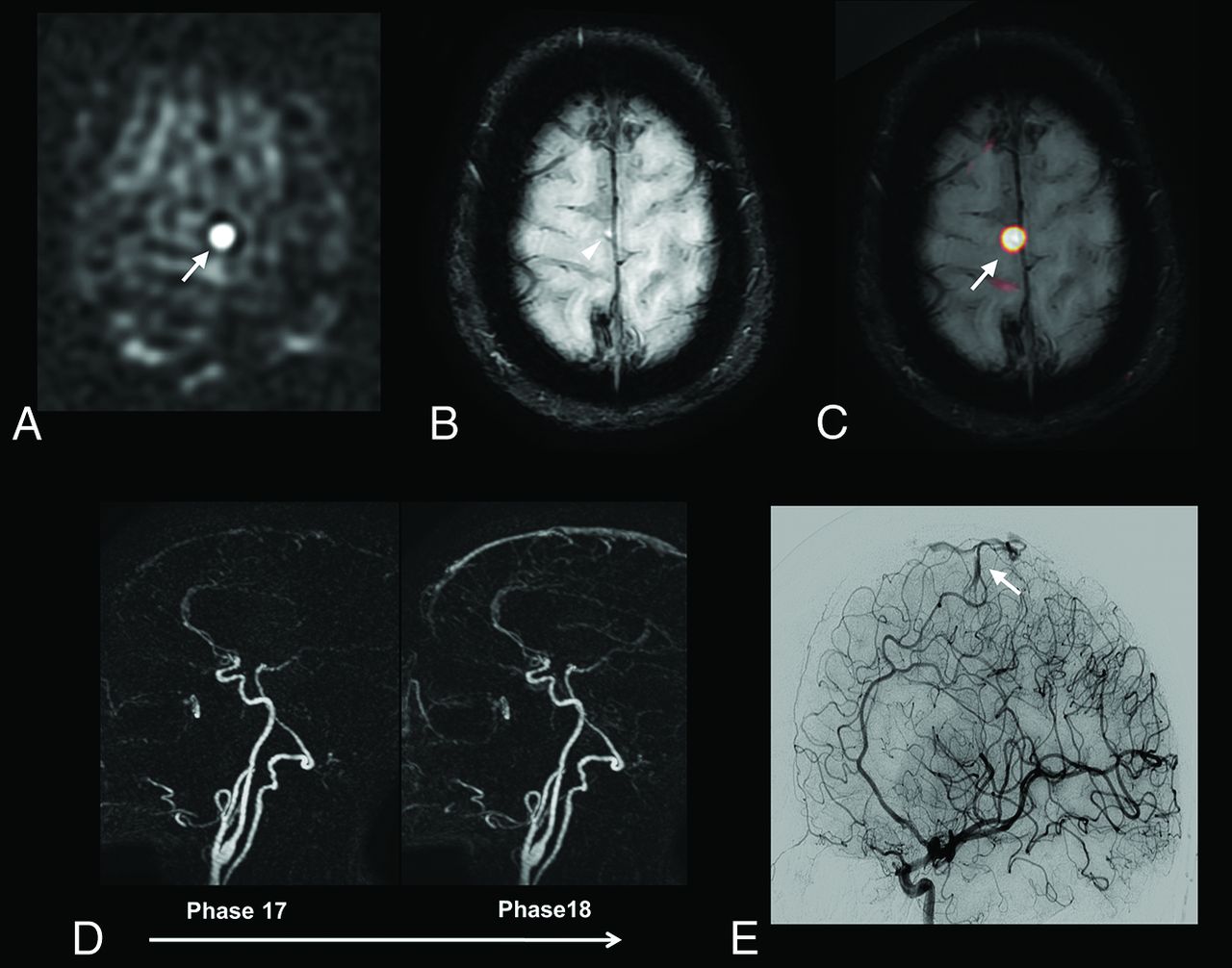
  • Fig 2.
    • Download figure
    • Open in new tab
    • Download powerpoint
    Fig 2.

    Patient 62 with a complex developmental venous anomaly. ASL raw data (A) and right internal carotid artery angiogram, venous phase, lateral view (B). Increased signal-intensity is visible on ASL images within the right frontal lobe and deep brain nuclei (A, arrows). ASL images were considered suggestive of AVS by 1 blinded reader. Developmental venous anomaly was correctly diagnosed (and thus absence of AVS) by using SWI and a combination of SWI and ASL (not shown). DSA confirms the diagnosis of developmental venous anomaly by revealing a classic umbrella-shaped aspect at the venous phase with medullary veins (B, arrowheads) draining into an enlarged collector (B, arrows), which further drains into the superficial Sylvian vein and cavernous sinus.

  • Fig 3.
    • Download figure
    • Open in new tab
    • Download powerpoint
    Fig 3.

    A 60-year-old patient with a right paracentral AVM. ASL raw data (A) demonstrates a strong hypersignal at the anterior part of the right paracentral region (A, arrow). The slight venous hypersignal related to AVS was initially missed by the blinded readers by using SWI alone (B, arrowhead) but was correctly identified by using ASL and SWI combined (C, ASL/SWI merged image, arrow). Findings of time-resolved 4D contrast-enhanced MRA (D) were considered negative by the blinded readers. DSA reveals a small pial AVM in the right paracentral region (E, arrow).
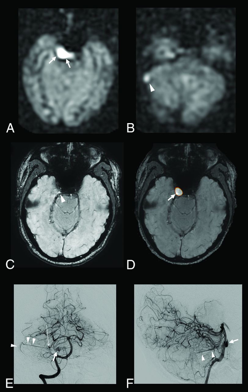
  • Fig 4.
    • Download figure
    • Open in new tab
    • Download powerpoint
    Fig 4.

    A 52-year-old patient with a right cerebellar AVM draining into the right transverse sinus and the right periclival plexus. ASL raw data demonstrate a strong hypersignal within the right periclival plexus (A, arrows) and the right lateral sinus (B, arrowhead). With SWI alone, the slight venous hypersignal within the right periclival plexus and related to AVS was initially missed by the blinded readers (C, arrowhead) but correctly identified by using ASL and SWI combined (D, ASL/SWI merged image, arrow). Findings of time-resolved and high-resolution contrast-enhanced MRA were negative. Anteroposterior (E) and lateral (F) views from the right vertebral conventional angiograms show a small nidus (E, long arrow) draining into the right lateral sinus through the right superior petrosal sinus (E and F, arrowheads) and into the periclival plexus through a lateropontine vein (E and F, arrow).

  • Fig 5.
    • Download figure
    • Open in new tab
    • Download powerpoint
    Fig 5.

    Patient 26 with a previously treated AVM and residual AVS according to DSA. SWI (A) and left internal carotid artery angiogram, arterial phase, lateral view (B). An increased signal intensity is visible with SWI within a deep vein adjacent to the treated nidus (A, arrows), suggestive of AVS. Of note, findings of ASL and contrast-enhanced MR images were considered negative (not shown). DSA confirms the presence of dysplastic vessels around the cast of the embolic agent (B, arrows) and an early opacification of venous drainage (B, arrowheads) coursing toward the deep venous system at the anterior and inferior pole of the embolized AVM.

Tables

  • Figures
    • View popup
    Table 1:

    Diagnosis of AVS (number of patients correctly diagnosed) using ASL, SWI, and conventional MRI

    MRI SequencesDSA
    No AVS (n = 29)AVS (n = 63)DAVF (n = 10)AVM (n = 53)
    SWI28 (97%)55 (87%)8 (80%)47 (89%)
    ASL26 (90%)60 (95%)8 (80%)52 (98%)
    ASL/SWI28 (97%)62 (98%)9 (90%)53 (100%)
    Conventional MRI28 (97%)57 (90%)9 (90%)48 (91%)
    • Note:—ASL/SWI indicates ASL and SWI combined; conventional MRI, conventional MRI protocol (including diffusion-weighted imaging, gradient-echo T2*, 3D FLAIR, TOF MRA, 4D and 3D contrast-enhanced MRA, and postcontrast 3D T1WI sequences).

    • View popup
    Table 2:

    Diagnostic accuracy parameters using ASL, SWI, and conventional MRI

    MRI SequencesDiagnostic Accuracy Parameters
    Se (95% CI)Sp (95% CI)PPV (95% CI)NPV (95% CI)AUC (95% CI)
    SWI0.87 (0.79–0.96)0.97 (0.90–1)0.98 (0.90–1)0.78 (0.61–0.90)0.92 (0.85–0.96)
    ASL0.95 (0.90–1)0.90 (0.78–1)0.95 (0.87–0.99)0.90 (0.73–0.98)0.92 (0.84–0.97)
    ASL/SWI0.98 (0.95–1)0.97 (0.90–1)0.98 (0.95–1)0.97 (0.90–1)0.97 (0.90–1)
    Conventional MRI0.90 (0.83–0.98)0.97 (0.90–1)0.98 (0.91–1)0.92 (0.65–0.93)0.93 (0.87–0.97)
    • Note:—Se indicates sensitivity; Sp, specificity; PPV, predictive positive value; NPV, negative predictive value.

PreviousNext
Back to top

In this issue

American Journal of Neuroradiology: 38 (1)
American Journal of Neuroradiology
Vol. 38, Issue 1
1 Jan 2017
  • Table of Contents
  • Index by author
  • Complete Issue (PDF)
Advertisement
Print
Download PDF
Email Article

Thank you for your interest in spreading the word on American Journal of Neuroradiology.

NOTE: We only request your email address so that the person you are recommending the page to knows that you wanted them to see it, and that it is not junk mail. We do not capture any email address.

Enter multiple addresses on separate lines or separate them with commas.
Intracranial Arteriovenous Shunting: Detection with Arterial Spin-Labeling and Susceptibility-Weighted Imaging Combined
(Your Name) has sent you a message from American Journal of Neuroradiology
(Your Name) thought you would like to see the American Journal of Neuroradiology web site.
CAPTCHA
This question is for testing whether or not you are a human visitor and to prevent automated spam submissions.
Cite this article
J. Hodel, X. Leclerc, E. Kalsoum, M. Zuber, R. Tamazyan, M.A. Benadjaoud, J.-P. Pruvo, M. Piotin, H. Baharvahdat, M. Zins, R. Blanc
Intracranial Arteriovenous Shunting: Detection with Arterial Spin-Labeling and Susceptibility-Weighted Imaging Combined
American Journal of Neuroradiology Jan 2017, 38 (1) 71-76; DOI: 10.3174/ajnr.A4961

Citation Manager Formats

  • BibTeX
  • Bookends
  • EasyBib
  • EndNote (tagged)
  • EndNote 8 (xml)
  • Medlars
  • Mendeley
  • Papers
  • RefWorks Tagged
  • Ref Manager
  • RIS
  • Zotero
0 Responses
Respond to this article
Share
Bookmark this article
Intracranial Arteriovenous Shunting: Detection with Arterial Spin-Labeling and Susceptibility-Weighted Imaging Combined
J. Hodel, X. Leclerc, E. Kalsoum, M. Zuber, R. Tamazyan, M.A. Benadjaoud, J.-P. Pruvo, M. Piotin, H. Baharvahdat, M. Zins, R. Blanc
American Journal of Neuroradiology Jan 2017, 38 (1) 71-76; DOI: 10.3174/ajnr.A4961
del.icio.us logo Twitter logo Facebook logo Mendeley logo
  • Tweet Widget
  • Facebook Like
  • Google Plus One
Purchase

Jump to section

  • Article
    • Abstract
    • ABBREVIATIONS:
    • Materials and Methods
    • Results
    • Discussion
    • Conclusions
    • Acknowledgments
    • Footnotes
    • References
  • Figures & Data
  • Info & Metrics
  • Responses
  • References
  • PDF

Related Articles

  • No related articles found.
  • PubMed
  • Google Scholar

Cited By...

  • Arterial Spin-Labeling MR Imaging in the Detection of Intracranial Arteriovenous Malformations in Patients with Hereditary Hemorrhagic Telangiectasia
  • Arterial Spin-Labeling MR Imaging for the Differential Diagnosis of Venous-Predominant AVMs and Developmental Venous Anomalies
  • Follow-Up MRI for Small Brain AVMs Treated by Radiosurgery: Is Gadolinium Really Necessary?
  • Intracranial Arteriovenous Shunting Detection with Arterial Spin-Labeling and Susceptibility-Weighted Imaging: Potential Pitfall of a Venous Predominant Parenchymal Arteriovenous Malformation
  • Reply:
  • Crossref (34)
  • Google Scholar

This article has been cited by the following articles in journals that are participating in Crossref Cited-by Linking.

  • Current state and guidance on arterial spin labeling perfusion MRI in clinical neuroimaging
    Thomas Lindner, Divya S. Bolar, Eric Achten, Frederik Barkhof, António J. Bastos‐Leite, John A. Detre, Xavier Golay, Matthias Günther, Danny J. J. Wang, Sven Haller, Silvia Ingala, Hans R. Jäger, Geon‐Ho Jahng, Meher R. Juttukonda, Vera C. Keil, Hirohiko Kimura, Mai‐Lan Ho, Maarten Lequin, Xin Lou, Jan Petr, Nandor Pinter, Francesca B. Pizzini, Marion Smits, Magdalena Sokolska, Greg Zaharchuk, Henk J. M. M. Mutsaerts
    Magnetic Resonance in Medicine 2023 89 5
  • Diagnostic value of alternative techniques to gadolinium-based contrast agents in MR neuroimaging—a comprehensive overview
    Anna Falk Delgado, Danielle Van Westen, Markus Nilsson, Linda Knutsson, Pia C. Sundgren, Elna-Marie Larsson, Alberto Falk Delgado
    Insights into Imaging 2019 10 1
  • Consensus statement on current and emerging methods for the diagnosis and evaluation of cerebrovascular disease
    Manus J Donahue, Eric Achten, Petrice M Cogswell, Frank-Erik De Leeuw, Colin P Derdeyn, Rick M Dijkhuizen, Audrey P Fan, Rashid Ghaznawi, Jeremy J Heit, M Arfan Ikram, Peter Jezzard, Lori C Jordan, Eric Jouvent, Linda Knutsson, Richard Leigh, David S Liebeskind, Weili Lin, Thomas W Okell, Adnan I Qureshi, Charlotte J Stagg, Matthias JP van Osch, Peter CM van Zijl, Jennifer M Watchmaker, Max Wintermark, Ona Wu, Greg Zaharchuk, Jinyuan Zhou, Jeroen Hendrikse
    Journal of Cerebral Blood Flow & Metabolism 2018 38 9
  • Arterial Spin Labeling: Techniques, Clinical Applications, and Interpretation
    Tatiana Iutaka, Marina Bellintani de Freitas, Samir Sari Omar, Felipe Alba Scortegagna, Kambiz Nael, Renato Hoffmann Nunes, Felipe Torres Pacheco, Antônio Carlos Martins Maia Júnior, Lázaro Luís Faria do Amaral, Antônio Jose da Rocha
    RadioGraphics 2023 43 1
  • Preliminary evidence for cerebral capillary shunting in adults with sickle cell anemia
    Meher R Juttukonda, Manus J Donahue, Larry T Davis, Melissa C Gindville, Chelsea A Lee, Niral J Patel, Adetola A Kassim, Sumit Pruthi, Jeroen Hendrikse, Lori C Jordan
    Journal of Cerebral Blood Flow & Metabolism 2019 39 6
  • Follow-Up MRI for Small Brain AVMs Treated by Radiosurgery: Is Gadolinium Really Necessary?
    X. Leclerc, O. Guillaud, N. Reyns, J. Hodel, O. Outteryck, F. Bala, N. Bricout, M. Bretzner, N. Ramdane, J.-P. Pruvo, L. Hacein-Bey, G. Kuchcinski
    American Journal of Neuroradiology 2020 41 3
  • Tips and tricks in the diagnosis of intracranial dural arteriovenous fistulas: A pictorial review
    Igor Gomes Padilha, Felipe Torres Pacheco, Alan Iuno Rios Araujo, Renato Hoffmann Nunes, Carlos Eduardo Baccin, Mario Luiz Marques Conti, Antônio Carlos Martins Maia Jr, Antônio José da Rocha
    Journal of Neuroradiology 2020 47 5
  • Evaluation of cerebral arteriovenous shunts: a comparison of parallel imaging time-of-flight magnetic resonance angiography (TOF-MRA) and compressed sensing TOF-MRA to digital subtraction angiography
    Akihiko Sakata, Yasutaka Fushimi, Tomohisa Okada, Satoshi Nakajima, Takuya Hinoda, Peter Speier, Michaela Schmidt, Christoph Forman, Kazumichi Yoshida, Hiroharu Kataoka, Susumu Miyamoto, Yuji Nakamoto
    Neuroradiology 2021 63 6
  • Clinical and Arterial Spin Labeling Brain MRI Features of Transitional Venous Anomalies
    Michael Zhang, Nicholas A. Telischak, Nancy J. Fischbein, Gary K. Steinberg, Michael Marks, Greg Zaharchuk, Jeremy J. Heit, Michael Iv
    Journal of Neuroimaging 2018 28 3
  • MRI and MR angiography evaluation of pulsatile tinnitus: A focused, physiology‐based protocol
    Daniel D. Cummins, Michael T. Caton, Vinil Shah, Karl Meisel, Christine Glastonbury, Matthew R. Amans
    Journal of Neuroimaging 2022 32 2

More in this TOC Section

  • Diagnostic Neuroradiology of Monoclonal Antibodies
  • Segmentation of Brain Metastases with BLAST
  • Cerebral ADC Changes in Fabry Disease
Show more ADULT BRAIN

Similar Articles

Advertisement

Indexed Content

  • Current Issue
  • Accepted Manuscripts
  • Article Preview
  • Past Issues
  • Editorials
  • Editor's Choice
  • Fellows' Journal Club
  • Letters to the Editor
  • Video Articles

Cases

  • Case Collection
  • Archive - Case of the Week
  • Archive - Case of the Month
  • Archive - Classic Case

More from AJNR

  • Trainee Corner
  • Imaging Protocols
  • MRI Safety Corner
  • Book Reviews

Multimedia

  • AJNR Podcasts
  • AJNR Scantastics

Resources

  • Turnaround Time
  • Submit a Manuscript
  • Submit a Video Article
  • Submit an eLetter to the Editor/Response
  • Manuscript Submission Guidelines
  • Statistical Tips
  • Fast Publishing of Accepted Manuscripts
  • Graphical Abstract Preparation
  • Imaging Protocol Submission
  • Evidence-Based Medicine Level Guide
  • Publishing Checklists
  • Author Policies
  • Become a Reviewer/Academy of Reviewers
  • News and Updates

About Us

  • About AJNR
  • Editorial Board
  • Editorial Board Alumni
  • Alerts
  • Permissions
  • Not an AJNR Subscriber? Join Now
  • Advertise with Us
  • Librarian Resources
  • Feedback
  • Terms and Conditions
  • AJNR Editorial Board Alumni

American Society of Neuroradiology

  • Not an ASNR Member? Join Now

© 2025 by the American Society of Neuroradiology All rights, including for text and data mining, AI training, and similar technologies, are reserved.
Print ISSN: 0195-6108 Online ISSN: 1936-959X

Powered by HighWire