Advertisement

Main menu

  • Home
  • Content
    • Current Issue
    • Accepted Manuscripts
    • Article Preview
    • Past Issue Archive
    • Video Articles
    • AJNR Case Collection
    • Case of the Week Archive
    • Case of the Month Archive
    • Classic Case Archive
  • Special Collections
    • AJNR Awards
    • Low-Field MRI
    • Alzheimer Disease
    • ASNR Foundation Special Collection
    • Photon-Counting CT
    • View All
  • Multimedia
    • AJNR Podcasts
    • AJNR SCANtastic
    • Trainee Corner
    • MRI Safety Corner
    • Imaging Protocols
  • For Authors
    • Submit a Manuscript
    • Submit a Video Article
    • Submit an eLetter to the Editor/Response
    • Manuscript Submission Guidelines
    • Statistical Tips
    • Fast Publishing of Accepted Manuscripts
    • Graphical Abstract Preparation
    • Imaging Protocol Submission
    • Author Policies
  • About Us
    • About AJNR
    • Editorial Board
    • Editorial Board Alumni
  • More
    • Become a Reviewer/Academy of Reviewers
    • Subscribers
    • Permissions
    • Alerts
    • Feedback
    • Advertisers
    • ASNR Home

User menu

  • Alerts
  • Log in

Search

  • Advanced search
American Journal of Neuroradiology

American Journal of Neuroradiology

ASHNR American Society of Functional Neuroradiology ASHNR American Society of Pediatric Neuroradiology ASSR
  • Alerts
  • Log in

Advanced Search

  • Home
  • Content
    • Current Issue
    • Accepted Manuscripts
    • Article Preview
    • Past Issue Archive
    • Video Articles
    • AJNR Case Collection
    • Case of the Week Archive
    • Case of the Month Archive
    • Classic Case Archive
  • Special Collections
    • AJNR Awards
    • Low-Field MRI
    • Alzheimer Disease
    • ASNR Foundation Special Collection
    • Photon-Counting CT
    • View All
  • Multimedia
    • AJNR Podcasts
    • AJNR SCANtastic
    • Trainee Corner
    • MRI Safety Corner
    • Imaging Protocols
  • For Authors
    • Submit a Manuscript
    • Submit a Video Article
    • Submit an eLetter to the Editor/Response
    • Manuscript Submission Guidelines
    • Statistical Tips
    • Fast Publishing of Accepted Manuscripts
    • Graphical Abstract Preparation
    • Imaging Protocol Submission
    • Author Policies
  • About Us
    • About AJNR
    • Editorial Board
    • Editorial Board Alumni
  • More
    • Become a Reviewer/Academy of Reviewers
    • Subscribers
    • Permissions
    • Alerts
    • Feedback
    • Advertisers
    • ASNR Home
  • Follow AJNR on Twitter
  • Visit AJNR on Facebook
  • Follow AJNR on Instagram
  • Join AJNR on LinkedIn
  • RSS Feeds

AJNR Awards, New Junior Editors, and more. Read the latest AJNR updates

Research ArticleNeurointervention

Suspected Metallic Embolism following Endovascular Treatment of Intracranial Aneurysms

R. Yasuda, M. Maeda, M. Umino, Y. Nakatsuka, Y. Umeda, N. Toma, H. Sakaida and H. Suzuki
American Journal of Neuroradiology September 2016, 37 (9) 1696-1699; DOI: https://doi.org/10.3174/ajnr.A4804
R. Yasuda
aFrom the Departments of Neurosurgery (R.Y., Y.N., Y.U., N.T., H.Sakaida, H.Suzuki)
  • Find this author on Google Scholar
  • Find this author on PubMed
  • Search for this author on this site
  • ORCID record for R. Yasuda
M. Maeda
bRadiology (M.U., M.M.), Mie University Graduate School of Medicine, Tsu, Mie, Japan.
  • Find this author on Google Scholar
  • Find this author on PubMed
  • Search for this author on this site
  • ORCID record for M. Maeda
M. Umino
bRadiology (M.U., M.M.), Mie University Graduate School of Medicine, Tsu, Mie, Japan.
  • Find this author on Google Scholar
  • Find this author on PubMed
  • Search for this author on this site
  • ORCID record for M. Umino
Y. Nakatsuka
aFrom the Departments of Neurosurgery (R.Y., Y.N., Y.U., N.T., H.Sakaida, H.Suzuki)
  • Find this author on Google Scholar
  • Find this author on PubMed
  • Search for this author on this site
  • ORCID record for Y. Nakatsuka
Y. Umeda
aFrom the Departments of Neurosurgery (R.Y., Y.N., Y.U., N.T., H.Sakaida, H.Suzuki)
  • Find this author on Google Scholar
  • Find this author on PubMed
  • Search for this author on this site
  • ORCID record for Y. Umeda
N. Toma
aFrom the Departments of Neurosurgery (R.Y., Y.N., Y.U., N.T., H.Sakaida, H.Suzuki)
  • Find this author on Google Scholar
  • Find this author on PubMed
  • Search for this author on this site
  • ORCID record for N. Toma
H. Sakaida
aFrom the Departments of Neurosurgery (R.Y., Y.N., Y.U., N.T., H.Sakaida, H.Suzuki)
  • Find this author on Google Scholar
  • Find this author on PubMed
  • Search for this author on this site
  • ORCID record for H. Sakaida
H. Suzuki
aFrom the Departments of Neurosurgery (R.Y., Y.N., Y.U., N.T., H.Sakaida, H.Suzuki)
  • Find this author on Google Scholar
  • Find this author on PubMed
  • Search for this author on this site
  • ORCID record for H. Suzuki
  • Article
  • Figures & Data
  • Info & Metrics
  • Responses
  • References
  • PDF
Loading

Abstract

SUMMARY: We describe a case series of suspected metallic embolism after coil embolization for intracranial aneurysms. Between January 2012 and December 2014, 110 intracranial aneurysms had been treated by coil embolization in our institution. In 6 cases, the postprocedural MR imaging revealed abnormal spotty lesions not detected on the preprocedural MR imaging. The lesions were also undetectable on the postprocedural CT scan. They were demonstrated as low-intensity spots on T1WI, T2WI, DWI, and T2*-weighted imaging. On DWI, they were accompanied by bright “halo,” and on T2*-weighted imaging, they showed a “blooming” effect. In 3 of the 6 cases, follow-up MR imaging was available and all the lesions remained and demonstrated no signal changes. Although histologic examination had not been performed, these neuroradiologic findings strongly supported the lesions being from metallic fragments. No specific responsible device was detected after reviewing all the devices used for the neuroendovascular treatment in the 6 cases.

Clinically asymptomatic emboli are a common complication of cerebral angiography and neuroendovascular procedures, occurring in approximately 20% of cases.1 Foreign body emboli after cerebral angiography are a much less known complication, and only a few isolated cases have been reported.2⇓–4 Recently, we have noticed abnormal signals suggesting metallic emboli on the postprocedural MR imaging in several cases of coil embolization for cerebral aneurysms. In this report, we describe the particular MR imaging findings of suspected metallic emboli following neuroendovascular procedures and discuss possible sources for these findings.

Case Series

After the retrospective review of our medical data base between January 2012 and December 2014 that contains 110 intracranial aneurysms treated by coil embolization, we encountered 6 cases that showed abnormal spotty lesions on the postprocedural MR imaging. The details of the clinical features of the 6 cases are summarized in Table 1. The devices used for the treatment in the 6 cases are summarized in Table 2. In all 6 cases, the lesions were low intensity on T1WI, T2WI, DWI, and T2*-weighted images. Additionally, they were accompanied by a hyperintense “halo” on DWI and showed a “blooming” effect on T2*-weighted images. In 5 of the 6 cases (cases 1–5), a postprocedural CT scan was obtained but none of the lesions were detected. Follow-up MR images were available in 3 of the 6 cases (cases 1, 5, and 6), and in all 3 cases, the lesions remained without any signal changes during the follow-up periods (range, 6–34 months). No patients showed any symptoms associated with the lesions.

View this table:
  • View inline
  • View popup
Table 1:

Patient characteristics

View this table:
  • View inline
  • View popup
Table 2:

Devices used for treatment in each case

Illustrative Cases

Case 1.

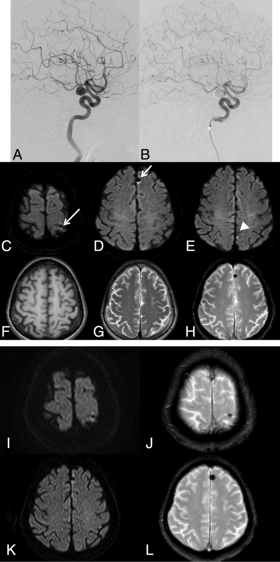
A 68-year-old woman was treated for a left ICA aneurysm (Fig 1A) with coil embolization. On the preprocedural MR imaging, no abnormal findings apart the aneurysm were identified. For the coil embolization procedure, a 5F guiding sheath was placed in the left ICA and a 1.7F microcatheter was inserted into the aneurysm by using a 0.014-inch microguidewire. The aneurysm was embolized with multiple platinum coils (Fig 1B). The MR images were obtained the next day by using a 1.5T MR imaging unit (Achieva Nova Dual; Philips Healthcare, Best, the Netherlands) with a 5-mm section thickness, and DWI revealed low-intensity spots accompanied by a hyperintense halo at the left medial frontal and left parietal regions (Fig 1C, -D). These signals were obviously different from the bright lesion in the left parietal lobe indicating thromboembolism (Fig 1E) that showed no signal changes on T2*-weighted images.

Fig 1.
  • Download figure
  • Open in new tab
  • Download powerpoint
Fig 1.

A left ICA aneurysm in case 1 (A) is embolized by using multiple coils (B). Postprocedural DWI shows abnormal low-intensity spots with a hyperintense halo (white arrow) in the left parietal (C) and frontal (D) lobes. These are different from a bright spot in the left parietal lobe (E), indicating thromboembolism. These abnormal spots are low intensity on T1WI (F) and T2WI (G) and show a blooming effect on T2*-weighted images (H). Follow-up MR imaging obtained 13 months after the procedure shows that all the abnormal signal spots remain without any signal changes (I–L).

While these lesions were shown as low-intensity spots on T1WI (Fig 1F) and T2WI (Fig 1G), they had a blooming effect on T2*-weighted images (Fig 1H). Head CT (Aquilion ONE CT scanner; Toshiba Medical Systems, Tokyo, Japan) obtained on the following day demonstrated no abnormal findings except a coil mass. The patient was discharged from our hospital 1 week after the coil embolization without any symptoms. Follow-up MR imaging was performed at 13 months after the procedure, and both abnormal spots remained without any signal changes (Fig 1I, -L). The patient remained asymptomatic at the last follow-up.

Case 5.

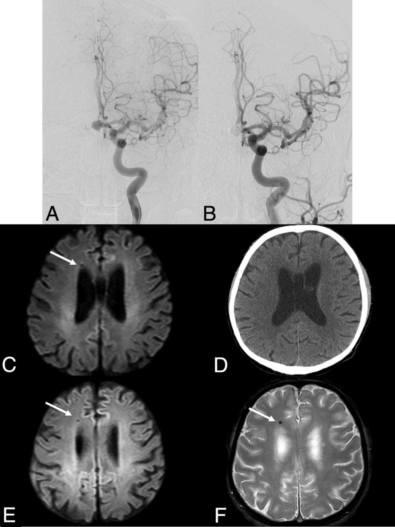
A 70-year-old man was referred to our hospital for treatment of his unruptured anterior communicating artery aneurysm (Fig 2A). Preprocedural MR imaging showed no abnormal findings apart from the aneurysm. The coil embolization was performed; a 6F guiding catheter was placed in his left ICA. After a balloon catheter was placed underneath the aneurysmal neck by using a 0.01-inch microguidewire, a 1.7F microcatheter was placed in the aneurysm by using another 0.014-inch microguidewire. During the embolization, the first coil uncoiled, and it was withdrawn by using a retrieval device. Because the hub of the microcatheter had to be cut to advance the retrieval device, another 1.7F microcatheter was used for the embolization. The aneurysm was finally embolized by using multiple platinum coils (Fig 2B). The MR imaging performed on postoperative day 1 by using a 1.5T machine (Achieva Nova Dual; Philips) with 5-mm section thickness demonstrated a low-intensity spot with a hyperintense halo on DWI in the right frontal lobe (Fig 2C). A head CT scan (Aquilion ONE; Toshiba) with 5-mm section thickness obtained on postoperative day 3 showed no abnormal findings except a coil mass (Fig 2D). The patient's postoperative course was good, and he was discharged from our hospital 1 week after the procedure without any symptoms. Follow-up MR imaging obtained 3 years after the procedure demonstrated that the lesion remained at the right frontal lobe without any signal changes (Fig 2E, -F). The patient remained asymptomatic at the last follow-up.

Fig 2.
  • Download figure
  • Open in new tab
  • Download powerpoint
Fig 2.

An anterior communicating artery aneurysm in case 5 (A) is embolized by using multiple coils (B). Postprocedural DWI shows an abnormal low-intensity spot with a hyperintense halo (white arrow) in the right frontal lobe (C), which is undetectable on the postprocedural CT (D). Follow-up MR imaging obtained 3 years after the procedure shows that the lesion remains without any signal changes (E and F).

Discussion

Because the new low-signal spotty lesions appeared just after the endovascular procedure and they were restricted to the vascular territory of the catheterized arteries, it is reasonable to believe they represented embolic phenomena.

Potential etiologies of the low-signal spotty lesions include air, thrombus, hemosiderin, calcium deposits, and metal. If they were air or thrombus, resolution of signal changes would be expected with time. Moreover, parenchymal ischemic changes due to an embolism could occur. Hemosiderin is also unlikely because it does not appear at this early stage just after coil embolization. Furthermore, if the lesions were calcium, they might be detected by CT. Metal results in a dark spot on MR imaging because it has no MR imaging signal. Additionally, it can cause substantial MR imaging artifacts,5 which could exhibit the hyperintense halo. Because only microscopic metal fragments are required to produce MR imaging artifacts,6 it is not surprising that the metal fragments were invisible on CT. Moreover, if microscopic metal fragments were present, it would be reasonable that patients had no embolic complications. Taking these observations into consideration, we believe that the spotty lesions found on the postoperative MR imaging in our cases were from metallic fragments.

Cerebral MR imaging findings suggesting embolic metal fragments are rare, and only 4 cases have been reported (Table 3).7⇓⇓–10 In addition to a mechanical prosthetic cardiac valve and robotic surgery, angiographic guidewires used during cardiac catheterization were suspected as an embolic source in 2 cases.9,10 To the best of our knowledge, this cases series is the first in reporting MR imaging metallic signal following neuroendovascular treatment of intracranial aneurysms. Besides the guidewires, various devices (ie, coils, balloons, and stents) composed of metal are used in neuroendovascular procedures, which can provide more frequent opportunities for metallic embolism compared with cardiac catheterization.

View this table:
  • View inline
  • View popup
Table 3:

Past cases of suspected metallic embolism

Our study has several limitations. First and most important, it was not pathologically confirmed that the lesion represented metallic emboli. Because our patients were completely free from symptoms, it was not reasonable to perform a brain biopsy. Second, we cannot determine which devices and which parts could be a source of metallic emboli in our cases. In neuroendovascular treatment, metallic emboli can be released at any step, including delivery; deployment or withdrawal of the devices; friction between the devices; and detachment of the coils. Coil uncoiling and withdrawal of the uncoiled coil were likely responsible for metallic emboli in case 5. However, in the remainder of cases, coil embolization was completed uneventfully. Although MR imaging susceptibility artifacts vary depending on metals,11 these were not metal-specific and there is no way to differentiate various metals on MR imaging. We have reviewed all the devices used during endovascular treatment in these cases (Table 3). No specific devices except one 0.035-inch guidewire (0.035-inch Radifocus Standard; Terumo, Tokyo, Japan) were used in all cases. It is unlikely, however, that the 0.035-inch guidewire is responsible because we have routinely used the guidewire in all endovascular procedures, including diagnostic angiography. Devices for intracranial use seem more likely than the 0.035-inch guidewire. Further investigations should be done in the near future.

Finally, the exact incidence of the abnormal MR imaging signals was unclear. Our retrospective review included 110 coiling procedures for aneurysm treatment. Although periprocedural MR imaging was available in almost all cases, many cases lacked pre- and/or postoperative DWI, T2*-weighted images, or SWI, and only 29 cases had adequate imaging. Prospective registration with a specific perioperative MR imaging protocol is ongoing.

Conclusions

This is the first report on the brain MR imaging findings suggesting that metallic emboli were found after coil embolization of cerebral aneurysms, to our knowledge. Particular DWI and T2*-weighted imaging findings of low intensity accompanied by a hyperintense halo should be kept in mind for patients undergoing coil embolization.

Footnotes

  • Paper previously presented, in part, at: 44th Annual Meeting of the Chubu Society of Neuroendovascular Therapy, August 8, 2015; Fukui, Japan.

References

  1. 1.↵
    1. Bendszus M,
    2. Koltzenburg M,
    3. Burger R, et al
    . Silent embolism in diagnostic cerebral angiography and neurointerventional procedures: a prospective study. Lancet 1999;354:1594–97 doi:10.1016/S0140-6736(99)07083-X pmid:10560674
    CrossRefPubMed
  2. 2.↵
    1. Mehta RI,
    2. Mehta RI,
    3. Solis OE, et al
    . Hydrophilic polymer emboli: an under-recognized iatrogenic cause of ischemia and infarct. Mod Pathol 2010;23:921–30 doi:10.1038/modpathol.2010.74 pmid:20305613
    CrossRefPubMed
  3. 3.↵
    1. Cruz JP,
    2. Marotta T,
    3. O'Kelly C, et al
    . Enhancing brain lesions after endovascular treatment of aneurysms. AJNR Am J Neuroradiol 2014;35:1954–58 doi:10.3174/ajnr.A3976 pmid:24874528
    Abstract/FREE Full Text
  4. 4.↵
    1. Minks D,
    2. Briley D,
    3. Schulz U, et al
    . Suspected cerebral foreign body granuloma following endovascular treatment of intracranial aneurysm: imaging features. Neuroradiology 2015;57:71–73 doi:10.1007/s00234-014-1440-1 pmid:25277246
    CrossRefPubMed
  5. 5.↵
    1. Hargreaves BA,
    2. Worters PW,
    3. Pauly KB, et al
    . Metal-induced artifacts in MRI. AJR Am J Roentgenol 2011;197:547–55 doi:10.2214/AJR.11.7364 pmid:21862795
    CrossRefPubMed
  6. 6.↵
    1. Alanen A,
    2. Bondenstam S,
    3. Komu M
    . Artifacts in MR imaging caused by small quantities of powdered iron. Acta Radiol 1995;36:92–95 pmid:7833178
    Abstract/FREE Full Text
  7. 7.↵
    1. Wingerchuk DM,
    2. Krecke KN,
    3. Fulgham JR
    . Multifocal brain MRI artifacts secondary to embolic metal fragments. Neurology 1997;49:1451–53 doi:10.1212/WNL.49.5.1451 pmid:9371940
    Abstract/FREE Full Text
  8. 8.↵
    1. Naumann M,
    2. Hofmann E,
    3. Toyka KV
    . Multifocal brain MRI hypointensities secondary to embolic metal fragments from a mechanical heart valve prosthesis: a possible source of epileptic seizures. Neurology 1998;51:1766–67 doi:10.1212/WNL.51.6.1766 pmid:9855548
    FREE Full Text
  9. 9.↵
    1. Jassal DS,
    2. Fast MD,
    3. McGinn G
    . Multifocal brain MRI hypointensities secondary to cardiac catheterization. Neurology 2000;54:2023–24 doi:10.1212/WNL.54.10.2023 pmid:10822453
    FREE Full Text
  10. 10.↵
    1. Roshal D,
    2. Snapp M,
    3. Friedman DP, et al
    . Iatrogenic brain and cervical cord magnetic resonance imaging susceptibility artifacts from metallic microemboli. Arch Neurol 2011;68:132–33 doi:10.1001/archneurol.2010.336 pmid:21220687
    CrossRefPubMed
  11. 11.↵
    1. Matsuura H,
    2. Inoue T,
    3. Konno H, et al
    . Quantification of susceptibility artifacts produced on high-field magnetic resonance images by various biomaterials used for neurosurgical implants: technical note. J Neurosurg 2002;97:1472–75 doi:10.3171/jns.2002.97.6.1472 pmid:12507151
    CrossRefPubMed
  • Received December 27, 2015.
  • Accepted after revision March 3, 2016.
  • © 2016 by American Journal of Neuroradiology
View Abstract
PreviousNext
Back to top

In this issue

American Journal of Neuroradiology: 37 (9)
American Journal of Neuroradiology
Vol. 37, Issue 9
1 Sep 2016
  • Table of Contents
  • Index by author
  • Complete Issue (PDF)
Advertisement
Print
Download PDF
Email Article

Thank you for your interest in spreading the word on American Journal of Neuroradiology.

NOTE: We only request your email address so that the person you are recommending the page to knows that you wanted them to see it, and that it is not junk mail. We do not capture any email address.

Enter multiple addresses on separate lines or separate them with commas.
Suspected Metallic Embolism following Endovascular Treatment of Intracranial Aneurysms
(Your Name) has sent you a message from American Journal of Neuroradiology
(Your Name) thought you would like to see the American Journal of Neuroradiology web site.
CAPTCHA
This question is for testing whether or not you are a human visitor and to prevent automated spam submissions.
Cite this article
R. Yasuda, M. Maeda, M. Umino, Y. Nakatsuka, Y. Umeda, N. Toma, H. Sakaida, H. Suzuki
Suspected Metallic Embolism following Endovascular Treatment of Intracranial Aneurysms
American Journal of Neuroradiology Sep 2016, 37 (9) 1696-1699; DOI: 10.3174/ajnr.A4804

Citation Manager Formats

  • BibTeX
  • Bookends
  • EasyBib
  • EndNote (tagged)
  • EndNote 8 (xml)
  • Medlars
  • Mendeley
  • Papers
  • RefWorks Tagged
  • Ref Manager
  • RIS
  • Zotero
0 Responses
Respond to this article
Share
Bookmark this article
Suspected Metallic Embolism following Endovascular Treatment of Intracranial Aneurysms
R. Yasuda, M. Maeda, M. Umino, Y. Nakatsuka, Y. Umeda, N. Toma, H. Sakaida, H. Suzuki
American Journal of Neuroradiology Sep 2016, 37 (9) 1696-1699; DOI: 10.3174/ajnr.A4804
del.icio.us logo Twitter logo Facebook logo Mendeley logo
  • Tweet Widget
  • Facebook Like
  • Google Plus One
Purchase

Jump to section

  • Article
    • Abstract
    • Discussion
    • Conclusions
    • Footnotes
    • References
  • Figures & Data
  • Info & Metrics
  • Responses
  • References
  • PDF

Related Articles

  • No related articles found.
  • PubMed
  • Google Scholar

Cited By...

  • Suspected Metallic Embolization Distal to Coiled Intracranial Aneurysms Detectable by Susceptibility-Weighted MR Imaging
  • Crossref (8)
  • Google Scholar

This article has been cited by the following articles in journals that are participating in Crossref Cited-by Linking.

  • Microbleeds after Carotid Artery Stenting: Small Embolism May Induce Cerebral Microbleeds
    Ai Ogawa Ito, Akihiro Shindo, Yuichiro Ii, Keita Matsuura, Ken-ichi Tabei, Masayuki Maeda, Maki Umino, Yume Suzuki, Masato Shiba, Naoki Toma, Hidenori Suzuki, Hidekazu Tomimoto
    Cerebrovascular Diseases Extra 2019 9 2
  • Magnetic and radio frequency dual-responsive shape-programmable robots for adaptive aneurysm embolization
    Jiancheng Liu, Shu Wang, Shunyuan Huang, Ke Zhang, Yulu li, Zhecheng Chen, Chenyang Huang, Yonghong Zhang, Shiwei Du, Tiantian Xu
    Cell Reports Physical Science 2024 5 9
  • Mimics of cerebral microbleeds after endovascular aneurysm treatment with the potential to imitate cerebral amyloid angiopathy
    Ulf Jensen-Kondering, Jan Höltje, Christian Erdmann, Peter Schramm, Alexander Neumann
    Journal of the Neurological Sciences 2025 470
  • Suspected Metallic Embolization Distal to Coiled Intracranial Aneurysms Detectable by Susceptibility-Weighted MR Imaging
    D.H. Yoo, Y.D. Cho, H.S. Lee, S.H. Kim, D. Jang, S.H. Lee, W.-S. Cho, H.-S. Kang, J.E. Kim, M.H. Han
    American Journal of Neuroradiology 2020 41 4
  • A Case of Suspected Metallic Embolism after Carotid Artery Stenting
    Tomoyuki Kishimoto, Ryuta Yasuda, Maki Umino, Fuki Goto, Naoki Toma, Masayuki Maeda, Hidenori Suzuki
    NMC Case Report Journal 2020 7 4
  • A Patient with Encephalopathy Following Coil Embolization of Cerebral Aneurysm and Extracranial Stent Placement
    Yoshiko Sagara, Hiro Kiyosue, Mika Okahara, Masatoshi Oga, Akihiko Kaga, Yosuke Kamenofuchi
    Journal of Neuroendovascular Therapy 2019 13 5
  • Post‐thrombectomy intracranial blooming artifact
    John Coward, Jillian Prier, Julian Duda, Anil Yallapragada
    Clinical Case Reports 2020 8 8
  • Unusual Cerebral Metallic Emboli
    Ankur Wadhwa, Piyush Ojha, Bijoy K. Menon, Manish Joshi
    Annals of Indian Academy of Neurology 2021 24 1

More in this TOC Section

  • Neuroform Atlas Stent for Intracranial Aneurysms
  • Transophthalmic Artery Embolization of Meningiomas
  • Factors Associated with Major Re-recanalization
Show more Neurointervention

Similar Articles

Advertisement

Indexed Content

  • Current Issue
  • Accepted Manuscripts
  • Article Preview
  • Past Issues
  • Editorials
  • Editor's Choice
  • Fellows' Journal Club
  • Letters to the Editor
  • Video Articles

Cases

  • Case Collection
  • Archive - Case of the Week
  • Archive - Case of the Month
  • Archive - Classic Case

More from AJNR

  • Trainee Corner
  • Imaging Protocols
  • MRI Safety Corner
  • Book Reviews

Multimedia

  • AJNR Podcasts
  • AJNR Scantastics

Resources

  • Turnaround Time
  • Submit a Manuscript
  • Submit a Video Article
  • Submit an eLetter to the Editor/Response
  • Manuscript Submission Guidelines
  • Statistical Tips
  • Fast Publishing of Accepted Manuscripts
  • Graphical Abstract Preparation
  • Imaging Protocol Submission
  • Evidence-Based Medicine Level Guide
  • Publishing Checklists
  • Author Policies
  • Become a Reviewer/Academy of Reviewers
  • News and Updates

About Us

  • About AJNR
  • Editorial Board
  • Editorial Board Alumni
  • Alerts
  • Permissions
  • Not an AJNR Subscriber? Join Now
  • Advertise with Us
  • Librarian Resources
  • Feedback
  • Terms and Conditions
  • AJNR Editorial Board Alumni

American Society of Neuroradiology

  • Not an ASNR Member? Join Now

© 2025 by the American Society of Neuroradiology All rights, including for text and data mining, AI training, and similar technologies, are reserved.
Print ISSN: 0195-6108 Online ISSN: 1936-959X

Powered by HighWire