
Article Figures & Data
Figures
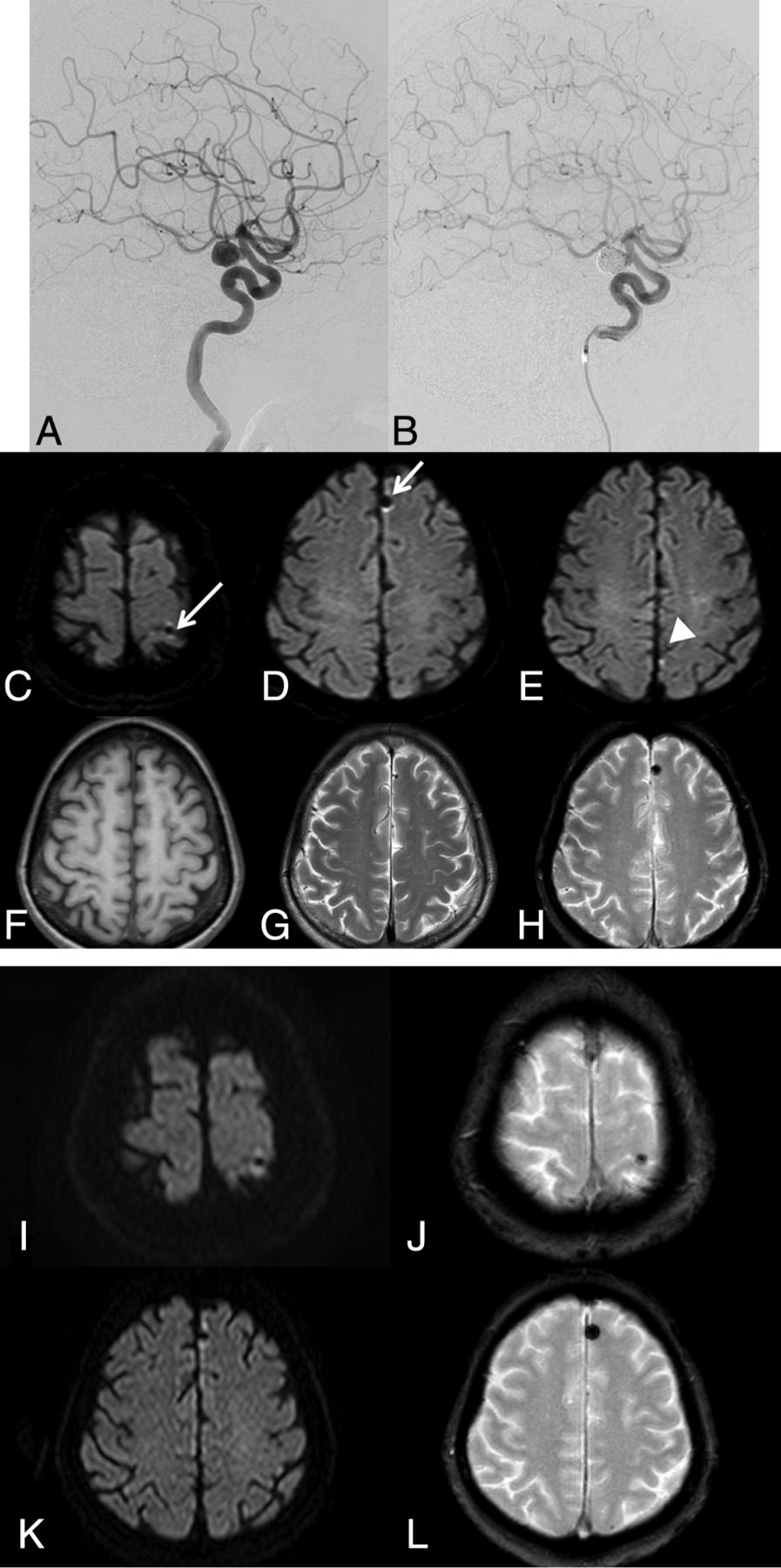
- Fig 1.
A left ICA aneurysm in case 1 (A) is embolized by using multiple coils (B). Postprocedural DWI shows abnormal low-intensity spots with a hyperintense halo (white arrow) in the left parietal (C) and frontal (D) lobes. These are different from a bright spot in the left parietal lobe (E), indicating thromboembolism. These abnormal spots are low intensity on T1WI (F) and T2WI (G) and show a blooming effect on T2*-weighted images (H). Follow-up MR imaging obtained 13 months after the procedure shows that all the abnormal signal spots remain without any signal changes (I–L).
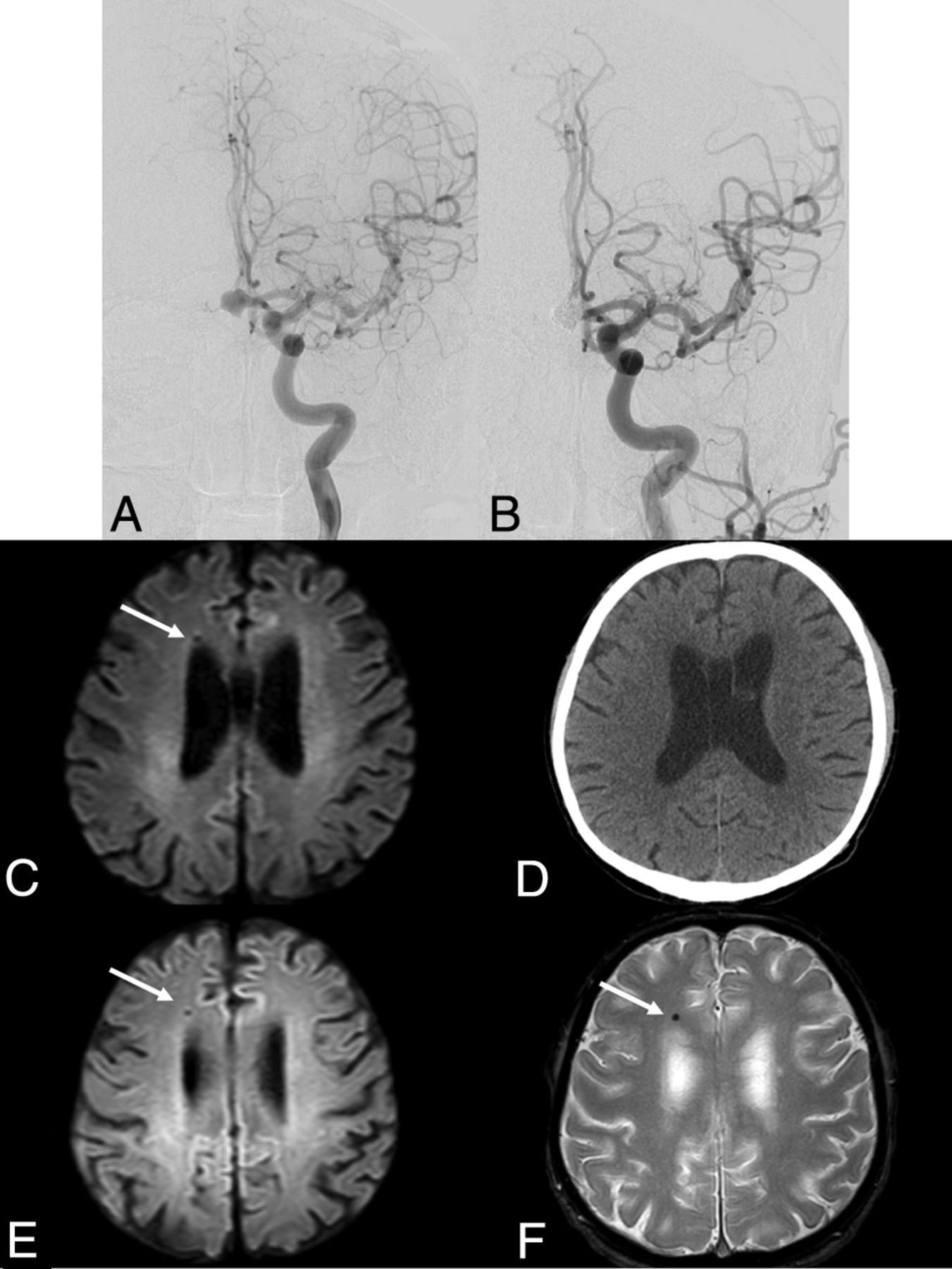
- Fig 2.
An anterior communicating artery aneurysm in case 5 (A) is embolized by using multiple coils (B). Postprocedural DWI shows an abnormal low-intensity spot with a hyperintense halo (white arrow) in the right frontal lobe (C), which is undetectable on the postprocedural CT (D). Follow-up MR imaging obtained 3 years after the procedure shows that the lesion remains without any signal changes (E and F).


Tables
Case No. Patient Age (yr)/Sex Location of Aneurysm Aneurysm Size (mm) Procedure Procedural Time (min) Postprodedural Symptoms 1 68/F Left ICA 10.2 × 8.2 × 7.3 Coil embolization 170 None 2 56/M Right vertebral artery 15.6 × 7.1 × 6.5 Internal trapping 135 None 3 80/F Right ICA 12.6 × 10.3 × 11.0 Coil embolization 150 None 4 45/F Right ICA 4.9 × 4.5 × 4.3 Coil embolization 158 None 5 70/M Anterior communicating artery 9.6 × 7.1 × 9.5 Coil embolization 145 None 6 41/F Left ICA 5.6 × 4.9 × 3.8 Coil embolization 150 None Devices 1 2 3 4 5 6 0.035-inch Radifocus Standarda ○ ○ ○ ○ ○ ○ 0.035-inch Radifocus Stiffa ○ 0.035-inch Quick Flex Standardb ○ 0.010–0.014-inch Tenroub ○ ○ ○ ○ ○ 0.014-inch CHIKAIc ○ ○ ○ ○ 0.010-inch X-Pediond ○ 0.010-inch SilverSpeedd ○ 0.014-inch Traxcesse ○ Excelsior SL-10f ○ ○ ○ ○ ○ Headway17e ○ ○ Prowler Select Plusg ○ Hydrogel coilse ○ ○ ED coilsb ○ ○ ○ ○ Target coilsf ○ ○ ○ Cashmereg ○ VFCe ○ Orbit Galaxyg ○ ○ ○ Deltapaqg ○ Deltaplushg ○ Matrix coilsf ○ Scepter XCe ○ ○ HyperFormd ○ Enterprise VRDg ○ Amplatz GooseNeck Snared ○ Author Patient/Sex T2WI/Proton T2*-Weighted Image CT Suspected Etiology Symptom Wingerchuk et al7 35/F Low intensity halo Low intensity blooming Normal Prosthetic cardiac valve None Naumann et al8 66/M Low intensity halo Not available Not available Prosthetic cardiac valve Seizure Jassal et al9 55/M Low intensity halo Not available Normal Coronary guidewire Headache Roshal et al10 55/F Low intensity Low intensity blooming, halo Normal Coronary guidewire, robotic surgery Headache


