Advertisement

Main menu

  • Home
  • Content
    • Current Issue
    • Accepted Manuscripts
    • Article Preview
    • Past Issue Archive
    • Video Articles
    • AJNR Case Collection
    • Case of the Week Archive
    • Case of the Month Archive
    • Classic Case Archive
  • Special Collections
    • AJNR Awards
    • Low-Field MRI
    • Alzheimer Disease
    • ASNR Foundation Special Collection
    • Photon-Counting CT
    • View All
  • Multimedia
    • AJNR Podcasts
    • AJNR SCANtastic
    • Trainee Corner
    • MRI Safety Corner
    • Imaging Protocols
  • For Authors
    • Submit a Manuscript
    • Submit a Video Article
    • Submit an eLetter to the Editor/Response
    • Manuscript Submission Guidelines
    • Statistical Tips
    • Fast Publishing of Accepted Manuscripts
    • Graphical Abstract Preparation
    • Imaging Protocol Submission
    • Author Policies
  • About Us
    • About AJNR
    • Editorial Board
    • Editorial Board Alumni
  • More
    • Become a Reviewer/Academy of Reviewers
    • Subscribers
    • Permissions
    • Alerts
    • Feedback
    • Advertisers
    • ASNR Home

User menu

  • Alerts
  • Log in

Search

  • Advanced search
American Journal of Neuroradiology

American Journal of Neuroradiology

ASHNR American Society of Functional Neuroradiology ASHNR American Society of Pediatric Neuroradiology ASSR
  • Alerts
  • Log in

Advanced Search

  • Home
  • Content
    • Current Issue
    • Accepted Manuscripts
    • Article Preview
    • Past Issue Archive
    • Video Articles
    • AJNR Case Collection
    • Case of the Week Archive
    • Case of the Month Archive
    • Classic Case Archive
  • Special Collections
    • AJNR Awards
    • Low-Field MRI
    • Alzheimer Disease
    • ASNR Foundation Special Collection
    • Photon-Counting CT
    • View All
  • Multimedia
    • AJNR Podcasts
    • AJNR SCANtastic
    • Trainee Corner
    • MRI Safety Corner
    • Imaging Protocols
  • For Authors
    • Submit a Manuscript
    • Submit a Video Article
    • Submit an eLetter to the Editor/Response
    • Manuscript Submission Guidelines
    • Statistical Tips
    • Fast Publishing of Accepted Manuscripts
    • Graphical Abstract Preparation
    • Imaging Protocol Submission
    • Author Policies
  • About Us
    • About AJNR
    • Editorial Board
    • Editorial Board Alumni
  • More
    • Become a Reviewer/Academy of Reviewers
    • Subscribers
    • Permissions
    • Alerts
    • Feedback
    • Advertisers
    • ASNR Home
  • Follow AJNR on Twitter
  • Visit AJNR on Facebook
  • Follow AJNR on Instagram
  • Join AJNR on LinkedIn
  • RSS Feeds

AJNR Awards, New Junior Editors, and more. Read the latest AJNR updates

Research ArticleADULT BRAIN
Open Access

Antiangiogenic Effect of Bevacizumab: Application of Arterial Spin-Labeling Perfusion MR Imaging in a Rat Glioblastoma Model

T.J. Yun, H.R. Cho, S.H. Choi, H. Kim, J.-K. Won, S.-W. Park, J.-h. Kim, C.-H. Sohn and M.H. Han
American Journal of Neuroradiology September 2016, 37 (9) 1650-1656; DOI: https://doi.org/10.3174/ajnr.A4800
T.J. Yun
aFrom the Department of Radiology (T.J.Y., H.R.C., S.H.C., H.K., S.-W.P., J.-h.K., C.-H.S., M.H.H.), Seoul National University College of Medicine, Republic of Korea
bDepartments of Radiology (T.J.Y., H.R.C., S.H.C., H.K., J.-h.K., C.-H.S., M.H.H.)
  • Find this author on Google Scholar
  • Find this author on PubMed
  • Search for this author on this site
  • ORCID record for T.J. Yun
H.R. Cho
aFrom the Department of Radiology (T.J.Y., H.R.C., S.H.C., H.K., S.-W.P., J.-h.K., C.-H.S., M.H.H.), Seoul National University College of Medicine, Republic of Korea
bDepartments of Radiology (T.J.Y., H.R.C., S.H.C., H.K., J.-h.K., C.-H.S., M.H.H.)
  • Find this author on Google Scholar
  • Find this author on PubMed
  • Search for this author on this site
  • ORCID record for H.R. Cho
S.H. Choi
aFrom the Department of Radiology (T.J.Y., H.R.C., S.H.C., H.K., S.-W.P., J.-h.K., C.-H.S., M.H.H.), Seoul National University College of Medicine, Republic of Korea
bDepartments of Radiology (T.J.Y., H.R.C., S.H.C., H.K., J.-h.K., C.-H.S., M.H.H.)
  • Find this author on Google Scholar
  • Find this author on PubMed
  • Search for this author on this site
  • ORCID record for S.H. Choi
H. Kim
aFrom the Department of Radiology (T.J.Y., H.R.C., S.H.C., H.K., S.-W.P., J.-h.K., C.-H.S., M.H.H.), Seoul National University College of Medicine, Republic of Korea
bDepartments of Radiology (T.J.Y., H.R.C., S.H.C., H.K., J.-h.K., C.-H.S., M.H.H.)
  • Find this author on Google Scholar
  • Find this author on PubMed
  • Search for this author on this site
  • ORCID record for H. Kim
J.-K. Won
cPathology (J.-K.W.)
  • Find this author on Google Scholar
  • Find this author on PubMed
  • Search for this author on this site
  • ORCID record for J.-K. Won
S.-W. Park
aFrom the Department of Radiology (T.J.Y., H.R.C., S.H.C., H.K., S.-W.P., J.-h.K., C.-H.S., M.H.H.), Seoul National University College of Medicine, Republic of Korea
eDepartment of Radiology (S.-W.P.), Seoul National University Boramae Medical Center, Seoul, Republic of Korea.
  • Find this author on Google Scholar
  • Find this author on PubMed
  • Search for this author on this site
  • ORCID record for S.-W. Park
J.-h. Kim
aFrom the Department of Radiology (T.J.Y., H.R.C., S.H.C., H.K., S.-W.P., J.-h.K., C.-H.S., M.H.H.), Seoul National University College of Medicine, Republic of Korea
bDepartments of Radiology (T.J.Y., H.R.C., S.H.C., H.K., J.-h.K., C.-H.S., M.H.H.)
  • Find this author on Google Scholar
  • Find this author on PubMed
  • Search for this author on this site
  • ORCID record for J.-h. Kim
C.-H. Sohn
aFrom the Department of Radiology (T.J.Y., H.R.C., S.H.C., H.K., S.-W.P., J.-h.K., C.-H.S., M.H.H.), Seoul National University College of Medicine, Republic of Korea
bDepartments of Radiology (T.J.Y., H.R.C., S.H.C., H.K., J.-h.K., C.-H.S., M.H.H.)
  • Find this author on Google Scholar
  • Find this author on PubMed
  • Search for this author on this site
  • ORCID record for C.-H. Sohn
M.H. Han
aFrom the Department of Radiology (T.J.Y., H.R.C., S.H.C., H.K., S.-W.P., J.-h.K., C.-H.S., M.H.H.), Seoul National University College of Medicine, Republic of Korea
bDepartments of Radiology (T.J.Y., H.R.C., S.H.C., H.K., J.-h.K., C.-H.S., M.H.H.)
dNeurosurgery (M.H.H.), Seoul National University Hospital, Seoul, Republic of Korea
  • Find this author on Google Scholar
  • Find this author on PubMed
  • Search for this author on this site
  • ORCID record for M.H. Han
  • Article
  • Figures & Data
  • Supplemental
  • Info & Metrics
  • Responses
  • References
  • PDF
Loading

Article Figures & Data

Figures

  • Tables
  • Fig 1.
    • Download figure
    • Open in new tab
    • Download powerpoint
    Fig 1.

    Experimental design showing the timeline of each group for U87 glioma cell inoculation, bevacizumab therapy, MR imaging, and sacrifice for brain harvest.
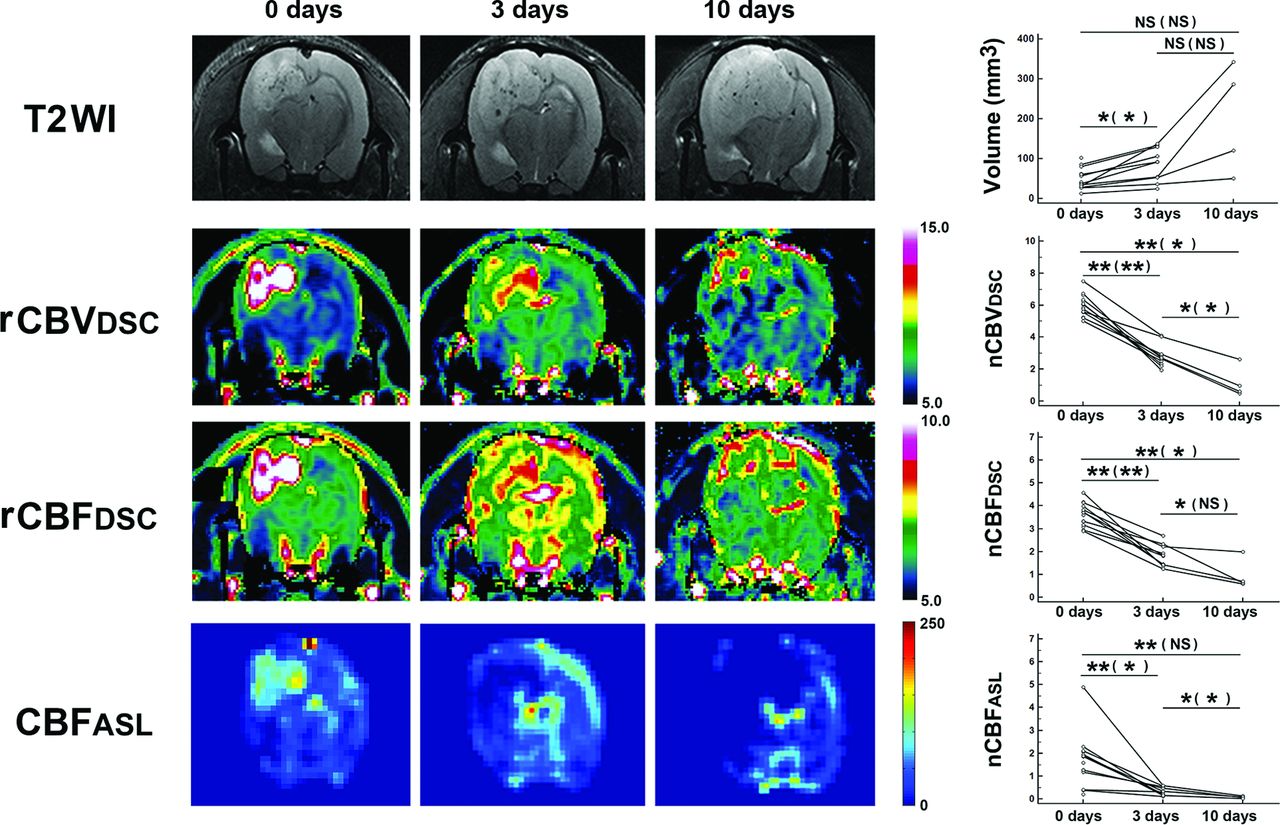
  • Fig 2.
    • Download figure
    • Open in new tab
    • Download powerpoint
    Fig 2.

    Quantification of tumor volume and perfusion parameters in all tumors. T2WI and perfusion maps were acquired from a rat belonging to the 10-day treatment group. Serial reductions in rCBV and rCBF based on DSC and CBF based on ASL are shown. Graphs in the right column show serial changes of tumor volume and perfusion parameters in all tumors. Scale units of rCBVDSC, rCBFDSC, and CBFASL are milliliters × 100 g−1, milliliters × 100 g−1 × min−1, and milliliters × 100 g−1 × min−1, respectively. Data are mean results from paired t tests (unpaired t tests). One asterisk indicates P < .05; 2 asterisks, P < .001; NS, not significant; nCBVDSC, nCBV based on DSC; nCBFDSC, nCBF based on DSC; nCBFASL, nCBF based on ASL.
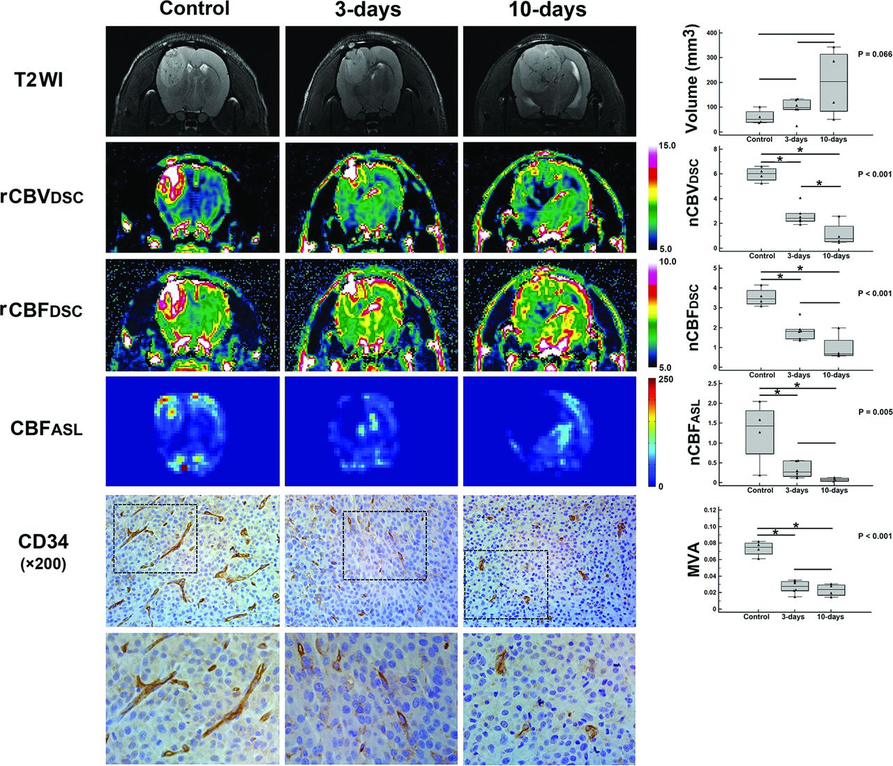
  • Fig 3.
    • Download figure
    • Open in new tab
    • Download powerpoint
    Fig 3.

    Quantification of tumor volume, perfusion parameters, and MVA in tumors with available histology. T2WI and perfusion maps were acquired from rats in the control group, 3-day treatment group, or 10-day treatment group. Differences in rCBV and rCBF based on DSC, CBF based on ASL, and MVA are shown. Graphs in the right column show differences in the tumor volume and hemodynamic parameters and MVA. In the lower images, the tumors stained immunohistochemically with anti-CD34 show positive brown cytoplasmic staining of the endothelial area and vessel lumen. Scale units of rCBVDSC, rCBFDSC, and CBFASL are milliliters × 100 g−1, milliliters × 100 g−1 × min−1, and milliliters × 100 g−1 × min−1, respectively. P values were based on 1-way analyses of variances. One asterisk indicates a significant (P < .05) difference from the Scheffe post hoc multiple comparisons.

Tables

  • Figures
    • View popup
    Table 1:

    Comparison of volume and perfusion parameters based on DSC and ASL MR imaging in all tumorsa

    Variables0 Days (n = 14)3 Days (n = 10)10 Days (n = 4)Paired t TestUnpaired t Test
    P ValuebP ValuecP ValuedP ValuebP ValuecP Valued
    Volume (mm3)40.0 (32.3–61.8)91.9 (53.9–124.1)203.7 (103.0–301.3).015.200.117.002.090.086
    nCBVDSC5.9 (5.7–6.3)2.7 (2.4–2.9)0.8 (0.6–1.4)<.001.004<.001<.001.002.005
    nCBFDSC3.7 (3.2–4.0)1.8 (1.4–2.2)0.7 (0.6–1.0)<.001.020<.001<.001.075.004
    nCBFASL1.7 (1.2–2.0)0.3 (0.2–0.5)0.1 (0.1–0.1)<.001.041<.001.004.029.050
    • Note:—nCBVDSC indicates nCBV based on DSC; nCBFDSC, nCBF based on DSC; nCBFASL, nCBF based on ASL.

    • ↵a Values are presented as medians (interquartile range, range from the 25th to the 75th percentile).

    • ↵b Results between 0 and 3 days.

    • ↵c Results between 3 and 10 days.

    • ↵d Results between 0 and 10 days.

    • View popup
    Table 2:

    Comparison and correlation of perfusion parameters based on DSC and ASL MR imaging with MVA in tumors with available histologya

    VariablesControl (n = 4)3-Day Treatment (n = 6)10-Day Treatment (n = 4)P ValuebDifferent Groupsc
    Volume (mm3)51.9 (40.8–72.1)99.7 (91.8–124.1)203.7 (103.0–301.3)0.066
    nCBVDSC6.0 (5.7–6.3)2.4 (2.2–2.7)0.8 (0.6–1.4)<.001(Control) (3 days) (3 days) (10 days) (Control) (10 days)
    nCBFDSC3.5 (3.3–3.7)1.8 (1.5–1.9)0.7 (0.6–1.0)<.001(Control) (3 days) (Control) (10 days)
    nCBFASL1.4 (1.0–1.7)0.3 (0.2–0.5)0.1 (0.1–0.1)0.005(Control) (3 days) (Control) (10 days)
    MVA (×10−2)7.5 (6.9–7.9)2.8 (2.3–3.3)2.4 (1.9–2.9)<.001(Control) (3 days) (Control) (10 days)
    • ↵a Values are presented as medians (interquartile range, range from the 25th to the 75th percentile).

    • ↵b Results from 1-way analyses of variances.

    • ↵c Results from Scheffe post hoc multiple comparisons.

PreviousNext
Back to top

In this issue

American Journal of Neuroradiology: 37 (9)
American Journal of Neuroradiology
Vol. 37, Issue 9
1 Sep 2016
  • Table of Contents
  • Index by author
  • Complete Issue (PDF)
Advertisement
Print
Download PDF
Email Article

Thank you for your interest in spreading the word on American Journal of Neuroradiology.

NOTE: We only request your email address so that the person you are recommending the page to knows that you wanted them to see it, and that it is not junk mail. We do not capture any email address.

Enter multiple addresses on separate lines or separate them with commas.
Antiangiogenic Effect of Bevacizumab: Application of Arterial Spin-Labeling Perfusion MR Imaging in a Rat Glioblastoma Model
(Your Name) has sent you a message from American Journal of Neuroradiology
(Your Name) thought you would like to see the American Journal of Neuroradiology web site.
CAPTCHA
This question is for testing whether or not you are a human visitor and to prevent automated spam submissions.
Cite this article
T.J. Yun, H.R. Cho, S.H. Choi, H. Kim, J.-K. Won, S.-W. Park, J.-h. Kim, C.-H. Sohn, M.H. Han
Antiangiogenic Effect of Bevacizumab: Application of Arterial Spin-Labeling Perfusion MR Imaging in a Rat Glioblastoma Model
American Journal of Neuroradiology Sep 2016, 37 (9) 1650-1656; DOI: 10.3174/ajnr.A4800

Citation Manager Formats

  • BibTeX
  • Bookends
  • EasyBib
  • EndNote (tagged)
  • EndNote 8 (xml)
  • Medlars
  • Mendeley
  • Papers
  • RefWorks Tagged
  • Ref Manager
  • RIS
  • Zotero
0 Responses
Respond to this article
Share
Bookmark this article
Antiangiogenic Effect of Bevacizumab: Application of Arterial Spin-Labeling Perfusion MR Imaging in a Rat Glioblastoma Model
T.J. Yun, H.R. Cho, S.H. Choi, H. Kim, J.-K. Won, S.-W. Park, J.-h. Kim, C.-H. Sohn, M.H. Han
American Journal of Neuroradiology Sep 2016, 37 (9) 1650-1656; DOI: 10.3174/ajnr.A4800
del.icio.us logo Twitter logo Facebook logo Mendeley logo
  • Tweet Widget
  • Facebook Like
  • Google Plus One
Purchase

Jump to section

  • Article
    • Abstract
    • ABBREVIATIONS:
    • Materials and Methods
    • Results
    • Discussion
    • Conclusions
    • Footnotes
    • References
  • Figures & Data
  • Supplemental
  • Info & Metrics
  • Responses
  • References
  • PDF

Related Articles

  • PubMed
  • Google Scholar

Cited By...

  • No citing articles found.
  • Crossref (10)
  • Google Scholar

This article has been cited by the following articles in journals that are participating in Crossref Cited-by Linking.

  • Perfusion MRI in treatment evaluation of glioblastomas: Clinical relevance of current and future techniques
    Bart R.J. van Dijken, Peter Jan van Laar, Marion Smits, Jan Willem Dankbaar, Roelien H. Enting, Anouk van der Hoorn
    Journal of Magnetic Resonance Imaging 2019 49 1
  • Increased Antiangiogenic Effect by Blocking CCL2-dependent Macrophages in a Rodent Glioblastoma Model: Correlation Study with Dynamic Susceptibility Contrast Perfusion MRI
    Hye Rim Cho, Nisha Kumari, Hien Thi Vu, Hyeonjin Kim, Chul-Kee Park, Seung Hong Choi
    Scientific Reports 2019 9 1
  • Assessment of Glioblastoma Response in the Era of Bevacizumab: Longstanding and Emergent Challenges in the Imaging Evaluation of Pseudoresponse
    Octavio D. Arevalo, Carolina Soto, Pejman Rabiei, Arash Kamali, Leomar Y. Ballester, Yoshua Esquenazi, Jay-Jiguang Zhu, Roy Francisco Riascos
    Frontiers in Neurology 2019 10
  • Development and characterization of sorafenib-loaded lipid nanocapsules for the treatment of glioblastoma
    Anne Clavreul, Emilie Roger, Milad Pourbaghi-Masouleh, Laurent Lemaire, Clément Tétaud, Philippe Menei
    Drug Delivery 2018 25 1
  • Imaging-guided precision medicine in glioblastoma patients treated with immune checkpoint modulators: research trend and future directions in the field of imaging biomarkers and artificial intelligence
    Mathieu Sinigaglia, Tarek Assi, Florent L. Besson, Samy Ammari, Myriam Edjlali, Whitney Feltus, Laura Rozenblum-Beddok, Binsheng Zhao, Lawrence H. Schwartz, Fatima-Zohra Mokrane, Laurent Dercle
    EJNMMI Research 2019 9 1
  • Cerebral and tumoral blood flow in adult gliomas: a systematic review of results from magnetic resonance imaging
    Mueez Waqar, Daniel Lewis, Erjon Agushi, Matthew Gittins, Alan Jackson, David Coope
    The British Journal of Radiology 2021 94 1125
  • EGFRvIII Peptide Nanocapsules and Bevacizumab Nanocapsules: A Nose-To-Brain Multitarget Approach Against Glioblastoma
    Aline de Cristo Soares Alves, Vladimir Lavayen, Amanda de Fraga Dias, Franciele Aline Bruinsmann, Juliete Nathali Scholl, Rodrigo Cé, Fernanda Visioli, Ana Maria Oliveira Battastini, Silvia Stanisçuaski Guterres, Fabrício Figueiró, Adriana Raffin Pohlmann
    Nanomedicine 2021 16 20
  • Music improves the therapeutic effects of bevacizumab in rats with glioblastoma: Modulation of drug distribution to the brain
    Oxana Semyachkina-Glushkovskaya, Sergey Diduk, Eroshova Anna, Dosadina Elina, Kruglov Artem, Alexander Khorovodov, Alexander Shirokov, Ivan Fedosov, Alexander Dubrovsky, Inna Blokhina, Andrey Terskov, Nikita Navolokin, Arina Evsukova, Daria Elovenko, Viktoria Adushkina, Jürgen Kurths
    Frontiers in Oncology 2022 12
  • Biodistribution and pharmacokinetics of [89Zr]-anti-VEGF mAbs using PET in glioblastoma rat models
    Lara García-Varela, Jessica Codesido, Alberto Perez-Pedrosa, María Muñoz-González, Emma Ramos-Docampo, David Rey-Bretal, Xurxo García-Otero, Noemí Gómez-Lado, Angela Turrero, Daniel Beiroa, Ana Isabel Rodríguez-Perez, Anxo Vidal, Anxo Fernández-Ferreiro, Virginia Pubul, Pablo Aguiar
    International Journal of Pharmaceutics 2024 652
  • Repeatability of perfusion measurements in adult gliomas using pulsed and pseudo-continuous arterial spin labelling MRI
    Amirah Faisal Alsaedi, David Lee Thomas, Enrico De Vita, Jasmina Panovska-Griffiths, Sotirios Bisdas, Xavier Golay
    Magnetic Resonance Materials in Physics, Biology and Medicine 2022 35 1

More in this TOC Section

ADULT BRAIN

  • Diagnostic Neuroradiology of Monoclonal Antibodies
  • Clinical Outcomes After Chiari I Decompression
  • Segmentation of Brain Metastases with BLAST
Show more ADULT BRAIN

FUNCTIONAL

  • Kurtosis and Epileptogenic Tubers: A Pilot Study
  • Glutaric Aciduria Type 1: DK vs. Conventional MRI
  • Multiparametric MRI in PEDS Pontine Glioma
Show more FUNCTIONAL

Similar Articles

Advertisement

Indexed Content

  • Current Issue
  • Accepted Manuscripts
  • Article Preview
  • Past Issues
  • Editorials
  • Editor's Choice
  • Fellows' Journal Club
  • Letters to the Editor
  • Video Articles

Cases

  • Case Collection
  • Archive - Case of the Week
  • Archive - Case of the Month
  • Archive - Classic Case

More from AJNR

  • Trainee Corner
  • Imaging Protocols
  • MRI Safety Corner
  • Book Reviews

Multimedia

  • AJNR Podcasts
  • AJNR Scantastics

Resources

  • Turnaround Time
  • Submit a Manuscript
  • Submit a Video Article
  • Submit an eLetter to the Editor/Response
  • Manuscript Submission Guidelines
  • Statistical Tips
  • Fast Publishing of Accepted Manuscripts
  • Graphical Abstract Preparation
  • Imaging Protocol Submission
  • Evidence-Based Medicine Level Guide
  • Publishing Checklists
  • Author Policies
  • Become a Reviewer/Academy of Reviewers
  • News and Updates

About Us

  • About AJNR
  • Editorial Board
  • Editorial Board Alumni
  • Alerts
  • Permissions
  • Not an AJNR Subscriber? Join Now
  • Advertise with Us
  • Librarian Resources
  • Feedback
  • Terms and Conditions
  • AJNR Editorial Board Alumni

American Society of Neuroradiology

  • Not an ASNR Member? Join Now

© 2025 by the American Society of Neuroradiology All rights, including for text and data mining, AI training, and similar technologies, are reserved.
Print ISSN: 0195-6108 Online ISSN: 1936-959X

Powered by HighWire