Advertisement

Main menu

  • Home
  • Content
    • Current Issue
    • Accepted Manuscripts
    • Article Preview
    • Past Issue Archive
    • Video Articles
    • AJNR Case Collection
    • Case of the Week Archive
    • Case of the Month Archive
    • Classic Case Archive
  • Special Collections
    • AJNR Awards
    • Low-Field MRI
    • Alzheimer Disease
    • ASNR Foundation Special Collection
    • Photon-Counting CT
    • View All
  • Multimedia
    • AJNR Podcasts
    • AJNR SCANtastic
    • Trainee Corner
    • MRI Safety Corner
    • Imaging Protocols
  • For Authors
    • Submit a Manuscript
    • Submit a Video Article
    • Submit an eLetter to the Editor/Response
    • Manuscript Submission Guidelines
    • Statistical Tips
    • Fast Publishing of Accepted Manuscripts
    • Graphical Abstract Preparation
    • Imaging Protocol Submission
    • Author Policies
  • About Us
    • About AJNR
    • Editorial Board
    • Editorial Board Alumni
  • More
    • Become a Reviewer/Academy of Reviewers
    • Subscribers
    • Permissions
    • Alerts
    • Feedback
    • Advertisers
    • ASNR Home

User menu

  • Alerts
  • Log in

Search

  • Advanced search
American Journal of Neuroradiology

American Journal of Neuroradiology

ASHNR American Society of Functional Neuroradiology ASHNR American Society of Pediatric Neuroradiology ASSR
  • Alerts
  • Log in

Advanced Search

  • Home
  • Content
    • Current Issue
    • Accepted Manuscripts
    • Article Preview
    • Past Issue Archive
    • Video Articles
    • AJNR Case Collection
    • Case of the Week Archive
    • Case of the Month Archive
    • Classic Case Archive
  • Special Collections
    • AJNR Awards
    • Low-Field MRI
    • Alzheimer Disease
    • ASNR Foundation Special Collection
    • Photon-Counting CT
    • View All
  • Multimedia
    • AJNR Podcasts
    • AJNR SCANtastic
    • Trainee Corner
    • MRI Safety Corner
    • Imaging Protocols
  • For Authors
    • Submit a Manuscript
    • Submit a Video Article
    • Submit an eLetter to the Editor/Response
    • Manuscript Submission Guidelines
    • Statistical Tips
    • Fast Publishing of Accepted Manuscripts
    • Graphical Abstract Preparation
    • Imaging Protocol Submission
    • Author Policies
  • About Us
    • About AJNR
    • Editorial Board
    • Editorial Board Alumni
  • More
    • Become a Reviewer/Academy of Reviewers
    • Subscribers
    • Permissions
    • Alerts
    • Feedback
    • Advertisers
    • ASNR Home
  • Follow AJNR on Twitter
  • Visit AJNR on Facebook
  • Follow AJNR on Instagram
  • Join AJNR on LinkedIn
  • RSS Feeds

AJNR Awards, New Junior Editors, and more. Read the latest AJNR updates

Research ArticleADULT BRAIN
Open Access

Cerebral CTA with Low Tube Voltage and Low Contrast Material Volume for Detection of Intracranial Aneurysms

Q.Q. Ni, G.Z. Chen, U.J. Schoepf, M.A.J. Klitsie, C.N. De Cecco, C.S. Zhou, S. Luo, G.M. Lu and L.J. Zhang
American Journal of Neuroradiology October 2016, 37 (10) 1774-1780; DOI: https://doi.org/10.3174/ajnr.A4803
Q.Q. Ni
aFrom the Department of Medical Imaging (Q.Q.N., G.Z.C., C.S.Z., S.L., G.M.L., L.J.Z.), Jinling Hospital, Medical School of Nanjing University, Nanjing, Jiangsu, China
  • Find this author on Google Scholar
  • Find this author on PubMed
  • Search for this author on this site
  • ORCID record for Q.Q. Ni
G.Z. Chen
aFrom the Department of Medical Imaging (Q.Q.N., G.Z.C., C.S.Z., S.L., G.M.L., L.J.Z.), Jinling Hospital, Medical School of Nanjing University, Nanjing, Jiangsu, China
  • Find this author on Google Scholar
  • Find this author on PubMed
  • Search for this author on this site
  • ORCID record for G.Z. Chen
U.J. Schoepf
bDepartment of Radiology and Radiological Science (U.J.S., M.A.J.K., C.N.D.C.), Medical University of South Carolina, Charleston, South Carolina.
  • Find this author on Google Scholar
  • Find this author on PubMed
  • Search for this author on this site
  • ORCID record for U.J. Schoepf
M.A.J. Klitsie
bDepartment of Radiology and Radiological Science (U.J.S., M.A.J.K., C.N.D.C.), Medical University of South Carolina, Charleston, South Carolina.
  • Find this author on Google Scholar
  • Find this author on PubMed
  • Search for this author on this site
  • ORCID record for M.A.J. Klitsie
C.N. De Cecco
bDepartment of Radiology and Radiological Science (U.J.S., M.A.J.K., C.N.D.C.), Medical University of South Carolina, Charleston, South Carolina.
  • Find this author on Google Scholar
  • Find this author on PubMed
  • Search for this author on this site
  • ORCID record for C.N. De Cecco
C.S. Zhou
aFrom the Department of Medical Imaging (Q.Q.N., G.Z.C., C.S.Z., S.L., G.M.L., L.J.Z.), Jinling Hospital, Medical School of Nanjing University, Nanjing, Jiangsu, China
  • Find this author on Google Scholar
  • Find this author on PubMed
  • Search for this author on this site
  • ORCID record for C.S. Zhou
S. Luo
aFrom the Department of Medical Imaging (Q.Q.N., G.Z.C., C.S.Z., S.L., G.M.L., L.J.Z.), Jinling Hospital, Medical School of Nanjing University, Nanjing, Jiangsu, China
  • Find this author on Google Scholar
  • Find this author on PubMed
  • Search for this author on this site
  • ORCID record for S. Luo
G.M. Lu
aFrom the Department of Medical Imaging (Q.Q.N., G.Z.C., C.S.Z., S.L., G.M.L., L.J.Z.), Jinling Hospital, Medical School of Nanjing University, Nanjing, Jiangsu, China
  • Find this author on Google Scholar
  • Find this author on PubMed
  • Search for this author on this site
  • ORCID record for G.M. Lu
L.J. Zhang
aFrom the Department of Medical Imaging (Q.Q.N., G.Z.C., C.S.Z., S.L., G.M.L., L.J.Z.), Jinling Hospital, Medical School of Nanjing University, Nanjing, Jiangsu, China
  • Find this author on Google Scholar
  • Find this author on PubMed
  • Search for this author on this site
  • ORCID record for L.J. Zhang
  • Article
  • Figures & Data
  • Supplemental
  • Info & Metrics
  • Responses
  • References
  • PDF
Loading

Article Figures & Data

Figures

  • Tables
  • Fig 1.
    • Download figure
    • Open in new tab
    • Download powerpoint
    Fig 1.

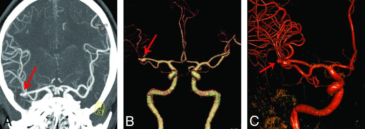
    Comparison of the 2 CTA protocols for detecting an aneurysm in the posterior communicating artery. A and B, An 80-kVp cerebral CTA with 30 mL of contrast agent in a 49-year-old woman. A volume-rendered digital subtraction CTA image (A) shows an aneurysm in the left posterior communicating artery (red arrow), which is confirmed by 3D-DSA (B). C and D, A 120-kVp cerebral CTA with 60 mL of contrast agent in a 66-year-old woman. C, Volume-rendered digital subtraction CTA image (C) shows an aneurysm in the right posterior communicating artery (red arrow), which is confirmed by 2D-DSA (D).

  • Fig 2.
    • Download figure
    • Open in new tab
    • Download powerpoint
    Fig 2.

    An 80-kVp cerebral CTA with 30 mL of contrast agent in a 45-year-old man. Maximum-intensity-projection image (A) and a volume-rendered digital subtraction CTA image (B) show an aneurysm in the right middle cerebral artery (red arrow), which is confirmed by 3D-DSA (C).

  • Fig 3.
    • Download figure
    • Open in new tab
    • Download powerpoint
    Fig 3.

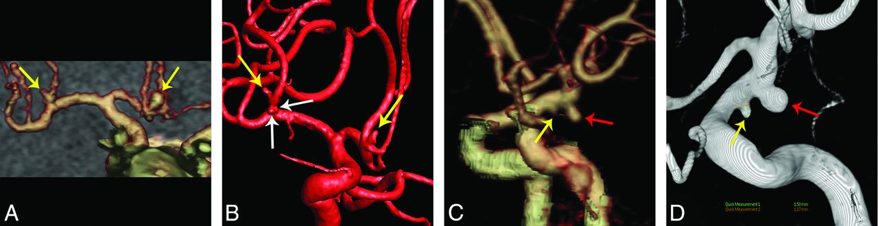
    False-negative aneurysms in the 2 CTA protocols. A and B, An 80-kVp cerebral CTA with 30 mL of contrast agent in a 50-year-old man. A volume-rendered digital subtraction CTA image (A) shows 2 true-positive aneurysms in the anterior communicating artery and right middle cerebral artery, respectively (yellow arrows), while another 2 small aneurysms with diameters of 1.1 and 0.6 mm were found in the right middle cerebral artery (white arrows) on 3D-DSA (B), which were not found in the CTA image. C and D, A 120-kVp cerebral CTA with 60 mL of contrast agent in a 46-year-old man. The volume-rendered digital subtraction CTA image (C) shows a true-positive aneurysm in the left anterior choroidal artery (red arrow) and a false-negative aneurysm in the left posterior communicating artery (yellow arrow). The aneurysm in the left posterior communicating artery (yellow arrow) was found at repeat interpretation. 3D-DSA shows the 2 aneurysms (D).

  • Fig 4.
    • Download figure
    • Open in new tab
    • Download powerpoint
    Fig 4.

    A 120-kVp cerebral CTA with 60 mL of contrast agent in a 70-year-old woman. A and B, Volume-rendered digital subtraction CTA images show a true-positive aneurysm in the top of basilar artery (red arrow), which was confirmed by 3D-DSA (C) and a false-positive aneurysm in the left middle cerebral artery (yellow arrow), which was not evident in 2D-DSA.

Tables

  • Figures
    • View popup
    Table 1:

    Aneurysm detection with cerebral CTA compared with a 3D-DSA reference standarda

    ApproachResults (No.)Statistical Analysis (%)
    TPTNFPFNSensitivitySpecificityPPVNPVAccuracy
    Per patient
        Group A60391296.8 (89.0–99.1)97.5 (87.1–99.6)98.4 (91.2–99.7)95.1 (83.9–98.7)97.0 (91.7–99.0)
        Group B58421198.3 (91.0–99.7)97.7 (87.9–99.6)98.3 (91.0–99.7)97.7 (87.9–99.6)98.0 (93.1–99.5)
    Per aneurysm
        Group A773931088.5 (81.6–95.4)92.9 (88.6–98.6)96.3 (91.3–100.0)79.6 (67.3–89.8)89.9 (84.5–94.6)
        Group B66423494.3 (88.6–98.6)93.3 (84.4–100.0)95.7 (89.9–100.0)91.3 (82.6–97.8)93.9 (88.7–98.3)
    • Note:—TP indicates true positive; TN, true negative; FP, false positive; FN, false negative.

    • ↵a The data in parentheses are 95% confidence intervals.

    • View popup
    Table 2:

    Aneurysm detection with cerebral CTA according to aneurysm sizea

    Aneurysm SizeResults (No.)Statistical Analysis (%)
    TPTNFPFNSensitivitySpecificityPPVNPVAccuracy
    <3 mm
        Group A24392875.0 (59.4–90.6)95.1 (87.8–100.0)92.3 (80.8–100.0)83.0 (72.3–93.6)86.3 (78.1–93.2)
        Group B17422481.0 (61.9–95.2)95.5 (88.6–100.0)89.5 (73.7–100.0)91.3 (82.6–97.8)90.8 (83.1–96.9)
    3–8 mm
        Group A44391295.7 (89.1–100.0)97.5 (92.5–100.0)97.8 (93.3–100.0)95.1 (87.8–100.0)96.5 (93.0–100.0)
        Group B384210100 (100.0–100.0)97.7 (93.0–100.0)97.4 (92.3–100.0)100 (100.0–100.0)98.8 (96.3–100.0)
    >8 mm
        Group A93900100 (100.0–100.0)100 (100.0–100.0)100 (100.0–100.0)100 (100.0–100.0)100 (100.0–100.0)
        Group B114200100 (100.0–100.0)100 (100.0–100.0)100 (100.0–100.0)100 (100.0–100.0)100 (100.0–100.0)
    • Note:—TP indicates true positive; TN, true negative; FP, false positive; FN, false negative.

    • ↵a The data in parentheses are 95% confidence intervals.

    • View popup
    Table 3:

    Aneurysm detection with cerebral CTA according to aneurysm locationa

    LocationResults (No.)Statistical Analysis (%)
    TPTNFPFNSensitivitySpecificityPPVNPVAccuracy
    Anterior circulation
        Group A50391886.2 (77.6–94.8)97.5 (92.5–100.0)98.0 (94.1–100.0)83.0 (70.2–93.6)90.8 (84.7–95.9)
        Group B28422293.3 (83.3–100.0)95.5 (88.5–100.0)93.3 (83.3–100.0)95.5 (88.6–100.0)94.6 (89.2–98.6)
    Posterior circulation
        Group A27392293.1 (82.8–100.0)95.1 (87.8–100.0)93.1 (82.8–100.0)95.1 (87.8–100.0)94.3 (88.6–98.6)
        Group B38421295.0 (87.5–100.0)97.7 (93.0–100.0)97.4 (92.3–100.0)95.5 (88.6–100.0)96.4 (91.6–100.0)
    • Note:—TP indicates true positive; TN, true negative; FP, false positive; FN, false negative.

    • ↵a The data in parentheses are 95% confidence intervals.

PreviousNext
Back to top

In this issue

American Journal of Neuroradiology: 37 (10)
American Journal of Neuroradiology
Vol. 37, Issue 10
1 Oct 2016
  • Table of Contents
  • Index by author
  • Complete Issue (PDF)
Advertisement
Print
Download PDF
Email Article

Thank you for your interest in spreading the word on American Journal of Neuroradiology.

NOTE: We only request your email address so that the person you are recommending the page to knows that you wanted them to see it, and that it is not junk mail. We do not capture any email address.

Enter multiple addresses on separate lines or separate them with commas.
Cerebral CTA with Low Tube Voltage and Low Contrast Material Volume for Detection of Intracranial Aneurysms
(Your Name) has sent you a message from American Journal of Neuroradiology
(Your Name) thought you would like to see the American Journal of Neuroradiology web site.
CAPTCHA
This question is for testing whether or not you are a human visitor and to prevent automated spam submissions.
Cite this article
Q.Q. Ni, G.Z. Chen, U.J. Schoepf, M.A.J. Klitsie, C.N. De Cecco, C.S. Zhou, S. Luo, G.M. Lu, L.J. Zhang
Cerebral CTA with Low Tube Voltage and Low Contrast Material Volume for Detection of Intracranial Aneurysms
American Journal of Neuroradiology Oct 2016, 37 (10) 1774-1780; DOI: 10.3174/ajnr.A4803

Citation Manager Formats

  • BibTeX
  • Bookends
  • EasyBib
  • EndNote (tagged)
  • EndNote 8 (xml)
  • Medlars
  • Mendeley
  • Papers
  • RefWorks Tagged
  • Ref Manager
  • RIS
  • Zotero
0 Responses
Respond to this article
Share
Bookmark this article
Cerebral CTA with Low Tube Voltage and Low Contrast Material Volume for Detection of Intracranial Aneurysms
Q.Q. Ni, G.Z. Chen, U.J. Schoepf, M.A.J. Klitsie, C.N. De Cecco, C.S. Zhou, S. Luo, G.M. Lu, L.J. Zhang
American Journal of Neuroradiology Oct 2016, 37 (10) 1774-1780; DOI: 10.3174/ajnr.A4803
del.icio.us logo Twitter logo Facebook logo Mendeley logo
  • Tweet Widget
  • Facebook Like
  • Google Plus One
Purchase

Jump to section

  • Article
    • Abstract
    • ABBREVIATIONS:
    • Materials and Methods
    • Results
    • Discussion
    • Conclusions
    • Footnotes
    • References
  • Figures & Data
  • Supplemental
  • Info & Metrics
  • Responses
  • References
  • PDF

Related Articles

  • PubMed
  • Google Scholar

Cited By...

  • Neurovascular Imaging with Ultra-High-Resolution Photon-Counting CT: Preliminary Findings on Image-Quality Evaluation
  • Crossref (12)
  • Google Scholar

This article has been cited by the following articles in journals that are participating in Crossref Cited-by Linking.

  • Toward human intervention-free clinical diagnosis of intracranial aneurysm via deep neural network
    Zi-Hao Bo, Hui Qiao, Chong Tian, Yuchen Guo, Wuchao Li, Tiantian Liang, Dongxue Li, Dan Liao, Xianchun Zeng, Leilei Mei, Tianliang Shi, Bo Wu, Chao Huang, Lu Liu, Can Jin, Qiping Guo, Jun-Hai Yong, Feng Xu, Tijiang Zhang, Rongpin Wang, Qionghai Dai
    Patterns 2021 2 2
  • Automatic risk prediction of intracranial aneurysm on CTA image with convolutional neural networks and radiomics analysis
    Yuan Xie, Shuyu Liu, Hen Lin, Min Wu, Feng Shi, Feng Pan, Lichi Zhang, Bin Song
    Frontiers in Neurology 2023 14
  • Computed Tomography of the Head
    Michael Dieckmeyer, Nico Sollmann, Karina Kupfer, Maximilian T. Löffler, Karolin J. Paprottka, Jan S. Kirschke, Thomas Baum
    Clinical Neuroradiology 2023 33 3
  • Neurovascular Imaging with Ultra-High-Resolution Photon-Counting CT: Preliminary Findings on Image-Quality Evaluation
    Adrienn Tóth, Justin A. Chetta, Milad Yazdani, M. Gisele Matheus, Jim O‘Doherty, Sameer V. Tipnis, M. Vittoria Spampinato
    American Journal of Neuroradiology 2024 45 10
  • Comparison of eye-lens doses imparted during interventional and non-interventional neuroimaging techniques for assessment of intracranial aneurysms
    N Guberina, U Dietrich, M Forsting, A Ringelstein
    Journal of NeuroInterventional Surgery 2018 10 2
  • Cerebral bone subtraction CT angiography using 80 kVp and sinogram-affirmed iterative reconstruction: contrast medium and radiation dose reduction with improvement of image quality
    Yasunori Nagayama, Takeshi Nakaura, Akinori Tsuji, Joji Urata, Mitsuhiro Furusawa, Hideaki Yuki, Kenichiro Hirarta, Seitaro Oda, Masafumi Kidoh, Daisuke Utsunomiya, Yasuyuki Yamashita
    Neuroradiology 2017 59 2
  • LIVE-Net: Comprehensive 3D vessel extraction framework in CT angiography
    Qi Sun, Jinzhu Yang, Sizhe Zhao, Chen Chen, Yang Hou, Yuliang Yuan, Shuang Ma, Yan Huang
    Computers in Biology and Medicine 2023 159
  • Digital subtraction CT angiography for the detection of posterior inferior cerebellar artery aneurysms: comparison with digital subtraction angiography
    Guo Zhong Chen, Song Luo, Chang Sheng Zhou, Long Jiang Zhang, Guang Ming Lu
    European Radiology 2017 27 9
  • Radiation dose and image quality in intraoperative CT (iCT) angiography of the brain with stereotactic head frames
    Robert Forbrig, Lucas L. Geyer, Robert Stahl, Jun Thorsteinsdottir, Christian Schichor, Friedrich-Wilhelm Kreth, Maximilian Patzig, Moriz Herzberg, Thomas Liebig, Franziska Dorn, Christoph G. Trumm
    European Radiology 2019 29 6
  • Iodinated Contrast Agents Reduce the Efficacy of Intravenous Recombinant Tissue-Type Plasminogen Activator in Acute Ischemic Stroke Patients: a Multicenter Cohort Study
    Zhicai Chen, Chao Xu, Wansi Zhong, Xiaoxian Gong, Haitao Hu, Xuting Zhang, Yi Chen, Qingqing Li, Zhongyu Luo, Zhuojian Chen, Min Lou
    Translational Stroke Research 2021 12 4

More in this TOC Section

  • Diagnostic Neuroradiology of Monoclonal Antibodies
  • Clinical Outcomes After Chiari I Decompression
  • Segmentation of Brain Metastases with BLAST
Show more ADULT BRAIN

Similar Articles

Advertisement

Indexed Content

  • Current Issue
  • Accepted Manuscripts
  • Article Preview
  • Past Issues
  • Editorials
  • Editor's Choice
  • Fellows' Journal Club
  • Letters to the Editor
  • Video Articles

Cases

  • Case Collection
  • Archive - Case of the Week
  • Archive - Case of the Month
  • Archive - Classic Case

More from AJNR

  • Trainee Corner
  • Imaging Protocols
  • MRI Safety Corner
  • Book Reviews

Multimedia

  • AJNR Podcasts
  • AJNR Scantastics

Resources

  • Turnaround Time
  • Submit a Manuscript
  • Submit a Video Article
  • Submit an eLetter to the Editor/Response
  • Manuscript Submission Guidelines
  • Statistical Tips
  • Fast Publishing of Accepted Manuscripts
  • Graphical Abstract Preparation
  • Imaging Protocol Submission
  • Evidence-Based Medicine Level Guide
  • Publishing Checklists
  • Author Policies
  • Become a Reviewer/Academy of Reviewers
  • News and Updates

About Us

  • About AJNR
  • Editorial Board
  • Editorial Board Alumni
  • Alerts
  • Permissions
  • Not an AJNR Subscriber? Join Now
  • Advertise with Us
  • Librarian Resources
  • Feedback
  • Terms and Conditions
  • AJNR Editorial Board Alumni

American Society of Neuroradiology

  • Not an ASNR Member? Join Now

© 2025 by the American Society of Neuroradiology All rights, including for text and data mining, AI training, and similar technologies, are reserved.
Print ISSN: 0195-6108 Online ISSN: 1936-959X

Powered by HighWire