
Article Figures & Data
Figures
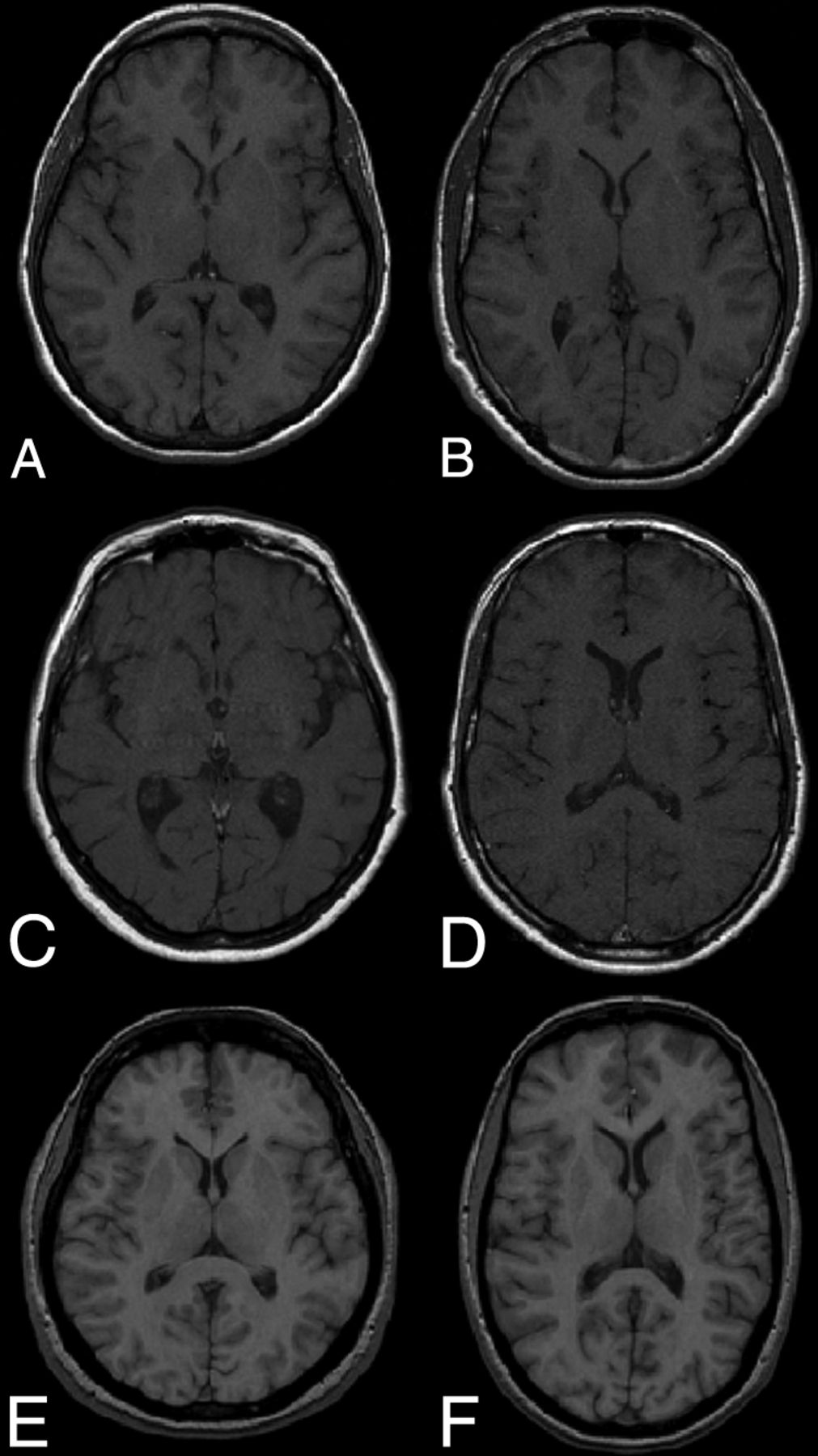
- Fig 1.
T1-weighted images from the 3 hospitals and scanners involved in the study: 1.5T Magnetom Symphony Quantum (Siemens) from H1 (first row), 1.5T Intera (R12) (Philips) from H2 (middle row), and 1.5T Signa HDxt (GE Healthcare) from H3 (last row).
- Fig 2.
Our pipeline approach. From the 30 T1-weighted scans of patients with MS, nonbrain parts are stripped and brain voxels are corrected for intensity inhomogeneities. From the same corrected set (original), a new set is generated by removing WML masks from scans before segmentation (masked). The scans of both sets are segmented into 1 of the 3 tissue classes (GM, WM, and CSF). Lesion voxels are added as WM after segmentation on masked images.
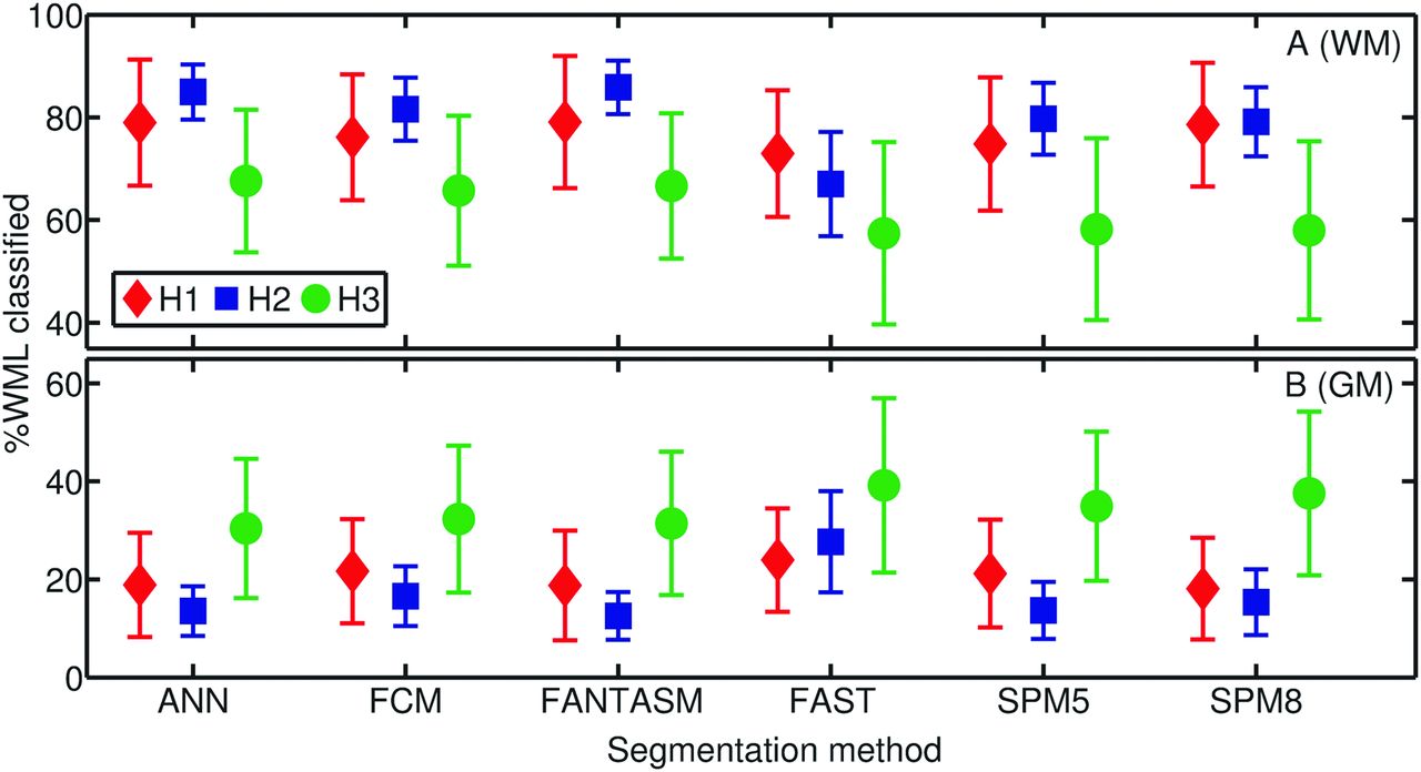
- Fig 3.
Percentage of voxels in WML regions having been classified as GM (top) and WM (bottom) for each segmentation method and hospital, H1 (♢), H2 (□) or H3 (○). Reported values are means and SDs.
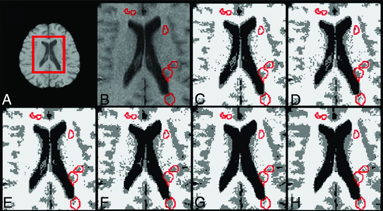
- Fig 4.
Classification output returned by each segmentation method on the same image. A, T1-weighted scan. B, Zoomed part of the scan with lesions outlined in red. Brain tissue segmentation outputs also with lesions outlined for ANN (C), FCM (D), FANTASM (E), FAST (F), SPM5 (G), and SPM8 (H). C–H, Segmented GM tissue is represented in gray; WM, in white; and CSF, in black.


Tables
- Table 1:
Average percentage of change in total tissue volume estimation between original and masked imagesa
Method H1 H2 H3 GM WM CSF GM WM CSF GM WM CSF ANN 0.33 ± 0.42 −0.23 ± 0.28 0.11 ± 0.11 1.59 ± 1.37 −0.56 ± 0.46 0.78 ± 0.76 0.25 ± 0.31 −0.16 ± 0.28 −0.09 ± 0.09 FCM 0.28 ± 0.37 −0.22 ± 0.29 0.09 ± 0.11 2.28 ± 2.26 −0.90 ± 0.83 0.94 ± 0.90 0.28 ± 0.23 −0.25 ± 0.20 0.08 ± 0.09 FANTASM 0.23 ± 0.26 −0.18 ± 0.21 0.08 ± 0.08 1.34 ± 1.13 −0.49 ± 0.37 0.80 ± 0.73 0.26 ± 0.22 −0.24 ± 0.19 0.07 ± 0.08 FAST 0.29 ± 0.36 −0.29 ± 0.36 0.12 ± 0.13 1.92 ± 1.59 −1.28 ± 1.03 0.47 ± 0.39 0.34 ± 0.28 −0.37 ± 0.31 0.12 ± 0.17 SPM5 0.20 ± 0.30 −0.21 ± 0.20 −0.14 ± 0.54 0.10 ± 2.68 −1.04 ± 3.01 0.53 ± 0.51 0.04 ± 0.17 −0.18 ± 0.36 0.15 ± 0.23 SPM8 0.08 ± 0.09 −0.08 ± 0.08 −0.04 ± 0.18 0.55 ± 0.34 −0.93 ± 0.55 0.54 ± 0.42 0.09 ± 0.15 −0.23 ± 0.25 0.17 ± 0.23 ↵a The results are divided by tissue and hospital. Reported values are the means ± SD. Positive values indicate a tissue overestimation on original images compared with masked.
- Table 2:
Pearson correlation coefficients between method differences in total volume estimation and WML sizea
Method GM WM CSF H1 ANN 0.94 −0.90 0.89 FCM 0.93 −0.89 0.83 FANTASM 0.87 −0.80 0.78 FAST 0.97 −0.97 0.96 SPM5 0.58b −0.89 −0.21b SPM8 0.92 −0.63 −0.69 H2 ANN 0.91 −0.88 0.93 FCM 0.92 −0.94 0.92 FANTASM 0.89 −0.87 0.84 FAST 0.95 −0.96 0.82 SPM5 −0.35b −0.06b 0.72 SPM8 0.76 −0.79 0.57b H3 ANN 0.56b −0.55b 0.88 FCM 0.77 −0.84 0.88 FANTASM 0.74 −0.82 0.85 FAST 0.88 −0.94 0.92 SPM5 −0.06b −0.03b 0.21b SPM8 0.56b −0.48b 0.09b - Table 3:
Average percentage change in the volume estimation of tissue outside the lesion regions between original and masked scansa
Method H1 H2 H3 GM WM CSF GM WM CSF GM WM CSF ANN 0.15 ± 0.26 −0.10 ± 0.18 0.07 ± 0.08 0.70 ± 0.61 −0.31 ± 0.24 0.67 ± 0.69 −0.01 ± 0.28 0.04 ± 0.24 −0.12 ± 0.08 FCM 0.09 ± 0.16 −0.07 ± 0.13 0.05 ± 0.08 1.27 ± 1.69 −0.56 ± 0.62 0.82 ± 0.81 0.01 ± 0.03 −0.03 ± 0.05 0.05 ± 0.07 FANTASM 0.06 ± 0.05 −0.05 ± 0.05 0.03 ± 0.05 0.48 ± 0.48 −0.25 ± 0.18 0.68 ± 0.63 0.00 ± 0.04 −0.02 ± 0.05 0.04 ± 0.07 FAST 0.08 ± 0.14 −0.09 ± 0.14 0.07 ± 0.08 0.56 ± 0.87 −0.45 ± 0.64 0.22 ± 0.33 0.02 ± 0.07 −0.06 ± 0.13 0.08 ± 0.16 SPM5 0.06 ± 0.25 0.02 ± 0.13 −0.19 ± 0.54 −0.29 ± 2.61 −0.47 ± 2.91 0.21 ± 0.32 −0.20 ± 0.24 0.23 ± 0.34 0.06 ± 0.15 SPM8 −0.03 ± 0.06 0.09 ± 0.15 −0.10 ± 0.23 0.13 ± 0.30 −0.29 ± 0.33 0.25 ± 0.26 −0.15 ± 0.12 0.14 ± 0.15 0.10 ± 0.20 ↵a The results are divided by hospital and tissue. Reported values are the means ± SD. Positive values indicate a tissue overestimation on original images compared with masked.
- Table 4:
Pearson correlation coefficients among method differences in volume estimation of tissue outside the lesion regions and WML sizea
Method GM WM CSF H1 ANN 0.77 −0.74 0.83 FCM 0.82 −0.80 0.71 FANTASM 0.80 −0.73 0.66 FAST 0.86 −0.93 0.97 SPM5 0.11 0.51b −0.30b SPM8 −0.57b 0.95 −0.77 H2 ANN 0.85 −0.92 0.93 FCM 0.71 −0.84 0.94 FANTASM 0.66 −0.82 0.87 FAST 0.33b −0.46b 0.62b SPM5 −0.43b 0.18b 0.65b SPM8 0.16b −0.37b 0.30b H3 ANN 0.07 −0.16b 0.79 FCM 0.50 −0.77 0.89 FANTASM 0.17 −0.57b 0.87 FAST 0.45 −0.73 0.89 SPM5 −0.78b 0.72 0.14b SPM8 −0.64b 0.72 −0.01b


Jump to section
Related Articles
Cited By...
- No citing articles found.