
Article Figures & Data
Figures
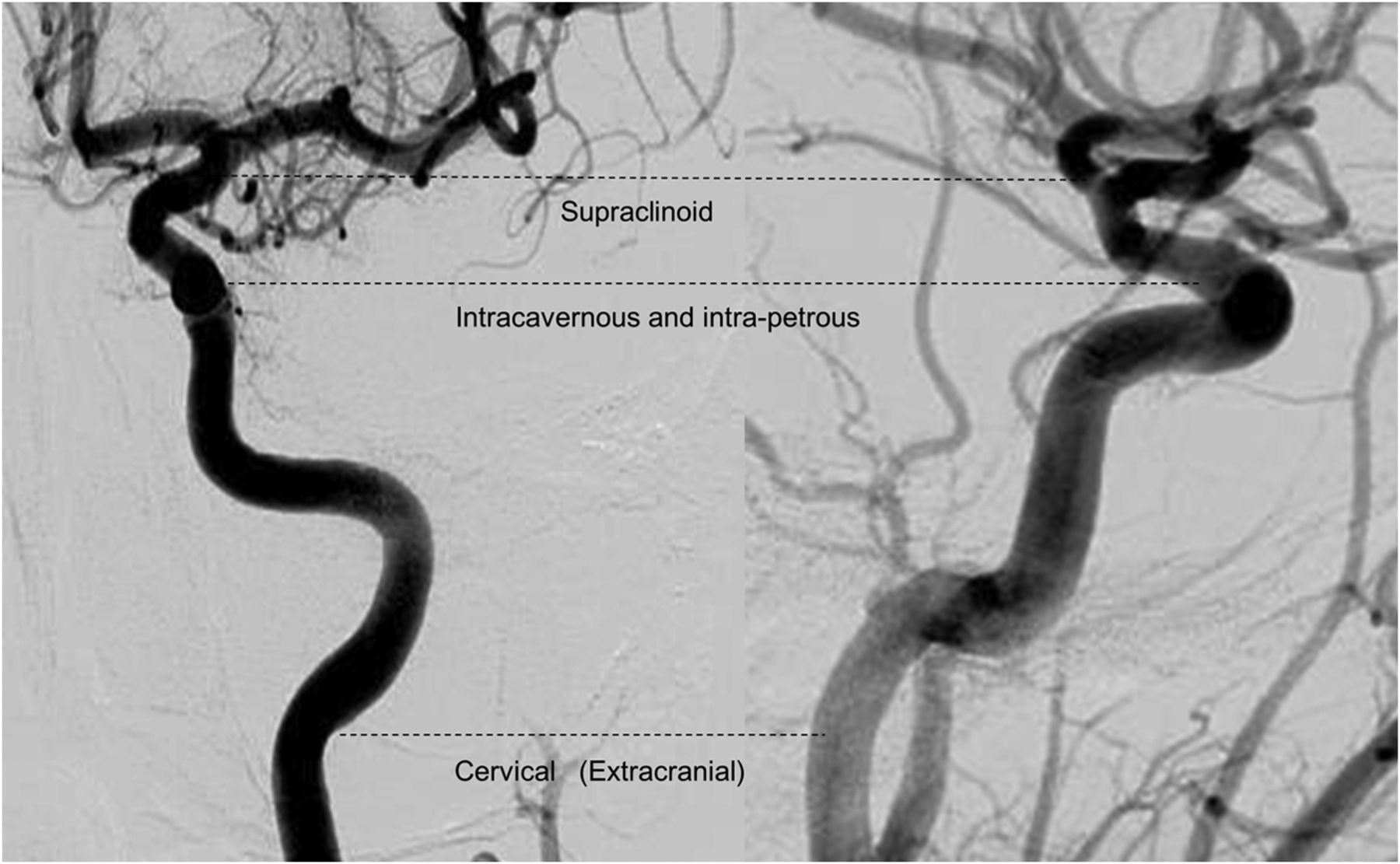
- Fig 1.
ICA segmentation used for the occlusion-level definition derived from Gibo et al, 1981.31 The internal carotid artery is divided into supraclinoid ICA, intracavernous and intrapetrous ICA, and cervical/extracranial ICA.
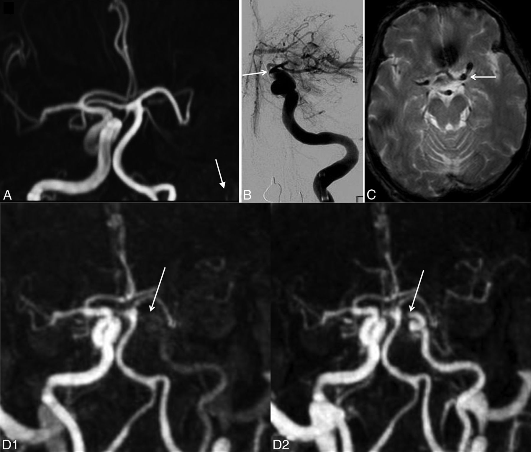
- Fig 2.
In patient 11, agreement between TR-CE MRA and DSA and discordance with TOF. A, 3D-TOF frontal MIP image shows complete occlusion of the left ICA from the IICA to distal segments of the left MCA. No data were available on the patency of the ICA proximal to the IICA segment (white arrow). B, Frontal DSA shows opacification of the left ICA up to the supraclinoid segment (white arrow). C, GRE T2 axial image displays a susceptibility vessel sign in the M1 segment of the left MCA. D, Frontal MIP TR-CE MRA shows patency of the left ICA up to the SCICA in the last phases (white arrows).
- Fig 3.
In patient 1, disagreement between TR-CE MRA and DSA and discordance with TOF. A, 3D-TOF frontal MIP image shows complete occlusion of the right ICA from the IICA to distal segments of the left MCA, with a doubt about the patency of the ICA proximal to the IICA segment (white arrow). B, Frontal DSA shows opacification of the right ICA up to the M1 segment (white arrow). C, Frontal MIP TR-CE MRA shows patency of the right ICA up to the SCICA in the last phases (white arrows).
- Fig 4.
Examples of occlusion and stenosis of the extracranial arteries.TR-CE MRA and its corresponding frontal DSA image. In patient 7 (A and B), occlusion of the left ICA, probably of atheromatous etiology (white arrow) on TR-CE MRA (A) and DSA (B). In patient 24 (C and D), severe stenosis (>50%) of the right ICA, on TR-CE MRA (C) and DSA (D), presumed due to an arterial dissection (white arrow).


Tables
TR-CE MRA 3D-TOF GRE T2 Receive coil Body coil 8-Channel brain coil 8-Channel brain coil TR/TE (ms) 4.0/1.25 23/3.5 757/16.1 Flip angle 25° 15° 18° Acquisition plane Coronal Axial Axial FOV (mm) 320 × 320 200 × 200 230 × 230 Acquisition matrix 268 × 267 312 × 206 512 × 512 Section thickness (after interpolation) 2.6 (1.3) 1.6 (0.8) 5 Acquired voxel size (mm) 1.2 × 1.2 × 2.6 0.64 × 0.97 × 1.6 0.45 × 0.45 × 5 Reconstructed voxel size (mm) 0.8 × 0.8 × 1.3 0.39 × 0.39 × 0.8 No. of sections 77 87 24 Anteroposterior coverage (mm) 100 k-t BLAST undersampling factor 4 – – No. of dynamics 12 1 – Phase acquisition times 8.7 sec – – Total acquisition times 1 minute 44 seconds 2 minutes 36 seconds 1 minute 37 seconds - Table 2:
Identification of intracranial occlusion location with 3D-TOF, GRE T2, and TR-CE MRA, compared with DSA as the standard referencea
Occlusion Location DSA (No.) (%) 3D-TOF MRA (No.) (%) GRE T2 (No.) (%) TR-CE MRA (No.) (%) No occlusion (no susceptibility vessel sign for GRE T2) 0 (0%) 0 (0%) 6 (17.6%) 0 (0%) ExCICA 3 (8.8%) 5 (14.7%) 0 (0%) 3 (8.8%) IICA 0 (0%) 5 (14.7%) 0 (0%) 0 (0%) SCICA 8 (23.5%) 3 (8.8%) 3 (8.8%) 9 (26.5%) M1 17 (50.0%) 20 (58.8%) 22 (64.7%) 19 (55.9%) M2 6 (17.6%) 1 (2.9%) 2 (5.9%) 3 (8.8%) κ 0.43 0.31 0.81 95% CI 0.26−0.60 0.15−0.48 0.60−1 (% of agreement) 61.8% 55.9% 88.2% ↵a Intracranial occlusion location with 3D-TOF, GRE T2, and TR-CE MRA, compared with DSA as the standard reference, showing the occlusion level detected as well as κ and percentage of agreement with DSA for each MRI sequence.


