Advertisement

Main menu

  • Home
  • Content
    • Current Issue
    • Accepted Manuscripts
    • Article Preview
    • Past Issue Archive
    • Video Articles
    • AJNR Case Collection
    • Case of the Week Archive
    • Case of the Month Archive
    • Classic Case Archive
  • Special Collections
    • AJNR Awards
    • Low-Field MRI
    • Alzheimer Disease
    • ASNR Foundation Special Collection
    • Photon-Counting CT
    • View All
  • Multimedia
    • AJNR Podcasts
    • AJNR SCANtastic
    • Trainee Corner
    • MRI Safety Corner
    • Imaging Protocols
  • For Authors
    • Submit a Manuscript
    • Submit a Video Article
    • Submit an eLetter to the Editor/Response
    • Manuscript Submission Guidelines
    • Statistical Tips
    • Fast Publishing of Accepted Manuscripts
    • Graphical Abstract Preparation
    • Imaging Protocol Submission
    • Author Policies
  • About Us
    • About AJNR
    • Editorial Board
    • Editorial Board Alumni
  • More
    • Become a Reviewer/Academy of Reviewers
    • Subscribers
    • Permissions
    • Alerts
    • Feedback
    • Advertisers
    • ASNR Home

User menu

  • Alerts
  • Log in

Search

  • Advanced search
American Journal of Neuroradiology

American Journal of Neuroradiology

ASHNR American Society of Functional Neuroradiology ASHNR American Society of Pediatric Neuroradiology ASSR
  • Alerts
  • Log in

Advanced Search

  • Home
  • Content
    • Current Issue
    • Accepted Manuscripts
    • Article Preview
    • Past Issue Archive
    • Video Articles
    • AJNR Case Collection
    • Case of the Week Archive
    • Case of the Month Archive
    • Classic Case Archive
  • Special Collections
    • AJNR Awards
    • Low-Field MRI
    • Alzheimer Disease
    • ASNR Foundation Special Collection
    • Photon-Counting CT
    • View All
  • Multimedia
    • AJNR Podcasts
    • AJNR SCANtastic
    • Trainee Corner
    • MRI Safety Corner
    • Imaging Protocols
  • For Authors
    • Submit a Manuscript
    • Submit a Video Article
    • Submit an eLetter to the Editor/Response
    • Manuscript Submission Guidelines
    • Statistical Tips
    • Fast Publishing of Accepted Manuscripts
    • Graphical Abstract Preparation
    • Imaging Protocol Submission
    • Author Policies
  • About Us
    • About AJNR
    • Editorial Board
    • Editorial Board Alumni
  • More
    • Become a Reviewer/Academy of Reviewers
    • Subscribers
    • Permissions
    • Alerts
    • Feedback
    • Advertisers
    • ASNR Home
  • Follow AJNR on Twitter
  • Visit AJNR on Facebook
  • Follow AJNR on Instagram
  • Join AJNR on LinkedIn
  • RSS Feeds

AJNR Awards, New Junior Editors, and more. Read the latest AJNR updates

Research ArticleSpine

Validation of Multisociety Combined Task Force Definitions of Abnormal Disk Morphology

C.H. Cho, L. Hsu, M.L. Ferrone, D.A. Leonard, M.B. Harris, A.A. Zamani and C.M. Bono
American Journal of Neuroradiology May 2015, 36 (5) 1008-1013; DOI: https://doi.org/10.3174/ajnr.A4212
C.H. Cho
aFrom the Departments of Radiology (C.H.C., L.H., A.A.Z.)
  • Find this author on Google Scholar
  • Find this author on PubMed
  • Search for this author on this site
L. Hsu
aFrom the Departments of Radiology (C.H.C., L.H., A.A.Z.)
  • Find this author on Google Scholar
  • Find this author on PubMed
  • Search for this author on this site
M.L. Ferrone
bOrthopedic Surgery (M.L.F., D.A.L., M.B.H., C.M.B.), Brigham and Women's Hospital, Harvard Medical School, Boston, Massachusetts.
  • Find this author on Google Scholar
  • Find this author on PubMed
  • Search for this author on this site
D.A. Leonard
bOrthopedic Surgery (M.L.F., D.A.L., M.B.H., C.M.B.), Brigham and Women's Hospital, Harvard Medical School, Boston, Massachusetts.
  • Find this author on Google Scholar
  • Find this author on PubMed
  • Search for this author on this site
M.B. Harris
bOrthopedic Surgery (M.L.F., D.A.L., M.B.H., C.M.B.), Brigham and Women's Hospital, Harvard Medical School, Boston, Massachusetts.
  • Find this author on Google Scholar
  • Find this author on PubMed
  • Search for this author on this site
A.A. Zamani
aFrom the Departments of Radiology (C.H.C., L.H., A.A.Z.)
  • Find this author on Google Scholar
  • Find this author on PubMed
  • Search for this author on this site
C.M. Bono
bOrthopedic Surgery (M.L.F., D.A.L., M.B.H., C.M.B.), Brigham and Women's Hospital, Harvard Medical School, Boston, Massachusetts.
  • Find this author on Google Scholar
  • Find this author on PubMed
  • Search for this author on this site
  • Article
  • Figures & Data
  • Supplemental
  • Info & Metrics
  • Responses
  • References
  • PDF
Loading

Article Figures & Data

Figures

  • Tables
  • Fig 1.
    • Download figure
    • Open in new tab
    • Download powerpoint
    Fig 1.

    Case 2. Sagittal T2-weighted image shows a thin connection (arrow) between the level of abnormal disk and the disk fragment. With imaging, this is classified as extrusion. However, it was classified intraoperatively as extrusion and sequestration.

  • Fig 2.
    • Download figure
    • Open in new tab
    • Download powerpoint
    Fig 2.

    Case 4. Sagittal T2-weighted image shows the height of the abnormal disk herniation (bold arrows) compared with the disk height (thin arrows). By imaging, this is classified as extrusion. However, it was classified intraoperatively as protrusion.
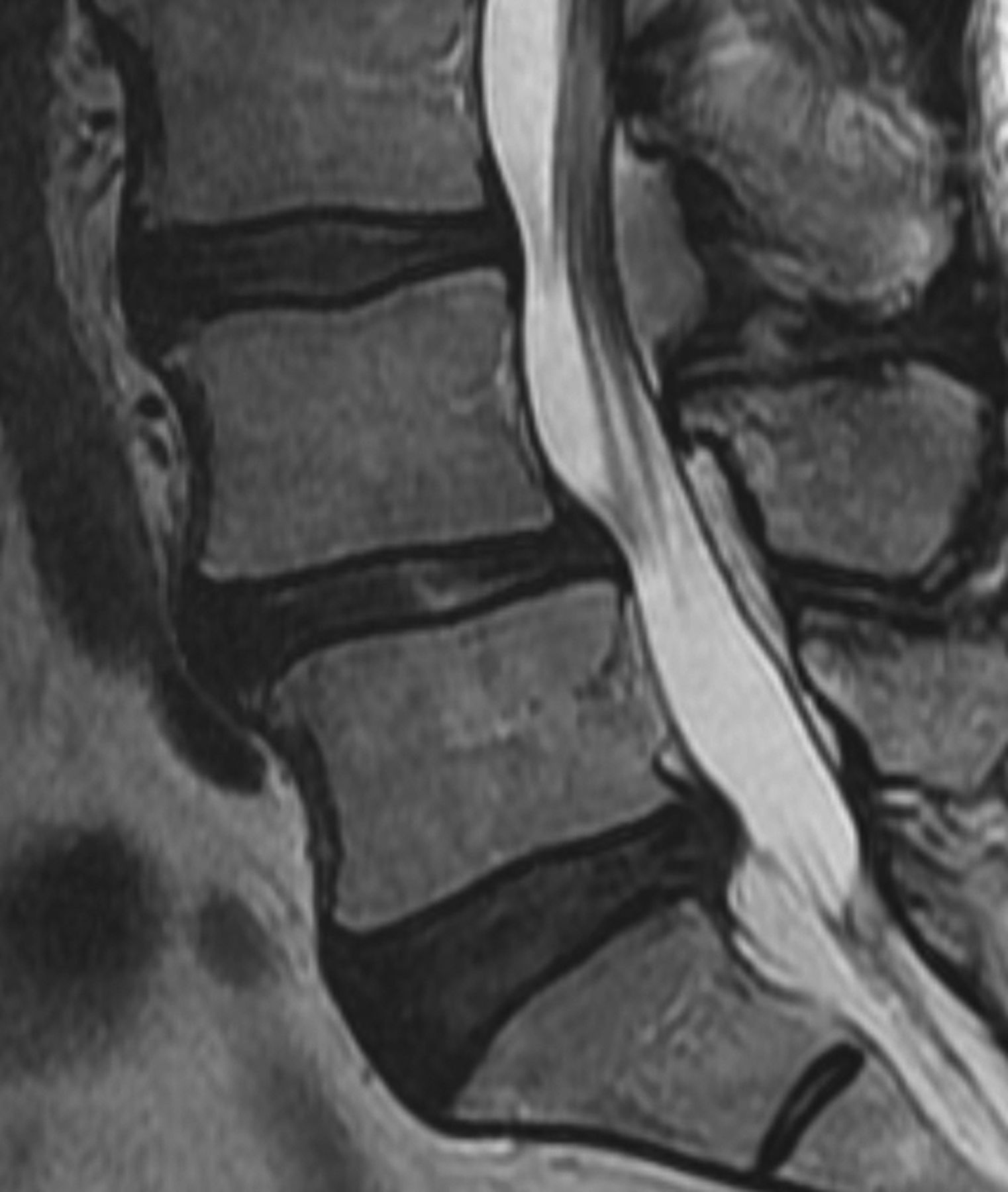
  • Fig 3.
    • Download figure
    • Open in new tab
    • Download powerpoint
    Fig 3.

    Case 9. Sagittal T2-weighted image shows decreased signal of the disk material, which makes differentiation of the disk from the annular margin difficult.

  • Fig 4.
    • Download figure
    • Open in new tab
    • Download powerpoint
    Fig 4.

    Case 15. Sagittal T2-weighted image shows disruption of outer annulus (arrow) even on retrospective interpretation; thus, the imaging classification in this case is extrusion. However, it was classified intraoperatively as a protrusion.

Tables

  • Figures
    • View popup
    Table 1:

    Discrepancy cases between imaging and surgery, with a description of the imaging findings

    No.Disk LevelImagingSurgicalDescription
    14/5ExtrusionProtrusionOn sagittal images, the height of the abnormal disk is slightly greater than the disk height
    22/3ExtrusionExtrusion and sequestrationAlthough imaging suggests a sequestered fragment, there is a thin continuity between the disk fragments visible on MR imaging (Fig 1)
    34/5ExtrusionProtrusionOn sagittal images, the height of the abnormal disk is slightly greater and very close in distance to the maximum disk height
    44/5ExtrusionProtrusionOn sagittal images, the height of the abnormal disk is very close in distance to the maximum disk height (Fig 2)
    54/5ExtrusionExtrusion and sequestrationSagittal images show a connection between disk fragments
    64/5ExtrusionProtrusionOn sagittal images, the abnormal disk appears contained by the outer annulus, but the height of the annulus is slightly greater than the disk height
    74/5ExtrusionProtrusionOn sagittal images, the height of the abnormal disk is slightly greater than the disk height
    85/1ExtrusionProtrusionOn sagittal images, the height of the abnormal disk is slightly greater than the disk height
    95/1ProtrusionExtrusionOn the sagittal view, the low signal of the disk and annulus is very difficult to differentiate (Fig 3)
    104/5ExtrusionProtrusionOn sagittal images, the height of the abnormal disk is very similar to the disk height
    114/5ProtrusionExtrusionOn the sagittal view, the low signal of the disk and annulus is very difficult to differentiate
    124/5ProtrusionExtrusionOn sagittal images, the entire annulus is not clearly defined
    135/1ProtrusionExtrusionOn sagittal images, the abnormal disk height is similar to the maximum disk height
    144/5ExtrusionProtrusionOn sagittal images, the abnormal disk height is slightly greater than the maximum disk height
    155/1ExtrusionProtrusionOn the sagittal view, abnormal disk height is much greater than the normal disk height and the outer annulus appears disrupted; in retrospect, the diagnosis is still extrusion by imaging criteria (Fig 4)
    164/5ExtrusionSequestrationImaging shows a thin connection between the parent disk and disk the fragment
    • View popup
    Table 2:

    2 × 2 contingency table for a ruptured-versus-intact annulus as determined surgically and on imaging

    Imaging AssessmentSurgical AssessmentTotals
    (Criterion Standard)
    Annulus RupturedaIntact Annulusb
    Annulus ruptureda37941
    Intact annulusb4413
    Totals46854
    • ↵a Extrusions and sequestrations.

    • ↵b Protrusions only.

PreviousNext
Back to top

In this issue

American Journal of Neuroradiology: 36 (5)
American Journal of Neuroradiology
Vol. 36, Issue 5
1 May 2015
  • Table of Contents
  • Index by author
  • Complete Issue (PDF)
Advertisement
Print
Download PDF
Email Article

Thank you for your interest in spreading the word on American Journal of Neuroradiology.

NOTE: We only request your email address so that the person you are recommending the page to knows that you wanted them to see it, and that it is not junk mail. We do not capture any email address.

Enter multiple addresses on separate lines or separate them with commas.
Validation of Multisociety Combined Task Force Definitions of Abnormal Disk Morphology
(Your Name) has sent you a message from American Journal of Neuroradiology
(Your Name) thought you would like to see the American Journal of Neuroradiology web site.
CAPTCHA
This question is for testing whether or not you are a human visitor and to prevent automated spam submissions.
Cite this article
C.H. Cho, L. Hsu, M.L. Ferrone, D.A. Leonard, M.B. Harris, A.A. Zamani, C.M. Bono
Validation of Multisociety Combined Task Force Definitions of Abnormal Disk Morphology
American Journal of Neuroradiology May 2015, 36 (5) 1008-1013; DOI: 10.3174/ajnr.A4212

Citation Manager Formats

  • BibTeX
  • Bookends
  • EasyBib
  • EndNote (tagged)
  • EndNote 8 (xml)
  • Medlars
  • Mendeley
  • Papers
  • RefWorks Tagged
  • Ref Manager
  • RIS
  • Zotero
0 Responses
Respond to this article
Share
Bookmark this article
Validation of Multisociety Combined Task Force Definitions of Abnormal Disk Morphology
C.H. Cho, L. Hsu, M.L. Ferrone, D.A. Leonard, M.B. Harris, A.A. Zamani, C.M. Bono
American Journal of Neuroradiology May 2015, 36 (5) 1008-1013; DOI: 10.3174/ajnr.A4212
del.icio.us logo Twitter logo Facebook logo Mendeley logo
  • Tweet Widget
  • Facebook Like
  • Google Plus One
Purchase

Jump to section

  • Article
    • Abstract
    • Materials and Methods
    • Results
    • Discussion
    • Conclusions
    • Footnotes
    • REFERENCES
  • Figures & Data
  • Supplemental
  • Info & Metrics
  • Responses
  • References
  • PDF

Related Articles

  • PubMed
  • Google Scholar

Cited By...

  • No citing articles found.
  • Crossref
  • Google Scholar

This article has not yet been cited by articles in journals that are participating in Crossref Cited-by Linking.

More in this TOC Section

  • Bern Score Validity for SIH
  • MP2RAGE 7T in MS Lesions of the Cervical Spine
  • Resisted Inspiration for CSF-Venous Fistula
Show more Spine

Similar Articles

Advertisement

Indexed Content

  • Current Issue
  • Accepted Manuscripts
  • Article Preview
  • Past Issues
  • Editorials
  • Editor's Choice
  • Fellows' Journal Club
  • Letters to the Editor
  • Video Articles

Cases

  • Case Collection
  • Archive - Case of the Week
  • Archive - Case of the Month
  • Archive - Classic Case

More from AJNR

  • Trainee Corner
  • Imaging Protocols
  • MRI Safety Corner
  • Book Reviews

Multimedia

  • AJNR Podcasts
  • AJNR Scantastics

Resources

  • Turnaround Time
  • Submit a Manuscript
  • Submit a Video Article
  • Submit an eLetter to the Editor/Response
  • Manuscript Submission Guidelines
  • Statistical Tips
  • Fast Publishing of Accepted Manuscripts
  • Graphical Abstract Preparation
  • Imaging Protocol Submission
  • Evidence-Based Medicine Level Guide
  • Publishing Checklists
  • Author Policies
  • Become a Reviewer/Academy of Reviewers
  • News and Updates

About Us

  • About AJNR
  • Editorial Board
  • Editorial Board Alumni
  • Alerts
  • Permissions
  • Not an AJNR Subscriber? Join Now
  • Advertise with Us
  • Librarian Resources
  • Feedback
  • Terms and Conditions
  • AJNR Editorial Board Alumni

American Society of Neuroradiology

  • Not an ASNR Member? Join Now

© 2025 by the American Society of Neuroradiology All rights, including for text and data mining, AI training, and similar technologies, are reserved.
Print ISSN: 0195-6108 Online ISSN: 1936-959X

Powered by HighWire