
Article Figures & Data
Figures
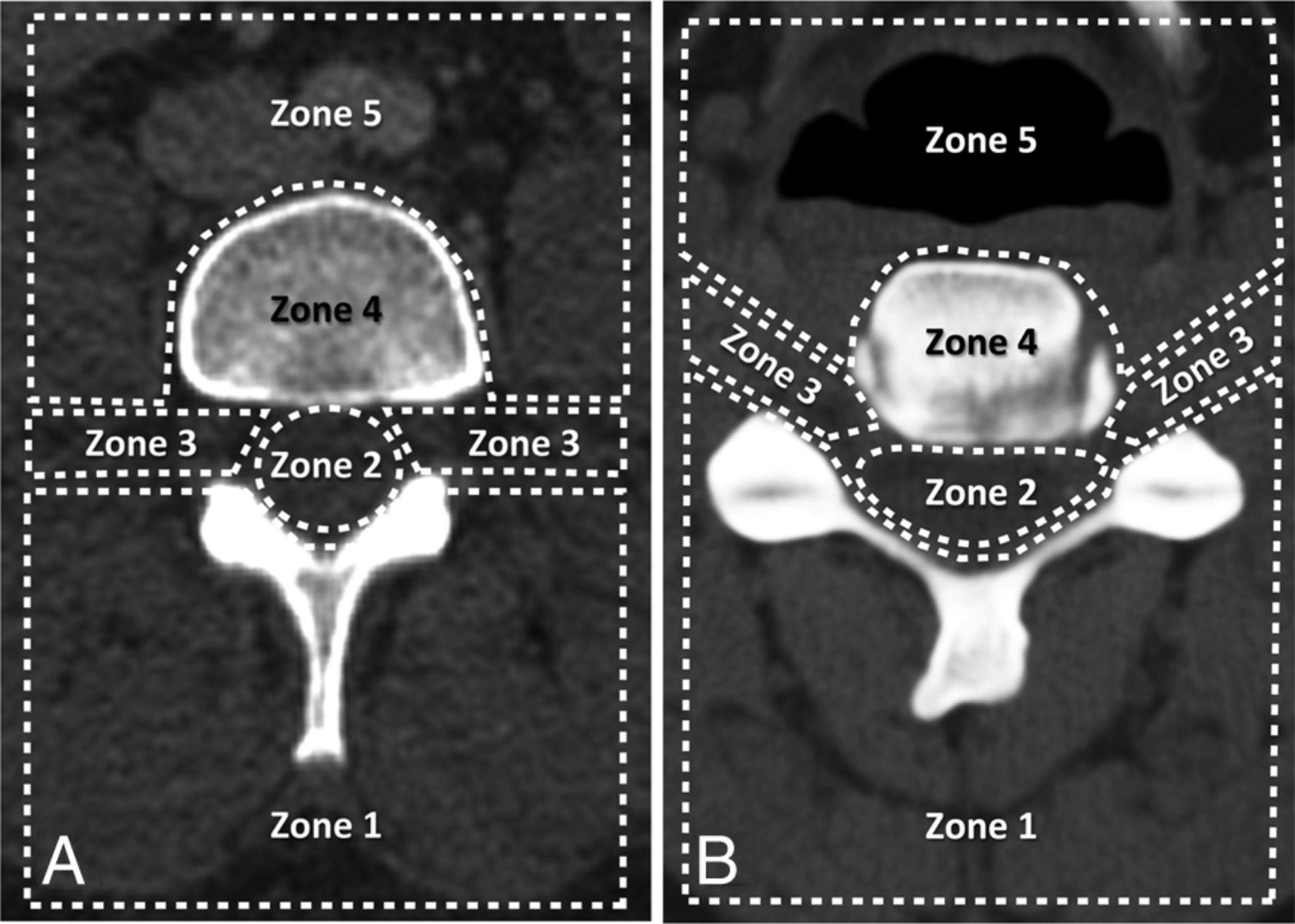
- Fig 1.
Classification scheme for the location of visualized vessels seen during inadvertent intravascular injection in the lumbar (A) and cervical (B) spine. Regions include the following: posterior paraspinal soft tissues (zone 1), spinal canal (zone 2), foraminal region (zone 3), vertebral body (zone 4), and anterior paraspinal soft tissues (zone 5).
- Fig 2.
Intravascular injection during lumbar TFESI. Preinjection (A), immediate postinjection (B), and delayed postinjection (C) images demonstrate a vessel in the left foraminal zone (arrow) that washes out on the delayed image.
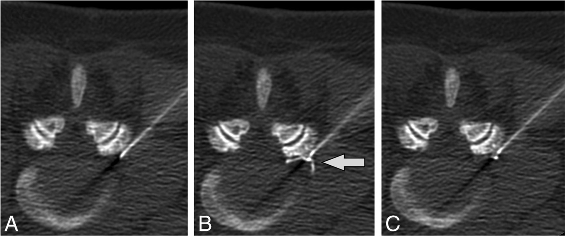
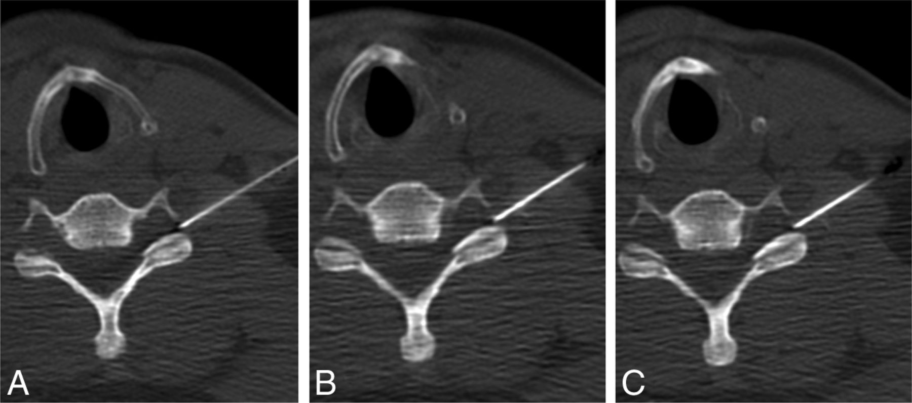
- Fig 3.
A case of intravascular injection is demonstrated on preinjection (A), immediate postinjection (B), first delayed (C) images, and an additional delayed (D) image. On the immediate postinjection image, contrast is seen in the ascending lumbar vein (arrow) and the inferior vena cava (arrowhead). There is washout of contrast from the inferior vena cava on the first delayed image and from the ascending lumbar vein on the second delayed image.
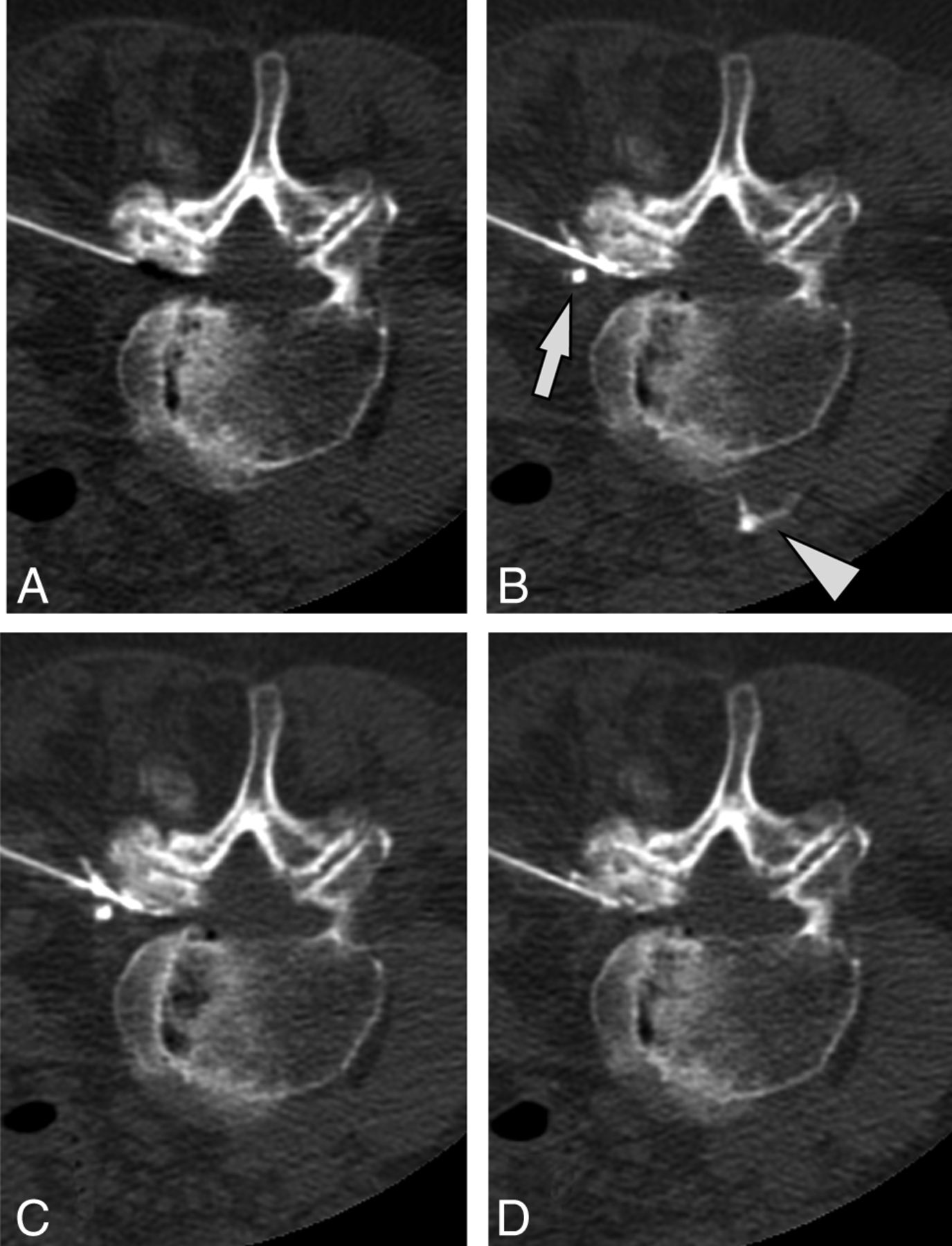
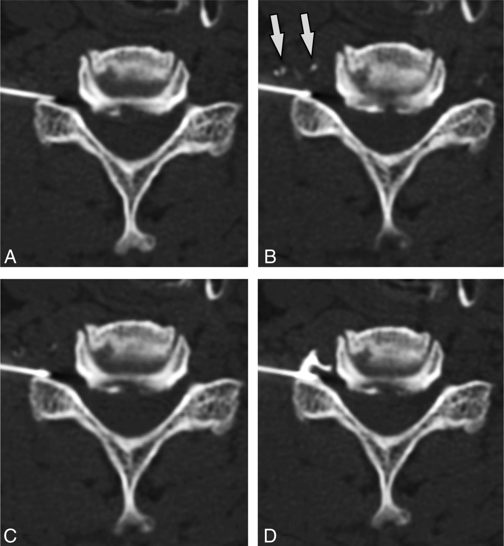
- Fig 4.
Intravascular contrast seen both close to and more remote from the needle tip. Preinjection (A), immediate postinjection (B), and delayed postinjection (C) images show intravascular contrast at the needle tip (arrowhead) and further away (arrows) in the sacral epidural venous plexus. Note that these vessels are not seen on the delayed washout image. No epidural contrast is seen.
- Fig 5.
Immediate contrast washout. Preinjection (A), immediate postinjection (B), and delayed postinjection (C) images obtained after injection of 1.0-mL contrast. Neither vascular nor epidural contrast is seen. A preceding injection with 0.2-mL contrast showed similar negative findings, and images obtained cranial and caudal to the needle tip showed no contrast (images not shown).
- Fig 6.
Delayed contrast washout pattern. Preinjection (A), immediate postinjection (B), and delayed postinjection (C) images demonstrate intravascular contrast (arrows) that progressively washes out on delayed images. After needle repositioning (D), re-injection of contrast shows only epidural contrast spread.
- Fig 7.
Intravascular injection during cervical ILESI. Preinjection (A), immediate postinjection (B), and delayed postinjection (C) images demonstrate a vessel in the left neural foramen (arrowhead) that washes out on the delayed image.
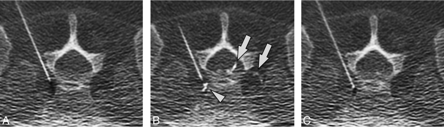
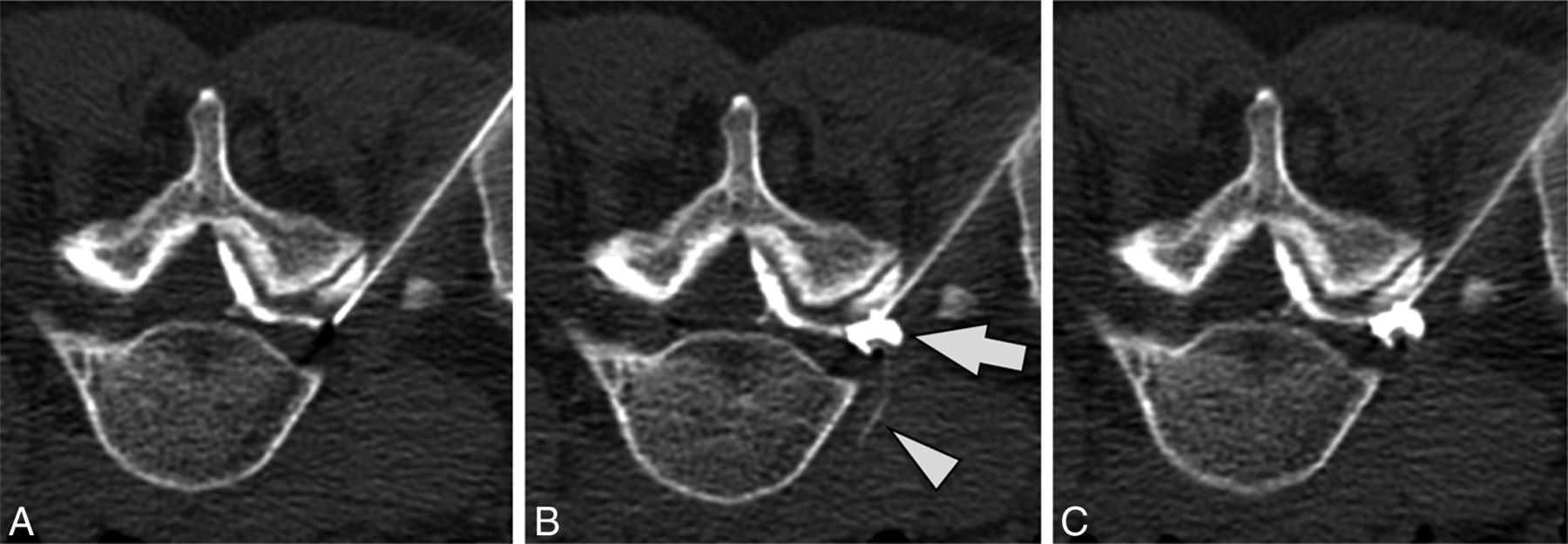
- Fig 8.
Simultaneous epidural and intravascular injection. Preinjection (A), immediate postinjection (B), and delayed postinjection (C) images demonstrate a vessel extending into the anterior paraspinal soft tissues (arrowhead) that washes out on the delayed image. Epidural contrast in the neural foramen (arrow) persists on the delayed image. Note that there is contrast in the epidural space of the spinal canal on the preinjection image due to prior injection at an adjacent level.


Tables
Score Vessel Suggestive Features 5 Definite vein Flow into a specific, anatomically identifiable venous structure 4 Probable vein Flow into region of known venous structure, flow predominantly away from spinal canal, delayed washout 3 Indeterminate Not meeting criteria for other categories 2 Probable artery Flow into region of known arterial structure, flow predominantly toward the spinal canal, rapid washout 1 Definite artery Flow into a specific, anatomically identifiable arterial structure Injection Type Anatomic Zone of Identified Vessel No. of Injections 1 2 3 4 5 0 Lumbar ILESI 25% 100% 25% 0% 0% 0% 4 Lumbar TFESI 14% 18% 86% 5% 36% 0% 22 Cervical ILESI 0% 100% 50% 0% 50% 0% 4 Cervical TFESI 29% 0% 71% 0% 14% 14% 7 ↵a Percentages across each injection type may sum to >100% because vessels may be visualized in >1 location simultaneously.
n % Distance from needle tip to vessel <1 cm only 11 30% >1 cm only 2 5% Both <1 cm and >1 cm 23 62% Immediate washout (vessel not seen) 1 3% Combined epidural injection Vascular only 25 68% Epidural + vascular 12 32% Washout pattern None 0 0% Delayed 32 86% Immediate 5 14% Vessel type Definite venous (n = 5) 4 11% Probable venous (n = 4) 27 73% Indeterminate (n = 3) 4 11% Probable arterial (n = 2) 2 5% Definite arterial (n = 1) 0 0%


Jump to section
Related Articles
Cited By...
- Hypersensitivity Reactions to Fibrin Glue during Epidural Blood Patching
- CT Fluoroscopy-Guided Blood Patching of Ventral CSF Leaks by Direct Needle Placement in the Ventral Epidural Space Using a Transforaminal Approach
- CT-Fluoroscopic Cervical Transforaminal Epidural Steroid Injections: Extraforaminal Needle Tip Position Decreases Risk of Intravascular Injection