
Article Figures & Data
Figures
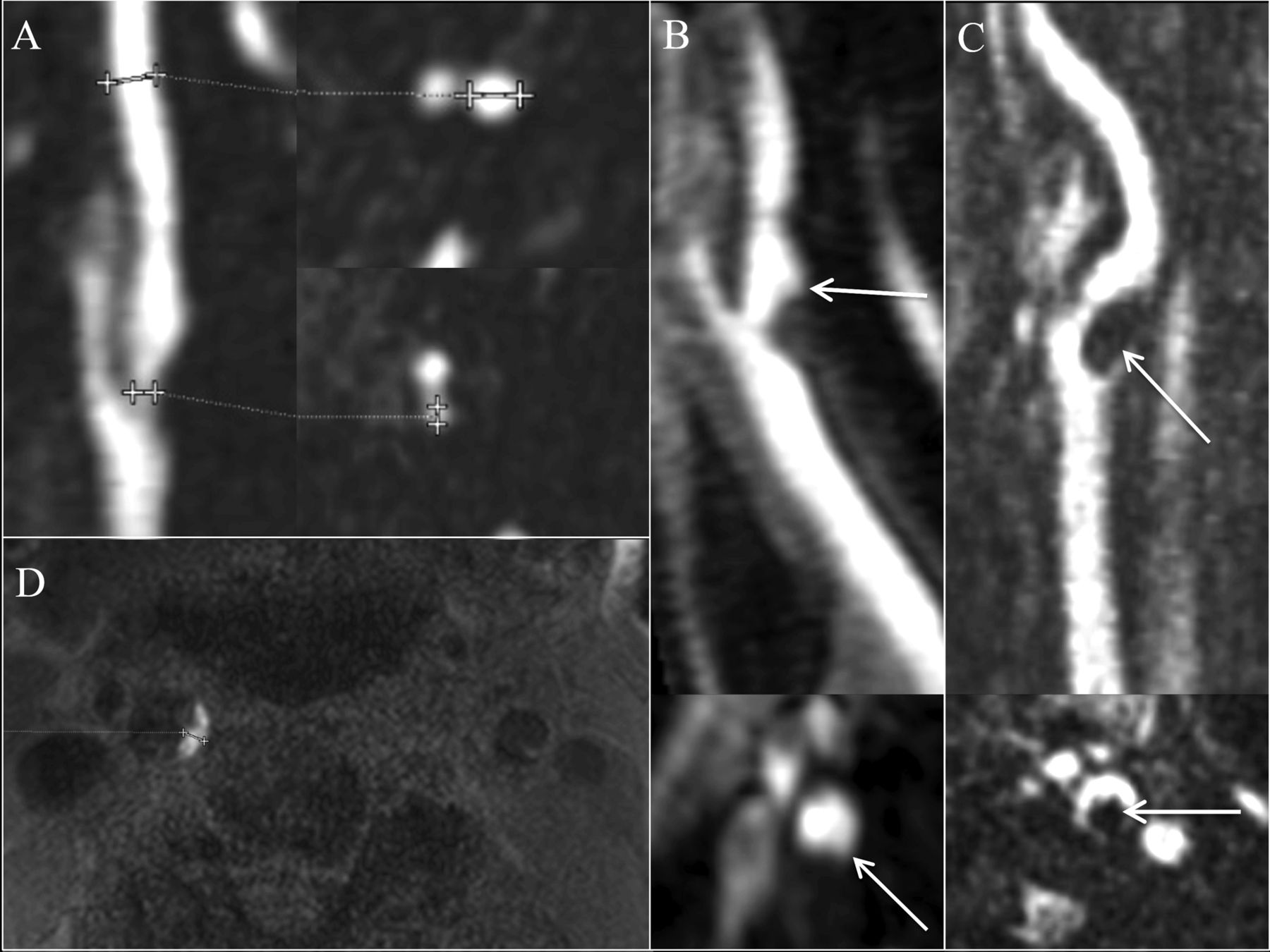
- Fig 1.
Carotid plaque imaging markers. Stenosis is measured by using percentage diameter stenosis [(a − b) / a] and millimeter stenosis (b) (A, cursors). The presence of ulceration is determined on contrast MRA images by using a 2-mm measurement threshold (B, arrow). Intraluminal thrombus is defined as a filling defect on contrast MRA images (C, arrow). IPH is defined by MPRAGE-positive plaque, by using a signal threshold of 2-fold signal intensity over the adjacent sternocleidomastoid muscle (D, right carotid artery is MPRAGE-positive; left side of image). Maximum plaque thickness is measured in the transverse plane on 3D MPRAGE image (D, cursors).
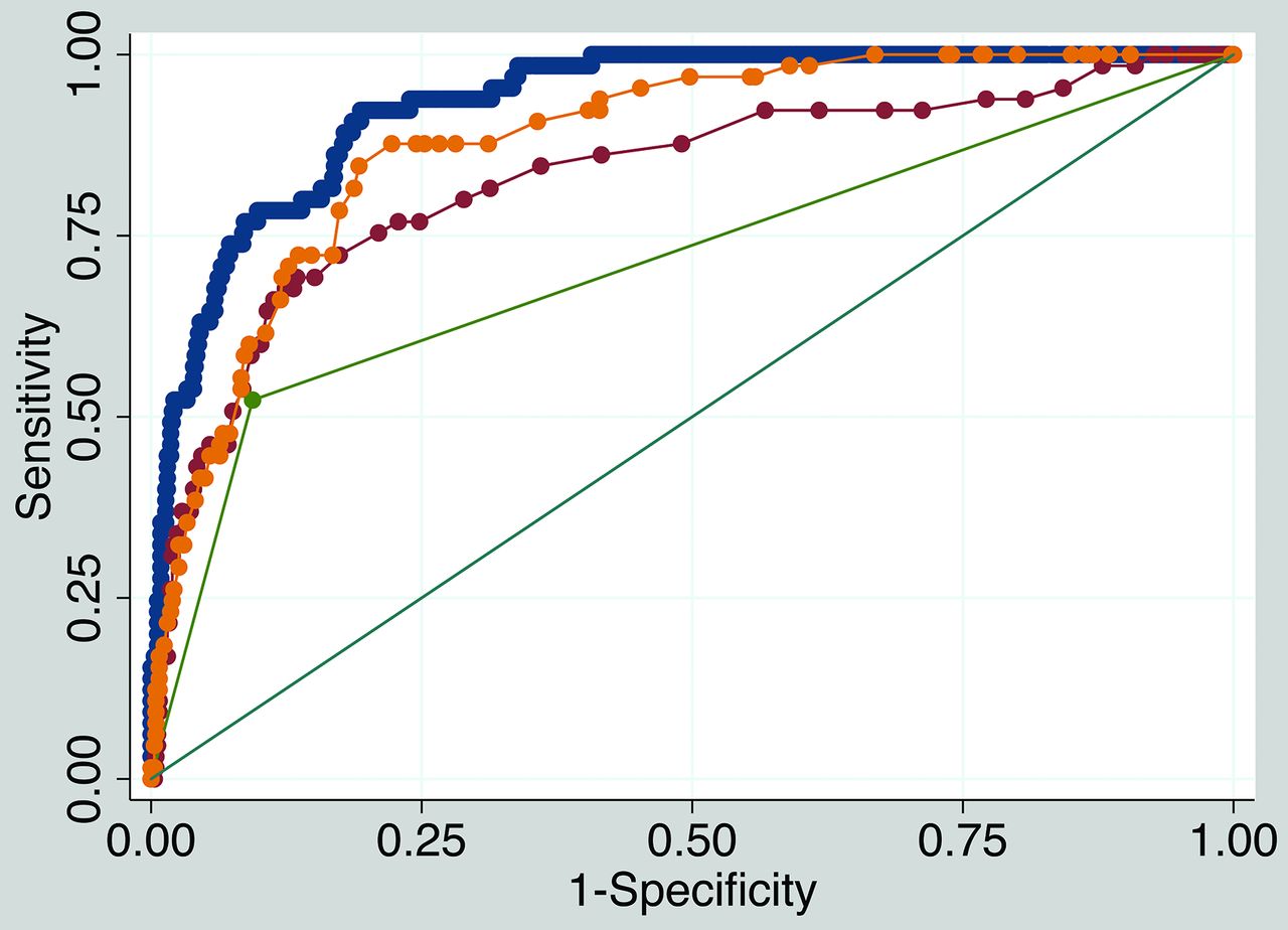
- Fig 2.
Receiver operating characteristic comparison analysis. The final model IPH discriminatory value is excellent (blue, AUC = 0.932). The final model discriminatory value (blue) is significantly higher than a model with maximum plaque thickness only (yellow, AUC = 0.881, P < .001), a model with millimeter stenosis only (red, AUC = 0.830, P < .001), and a model with ulceration only (green, AUC = 0.715, P < .001).


Tables
Carotid Stroke Predictor Demographics by Vessel Male sex (No./total No.) (%) 387/726 (53.3) Age (yr) (mean) (SD) 64.2 (15.6) BMI (mean) (SD) (kg/m2) 28.4 (6.4) Smoking (No./total No.) (%) Current smoker 138/726 (19.0) Prior smoker 158/726 (21.8) Never smoked 430/726 (59.2) Hypertension (No./total No.) (%) 499/726 (68.7) Hyperlipidemia (No./total No.) (%) 358/726 (49.3) Diabetes (No./total No.) (%) 227/726 (31.3) Cardiovascular medications Antihypertension (No./total No.) (%) 412/726 (56.8) Statins (No./total No.) (%) 316/726 (43.5) Antiplatelet (No./total No.) (%) 294/726 (40.5) Anticoagulation (No./total No.) (%) 74/726 (10.2) Carotid plaque imaging markers Stenosis (mean) (SD) (%) 12.2 (23.1) Mild stenosis (0%–49%) (No./total No.) (%) 647/726 (89.1) Moderate stenosis (50%–69%) (No./total No.) (%) 45/726 (6.2) Severe stenosis (70%–99%) (No./total No.) (%) 34/726 (4.7) Stenosis (mean) (SD) (mm) 4.1 (1.2) Maximum plaque thickness (mean) (SD) (mm) 3.0 (1.6) Ulceration (No./total No.) (%) 96/726 (13.2) Intraluminal thrombus (No./total No.) (%) 19/726 (2.6) Intraplaque hemorrhage (No./total No.) (%) 65/726 (9.0) Magnet strength = 3T (No./total No.) (%) 58/726 (8.0) Note:—BMI indicates body mass index.
Carotid-IPH Predictor IPH+ (n = 65) IPH− (n = 661) OR P Value 95% CI Cardiovascular risk factors Male sex (No./total No.) (%) 55/65 (70.0) 332/661 (50.2) 3.05 .104 0.79 11.7 Age (yr) (mean) (SD) 76.0 (9.7) 63.1 (15.6) 1.11 .003 1.04 1.19 BMI (yr) (mean) (kg/m2) 26.8 (4.0) 28.5 (6.6) 0.94 .325 0.84 1.06 Smoking (No./total No.) (%) Current smoker 12/65 (18.5) 126/661 (19.1) 1.52 .620 0.29 7.88 Prior smoker 24/65 (36.9) 134/661 (20.3) 1.74 .399 0.48 6.36 Hypertension (No./total No.) (%) 46/65 (70.8) 453/661 (68.5) 0.30 .126 0.06 1.41 Hyperlipidemia (No./total No.) (%) 46/65 (70.8) 312/661 (47.2) 1.08 .895 0.33 3.62 Diabetes (No./total No.) (%) 23/65 (35.4) 204/661 (30.9) 1.04 .948 0.33 3.30 Cardiovascular medications (No./total No.) (%) Antihypertension 43/65 (66.2) 369/661 (55.8) 2.70 .170 0.65 11.1 Statin 42/65 (64.6) 274/661 (41.5) 1.24 .749 0.33 4.70 Antiplatelet 38/65 (58.5) 256/661 (38.7) 1.07 .922 0.29 3.88 Anticoagulation 7/65 (10.8) 67/661 (10.1) 0.63 .628 0.10 4.00 Carotid plaque imaging markers Stenosis (mean) (SD) (%) 46.5 (30.1) 8.9 (19.3) .09 .409 0.0003 25.7 Stenosis (mean) (SD) (mm) 2.5 (1.5) 4.3 (1.0) 0.31 .052 0.09 1.01 Maximum plaque thickness (mean) (SD) (mm) 5.4 (1.9) 2.8 (1.4) 2.26 <.001 0.09 1.01 Ulceration (No./total No.) (%) 34/65 (52.3) 62/661 (9.4) 4.36 .017 1.30 14.7 Intraluminal thrombus (No./total No.) (%) 5/65 (7.7) 14/661 (2.1) 0.49 .496 0.06 3.85 Magnet strength = 3T (No./total No.) (%) 16/65 (24.6) 42/661 (6.4) 1.63 .544 0.34 7.82 Note:—BMI indicates body mass index.
Carotid IPH predictor OR P Value 95% CI Ulceration 4.25 .020 1.25 14.4 Male sex 3.23 .077 0.88 11.9 Maximum plaque thickness 2.20 <.001 1.50 3.22 Age 1.11 .001 1.05 1.18 Stenosis (mm) 0.46 <.001 0.30 0.71


Jump to section
Related Articles
Cited By...
- Reassessing the Carotid Artery Plaque "Rim Sign" on CTA: A New Analysis with Histopathologic Confirmation
- Carotid Vessel Wall Imaging on CTA
- Prevalence and Characteristics of Carotid Artery High-Risk Atherosclerotic Plaques in Chinese Patients With Cerebrovascular Symptoms: A Chinese Atherosclerosis Risk Evaluation II Study
- Prediction of Carotid Intraplaque Hemorrhage Using Adventitial Calcification and Plaque Thickness on CTA
- Blood Pressure Is a Major Modifiable Risk Factor Implicated in Pathogenesis of Intraplaque Hemorrhage: An In Vivo Magnetic Resonance Imaging Study