
Article Figures & Data
Figures
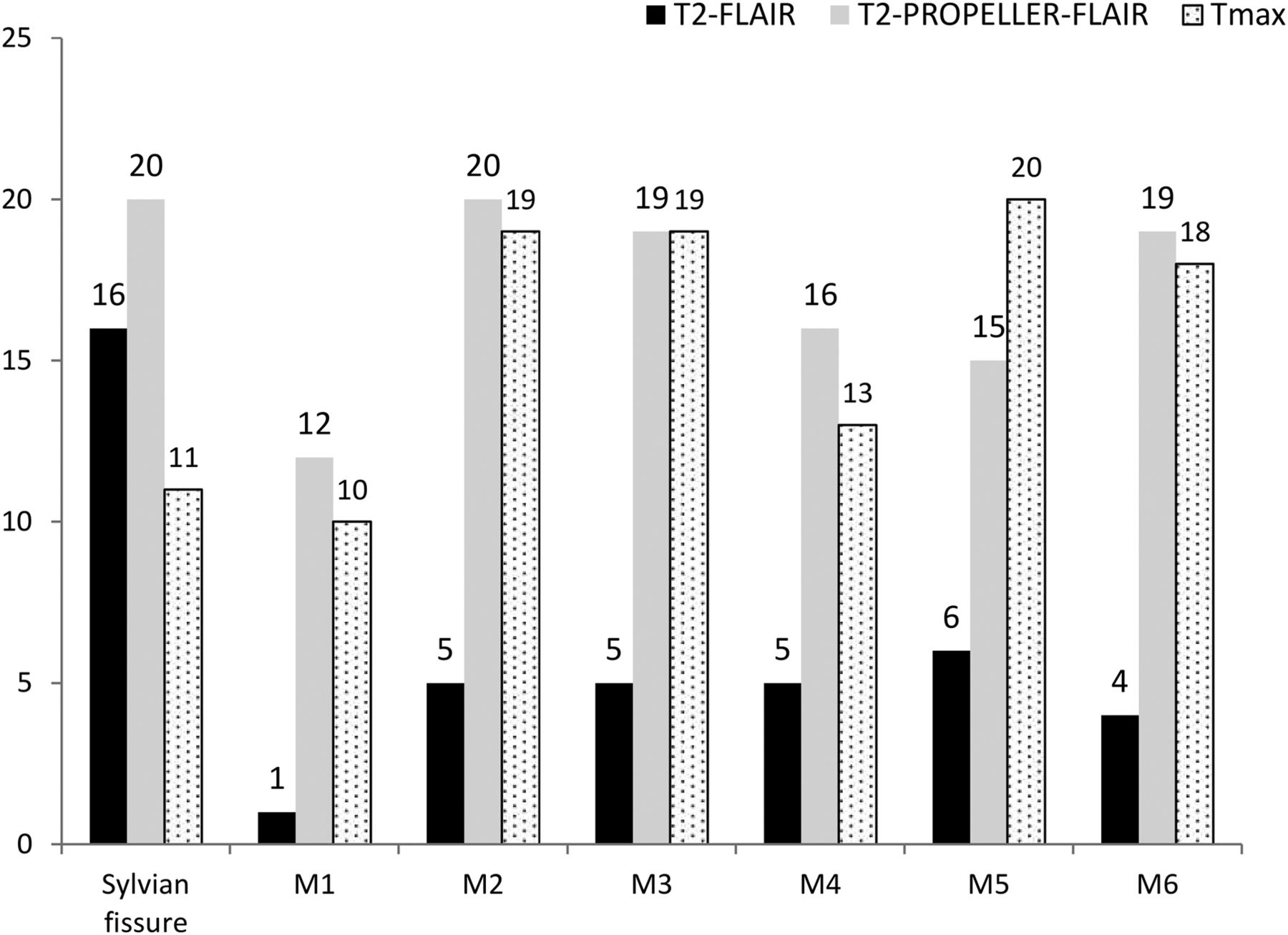
- Fig 1.
The distribution of FHVs and perfusion abnormality in each MCA-ASPECTS territory. The black, gray, and dotted bars represent the frequency of FHVs and perfusion abnormality for T2-FLAIR, T2-PROPELLER-FLAIR, and Tmax, respectively.
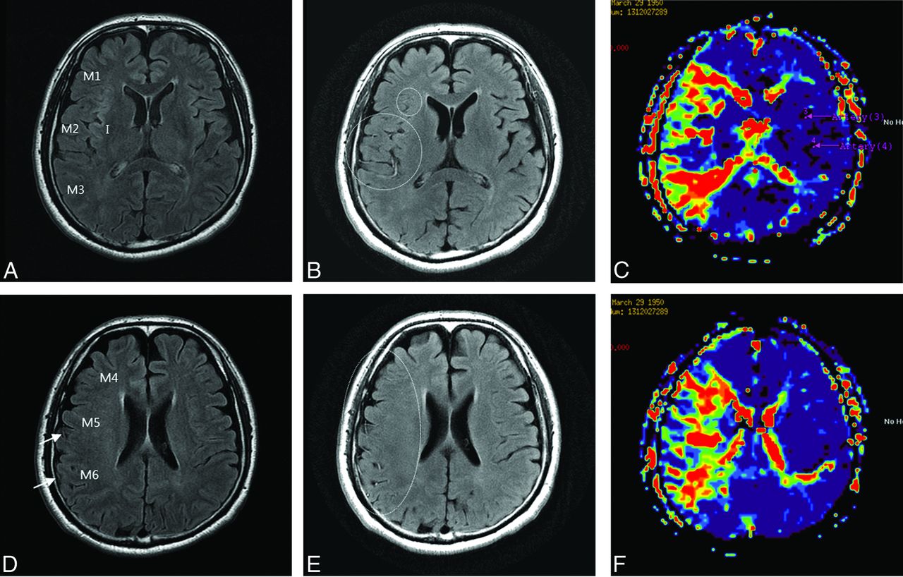
- Fig 2.
A 63-year-old man with right MCA territory infarction. FHVs on T2-FLAIR at the level of the basal ganglia (A) were not seen, whereas FHVs on T2-PROPELLER-FLAIR (B) were seen in the Sylvian fissure, M1, M2, and M3 (dotted circle). On the Tmax map (C), FHVs were well-matched with perfusion abnormality. FHVs on T2-FLAIR at the level of the ventricle above the basal ganglia (D) were seen in the M5 and M6 territory (arrow), whereas FHVs on T2-PROPELLER-FLAIR (E) were seen in all territories (dotted circle). On Tmax (F), FHVs on T2-PROPELLER-FLAIR were well-matched with perfusion abnormality.


Tables
Features No. of patients 35 Age (yr) (mean) 65.1 Female sex 12/35 (34) Hypertension 15/35 (42) Diabetes mellitus 11/35 (31) Median time interval from symptom onset to MRI (hr) (IQR) 23.4 (9.62–57.5) MRA findings Large-artery stenosis or occlusionb 28/35 (80) MCA horizontal segment 12 (34) MCA insular segment 5 (15) MCA cortical segment 1 (3) Distal ICA 3 (8) Proximal ICA 7 (20) Negative 7 (20) - Table 3:
The predictability of FHVs for large-artery stenosis on T2-FLAIR and T2-PROPELLER-FLAIRa
T2-FLAIR T2-PROPELLER-FLAIR P Value Arterial stenosis or occlusion on MRA 28/35 (80%) Incidence of FHV 19/35 (54%) 26/35 (74%) .06 Predictabilityb True-positive 19 26 True-negative 7 7 False-positive 0 0 False-negative 9 2 Sensitivity 68% 93% .03 Specificity 100% 100% - Table 4:
Associations among FHV scores, ischemic lesion volume, and perfusion abnormality volume in 9 patients with MCA horizontal segment occlusion
Ischemic DWI Lesion Volume (r)a Perfusion Abnormality Volume (r)a Median (IQR) (mL) 4.74 (1.76–11.57) 64.91 (60.33–70.31) FHV scores on T2-FLAIR 0.86, 0.01 0.28, 0.43 FHV scores on T2-PROPELLER-FLAIR 0.38, 0.34 0.79, 0.02 FHV mismatchb −0.79, 0.01 0.33, 0.42


