
Article Figures & Data
Figures
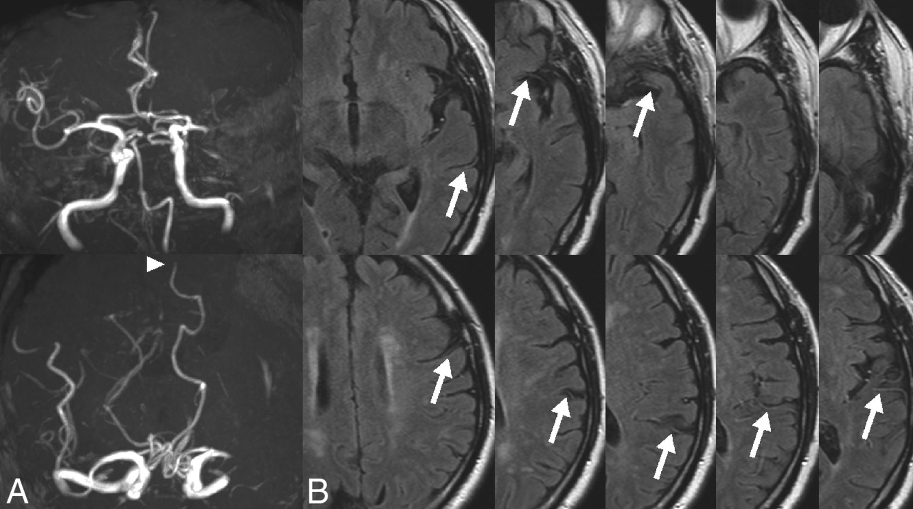
- Fig 1.
PCA laterality sign and hyperintense vessels on MR images. A, MRA shows an occlusion of the M1 portion of the left MCA and signal extent of the ipsilateral PCA (arrowhead). B, FLAIR MR imaging shows hyperintense vessels (arrows) in 8 of 10 axial sections.
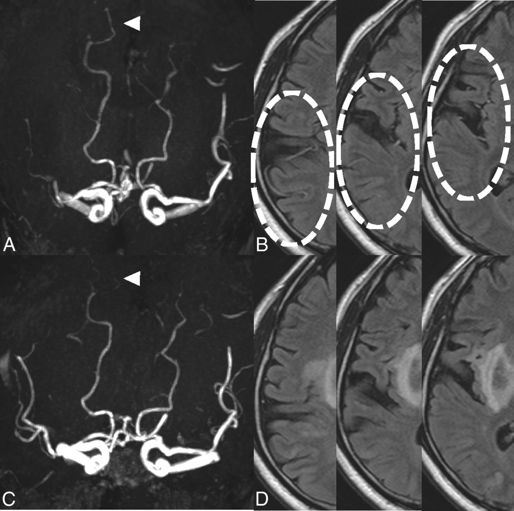
- Fig 2.
Reversion of collateral signs on MR images. MRA (A and C) and FLAIR MR imaging (B and D) of a representative patient who experienced early neurologic improvement after IV rtPA. PCA laterality (arrowheads) and hyperintense vessels (dotted circles) were observed before treatment (A and B) but disappeared after thrombolysis (C and D), indicating the reversion of collaterals.
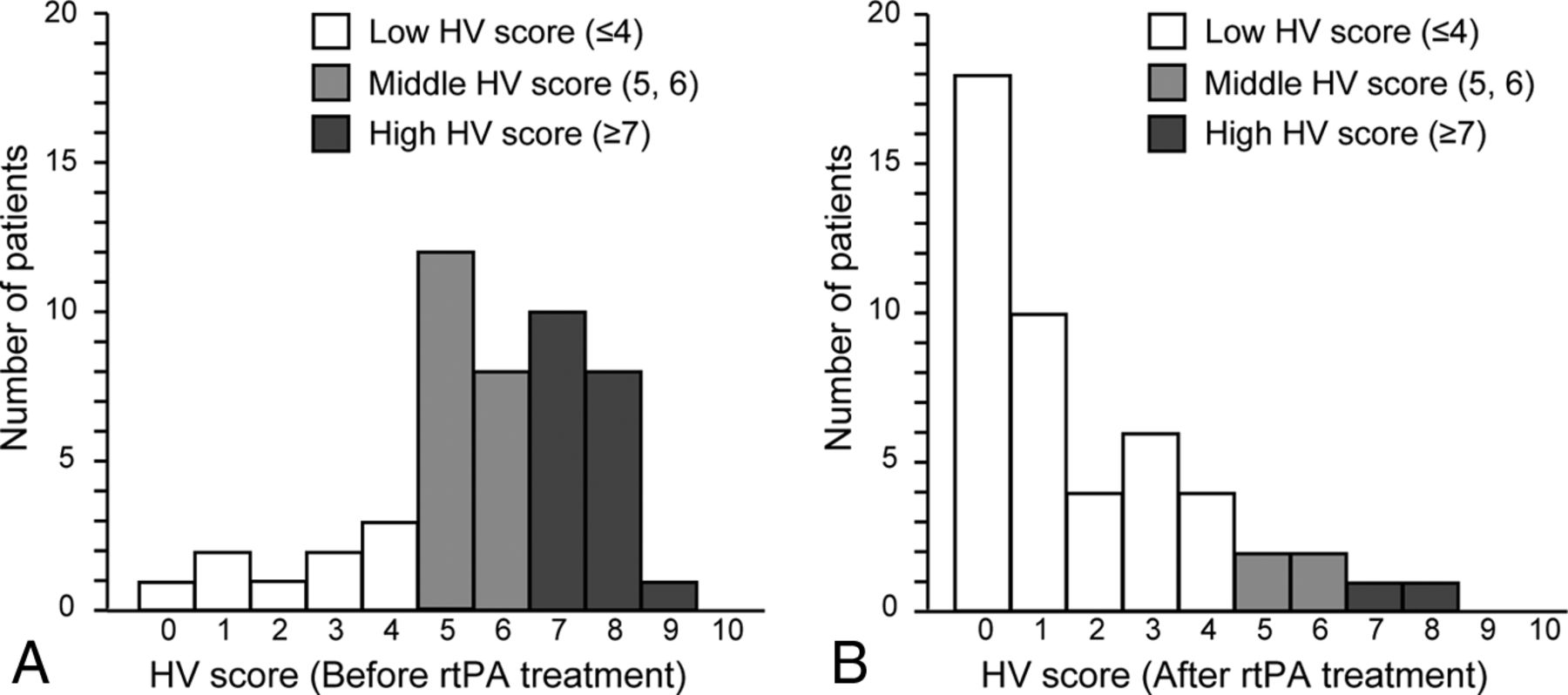
- Fig 3.
Distribution of the hyperintense vessel (HV) score before and after rtPA treatment. Most patients (39 of 48, 81%) were initially classified in the middle and high HV score group before treatment (A); however, the number of patients with a middle or high HV score dramatically decreased after thrombolysis (B).


Tables
Reversion of Collaterals P Value Yes (n = 25) No (n = 23) Age (median) (IQR) 78 (71–81) 79 (68–86) .52 Male sex (No.) (%) 13 (52) 13 (57) .78 mRS 0–1 before stroke (No.) (%) 25 (100) 22 (96) .48 Cardiovascular risk factors (No.) (%) Hypertension 15 (60) 13 (57) 1 Diabetes mellitus 7 (28) 3 (13) .29 Hyperlipidemia 4 (16) 7 (30) .31 Atrial fibrillation 21 (84) 13 (57) .057 Congestive heart failure 5 (20) 4 (17) 1 Previous stroke 6 (24) 6 (26) 1 Smoking 9 (36) 9 (41) .77 Past medication at stroke onset (No.) (%) Antiplatelet therapy 7 (28) 8 (35) .76 Anticoagulant therapy 5 (20) 4 (17) 1 Antihypertensive therapy 13 (52) 10 (44) .58 Statin therapy 2 (8) 4 (17) .41 Stroke etiology (No.) (%) Cardioembolism 19 (76) 11 (48) .07 Atherosclerosis 4 (16) 8 (35) .19 Other or undetermined 2 (8) 4 (17) .41 Severity of stroke at arrival (median) (IQR) Initial GCS 13 (11–14) 12 (10–14) .62 Initial DWI volume (mL) 21.6 (13.8–42.5) 22.9 (10.6–41.1) .63 DWI ASPECTS at arrival 8 (7–9) 8 (6–8) .72 Initial NIHSS score 17 (14–24) 16 (11–21) .35 Duration between 2 MRI scans (days) 7 (5–9) 6 (3–8) .2 Neurologic and radiologic outcome after rtPA (median) (IQR) 24-hr NIHSS 7 (2–12) 11 (6–17) .022b 7-day NIHSS 4 (1–8) 8 (3–14) .008c Hemorrhagic transformation 6 (24) 10 (43) .22 Successful recanalization 23 (92) 8 (35) <.001d Follow-up CT ASPECTS 8 (6.5–9) 6 (4–8) .017b M1 to M6 area in ASPECTS 5 (4–6) 3 (2–6) .021b C, I, L, IC area in ASPECTS 3 (2.5–4) 3 (2–3) .12 mRS 0–1 at 3 mo (No.) (%) 16 (64) 8 (35) .043b Note:—GCS indicates Glasgow Coma Scale; C, caudate nucleus; I, insular cortex; L, lenticular nucleus; IL, internal capsule.
↵a For continuous variables, the median and P values (Mann-Whitney U test) are shown. The resulting proportions and the P values (Fisher exact test with Yates correction, when appropriate) are shown.
↵b P < .05.
↵c P < .01.
↵d P < .001 was considered significant.
- Table 2:
Comparison of the presence and absence of early neurologic improvement after IV rtPA in patients with proximal middle cerebral artery occlusiona
Early Neurologic Improvement P Value Yes (n = 22) No (n = 26) Age (yr) ( median) (IQR) 78.5 (74–81) 78.5 (68–84) .92 Male sex (No.) (%) 10 (45) 16 (62) .38 mRS 0–1 before stroke (No.) (%) 22 (100) 25 (96) 1 NIHSS score at arrival (median) (mean) 17.9 ± 8.1 16.0 ± 5.7 .29 Systolic blood pressure (mean) 161.3 ± 29.6 155.0 ± 30.8 .91 Diastolic blood pressure (mean) 88.5 ± 26.1 78.3 ± 21.9 .19 Temperature (°C) (mean) 36.3 ± 0.4 36.2 ± 0.7 .75 Cardiovascular risk factors (No.) (%) Hypertension 14 (64) 14 (54) .57 Diabetes mellitus 7 (27) 4 (15) .48 Hyperlipidemia 5 (23) 6 (23) 1 Atrial fibrillation 19 (86) 15 (58) .054 Congestive heart failure 4 (18) 5 (19) 1 Previous stroke 5 (23) 7 (27) 1 Smoking 6 (27) 12 (48) .23 Past medication at stroke onset (No.) (%) Antiplatelet therapy 8 (36) 7 (27) .54 Anticoagulant therapy 4 (18) 5 (19) 1 Antihypertensive therapy 14 (64) 9 (35) .08 Statin therapy 2 (9) 4 (15) .67 Stroke etiology (No.) (%) Cardioembolism 17 (77) 13 (50) .074 Atherosclerosis 4 (18) 8 (31) .5 Other or undetermined 1 (5) 5 (19) .2 Imaging analysis Initial DWI volume (mL) (median) (IQR) 19.8 (11.5–42.3) 22.7(13.9–41.2) .26 DWI ASPECTS at arrival (median) (IQR) 8 (6.75–9) 8 (6.75–8) .33 MCA M1 occlusion (No.) (%) 18 (82) 17 (65) .33 Development of collaterals at arrival (No.) (%)c 15 (68) 9 (35) .042b Stroke outcome Follow-up CT ASPECTS (median) (IQR) 8 (6–9.25) 6 (4.75–8) .004d mRS 0–1 at 3 mo (No.) (%) 16 (73) 8 (31) .008d ↵a For continuous variables, the median and P values (Mann-Whitney U test) are shown. The resulting proportions and the P values (Fisher exact test with Yates correction, when appropriate) are shown.
↵b P < .05.
↵c The development of collaterals was defined as positive in the presence of PCA laterality and an HV score of ≥5 on initial MRI.
↵d P <.01 was considered significant.
- Table 3:
Univariate analyses and multivariate logistic regression analysis for the association of early neurologic improvement after IV rtPA in patients with proximal middle cerebral artery occlusion
Crude OR P Value Adjusted OR P Value (95% CI) (95% CI) Age (yr) 0.99 (0.95–1.06) .91 1.00 (0.94–1.06) .95 Male sex 0.52 (0.16–1.63) .26 History of atrial fibrillation 4.64 (1.20–23.36) .025a 5.32 (1.16–32.1) .031a NIHSS score at arrival 1.04 (0.88–1.04) .34 1.02 (0.89–1.09) .72 DWI ASPECTS at arrival 1.16 (0.80–1.73) .44 Time to rtPA administration 0.99 (0.97–1.02) .49 1.00 (0.97–1.03) .84 Development of collaterals at arrivalb 4.0 (1.25–14.27) .019a 4.82 (1.34–19.98) .015a


Jump to section
Related Articles
Cited By...
- Favourable arterial, tissue-level and venous collaterals correlate with early neurological improvement after successful thrombectomy treatment of acute ischaemic stroke
- Clinical prognosis of FLAIR hyperintense arteries in ischaemic stroke patients: a systematic review and meta-analysis
- The Association between FLAIR Vascular Hyperintensity and Stroke Outcome Varies with Time from Onset