Advertisement

Main menu

  • Home
  • Content
    • Current Issue
    • Accepted Manuscripts
    • Article Preview
    • Past Issue Archive
    • Video Articles
    • AJNR Case Collection
    • Case of the Week Archive
    • Case of the Month Archive
    • Classic Case Archive
  • Special Collections
    • AJNR Awards
    • Low-Field MRI
    • Alzheimer Disease
    • ASNR Foundation Special Collection
    • Photon-Counting CT
    • View All
  • Multimedia
    • AJNR Podcasts
    • AJNR SCANtastic
    • Trainee Corner
    • MRI Safety Corner
    • Imaging Protocols
  • For Authors
    • Submit a Manuscript
    • Submit a Video Article
    • Submit an eLetter to the Editor/Response
    • Manuscript Submission Guidelines
    • Statistical Tips
    • Fast Publishing of Accepted Manuscripts
    • Graphical Abstract Preparation
    • Imaging Protocol Submission
    • Author Policies
  • About Us
    • About AJNR
    • Editorial Board
    • Editorial Board Alumni
  • More
    • Become a Reviewer/Academy of Reviewers
    • Subscribers
    • Permissions
    • Alerts
    • Feedback
    • Advertisers
    • ASNR Home

User menu

  • Alerts
  • Log in

Search

  • Advanced search
American Journal of Neuroradiology

American Journal of Neuroradiology

ASHNR American Society of Functional Neuroradiology ASHNR American Society of Pediatric Neuroradiology ASSR
  • Alerts
  • Log in

Advanced Search

  • Home
  • Content
    • Current Issue
    • Accepted Manuscripts
    • Article Preview
    • Past Issue Archive
    • Video Articles
    • AJNR Case Collection
    • Case of the Week Archive
    • Case of the Month Archive
    • Classic Case Archive
  • Special Collections
    • AJNR Awards
    • Low-Field MRI
    • Alzheimer Disease
    • ASNR Foundation Special Collection
    • Photon-Counting CT
    • View All
  • Multimedia
    • AJNR Podcasts
    • AJNR SCANtastic
    • Trainee Corner
    • MRI Safety Corner
    • Imaging Protocols
  • For Authors
    • Submit a Manuscript
    • Submit a Video Article
    • Submit an eLetter to the Editor/Response
    • Manuscript Submission Guidelines
    • Statistical Tips
    • Fast Publishing of Accepted Manuscripts
    • Graphical Abstract Preparation
    • Imaging Protocol Submission
    • Author Policies
  • About Us
    • About AJNR
    • Editorial Board
    • Editorial Board Alumni
  • More
    • Become a Reviewer/Academy of Reviewers
    • Subscribers
    • Permissions
    • Alerts
    • Feedback
    • Advertisers
    • ASNR Home
  • Follow AJNR on Twitter
  • Visit AJNR on Facebook
  • Follow AJNR on Instagram
  • Join AJNR on LinkedIn
  • RSS Feeds

AJNR Awards, New Junior Editors, and more. Read the latest AJNR updates

Research ArticleNeurointervention

Building Multidevice Pipeline Constructs of Favorable Metal Coverage: A Practical Guide

M. Shapiro, E. Raz, T. Becske and P.K. Nelson
American Journal of Neuroradiology August 2014, 35 (8) 1556-1561; DOI: https://doi.org/10.3174/ajnr.A3902
M. Shapiro
aFrom the Departments of Radiology (M.S., E.R., T.B., P.K.N.), Bernard and Irene Schwartz Neurointerventional Radiology Section
bNeurology (M.S., T.B.)
  • Find this author on Google Scholar
  • Find this author on PubMed
  • Search for this author on this site
E. Raz
aFrom the Departments of Radiology (M.S., E.R., T.B., P.K.N.), Bernard and Irene Schwartz Neurointerventional Radiology Section
  • Find this author on Google Scholar
  • Find this author on PubMed
  • Search for this author on this site
T. Becske
aFrom the Departments of Radiology (M.S., E.R., T.B., P.K.N.), Bernard and Irene Schwartz Neurointerventional Radiology Section
bNeurology (M.S., T.B.)
  • Find this author on Google Scholar
  • Find this author on PubMed
  • Search for this author on this site
P.K. Nelson
aFrom the Departments of Radiology (M.S., E.R., T.B., P.K.N.), Bernard and Irene Schwartz Neurointerventional Radiology Section
cNeurosurgery (P.K.N.); New York University Langone Medical Center, New York, New York.
  • Find this author on Google Scholar
  • Find this author on PubMed
  • Search for this author on this site
  • Article
  • Figures & Data
  • Info & Metrics
  • Responses
  • References
  • PDF
Loading

Article Figures & Data

Figures

  • Tables
  • Fig 1.
    • Download figure
    • Open in new tab
    • Download powerpoint
    Fig 1.

    A, Representative image of a 4.25 × 20 mm Pipeline device inserted into plastic tubes of 0.5-mm incremental diameters. Variation in the degree of metal coverage is apparent. The configuration of cells is schematically depicted above the construct. Metal coverage can be calculated directly on the basis of measurements of the long and short diagonals of the rhombus and the diameter of each strand (30-μm), according to the formula above. Minimum coverage is seen when θ = 90°, corresponding to a square cell configuration. B, Scatterplot of tube diameter versus metal coverage for various device and “artery” combinations. All functions have a parabolic configuration. Note that absolute coverage values are higher for smaller diameter devices at each given tube diameter. The overall single-device coverage is, therefore, likely to be somewhat smaller for appropriately sized devices deployed in larger vessels.

  • Fig 2.
    • Download figure
    • Open in new tab
    • Download powerpoint
    Fig 2.

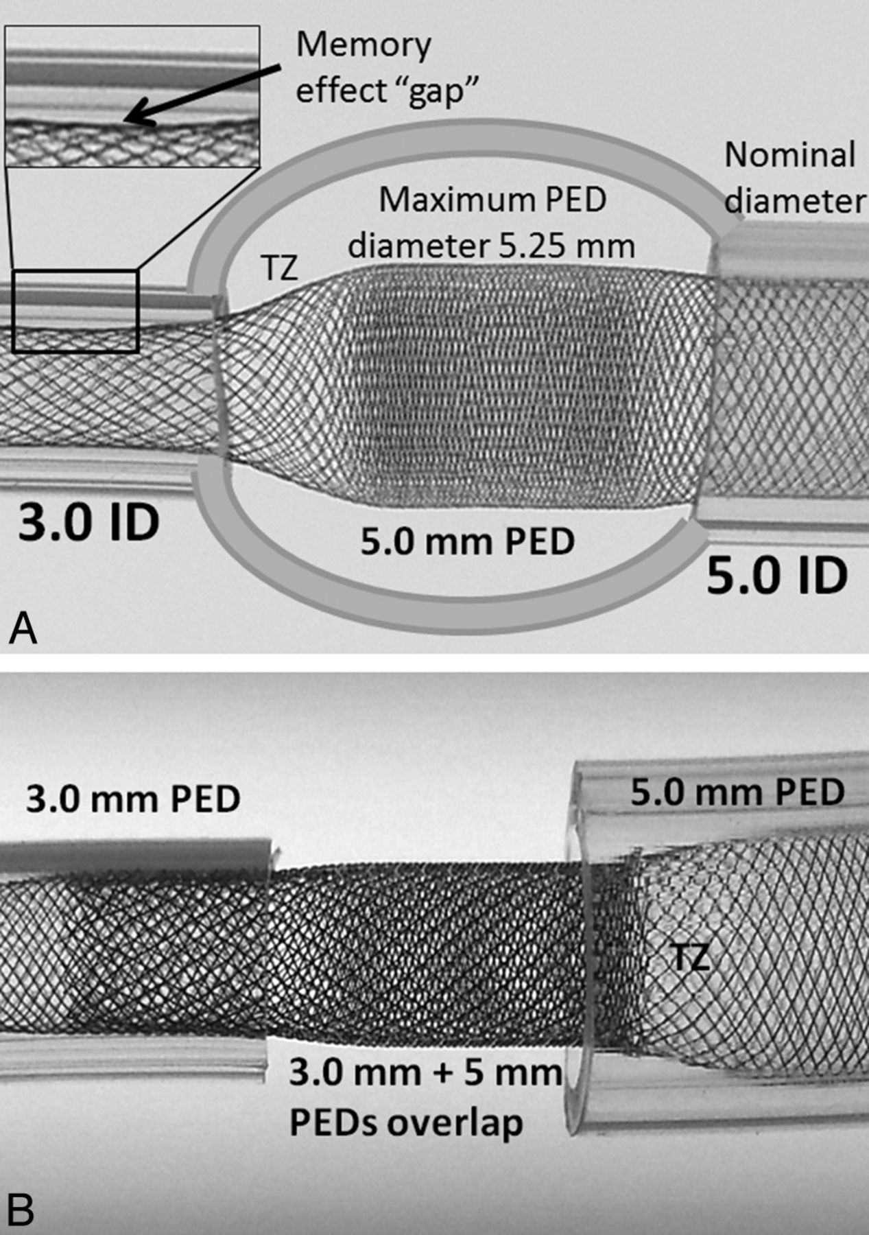
    Consequences of device oversizing and the proposed solution. A, A model of a fusiform aneurysm with 3.0- and 5.0-mm landing zones, bridged by a single 5 × 20 mm device. A transition zone (TZ) of minimum coverage is created as the device is constrained from its fully opened state into the 3-mm landing zone. Despite adequate length of the “landing zone” at the 3.0-mm end, the “shape memory” of the transition zone, TZ, nevertheless produces a “gap” where the device remains unapposed to the inner wall of the tube. B, To address these issues, 2 devices are required, each of which is appropriately sized for its recipient artery. The first 3.0-mm device is deployed from the 3.0-mm-diameter vessel into the 5-mm recipient vessel, following which a second 5.0-mm diameter device is telescoped into the first, with the 5.0-mm device anchored into its 5.0-mm vessel. Thus, the transition zone, TZ, is shifted outside the aneurysm, while the aneurysmal segment receives the benefit of double-coverage.

  • Fig 3.
    • Download figure
    • Open in new tab
    • Download powerpoint
    Fig 3.

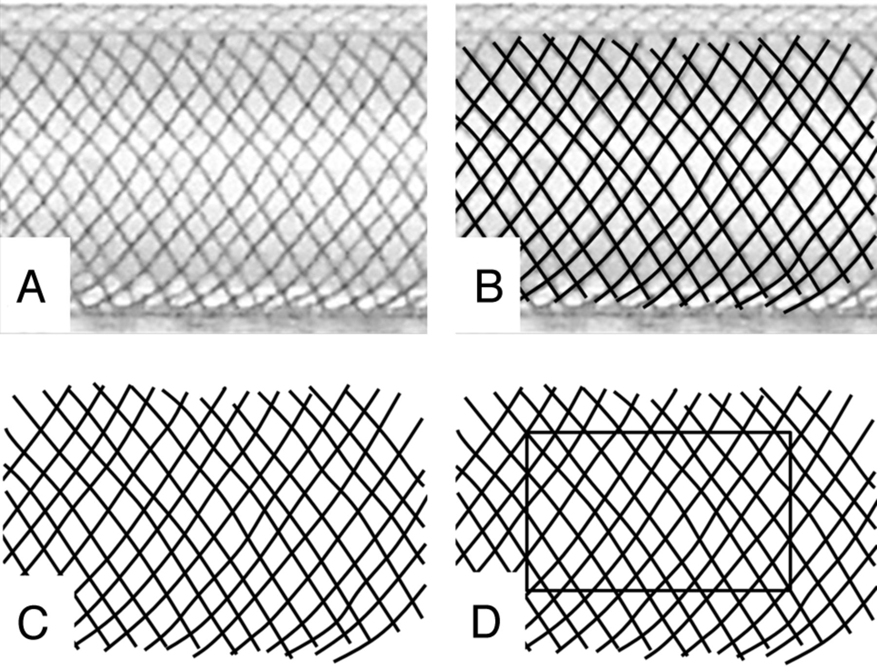
    Manual segmentation method of determining metal coverage. A, Photographic image of a 3.75-mm device deployed within a 3.5-mm plastic tube. A translucent fiberoptic rod is placed inside the construct to eliminate visualization of the “back” portion of the braids. B, Curved lines, with thickness corresponding to 30 μm (calibrated to a ruler placed alongside the construct) are traced along each braid. C, The underlying image is removed, leaving a black and white segmentation image. D, With the ImageJ measurement tool, the proportion of black pixels within a given rectangular area corresponds to the percentage of metal coverage, according to the formula listed in the “Materials and Methods” section.

  • Fig 4.
    • Download figure
    • Open in new tab
    • Download powerpoint
    Fig 4.

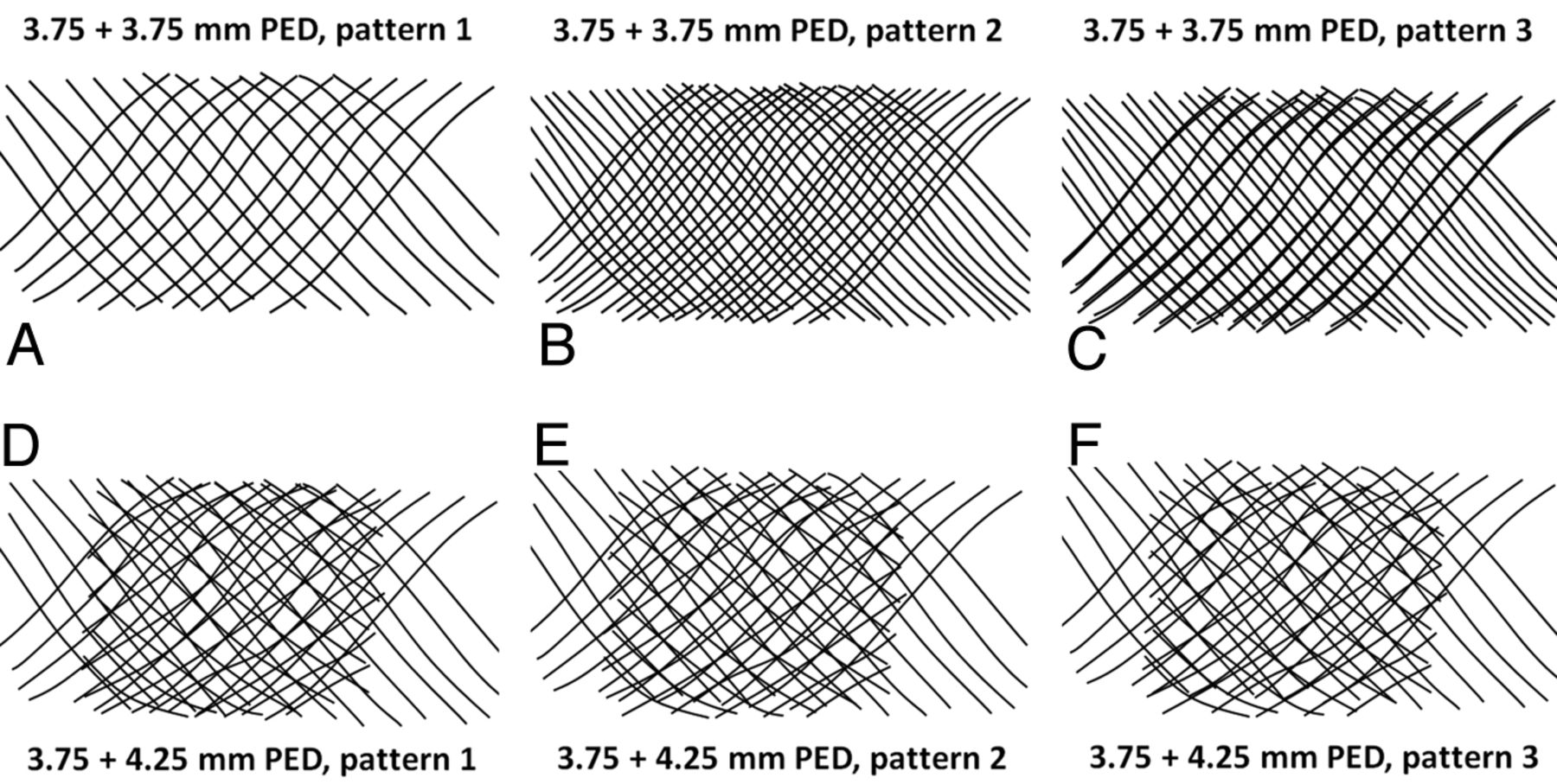
    Overlapping segmentation images illustrating patterns of double coverage with identical-diameter (A–C) and different-diameter devices (D–F). It can be readily seen from Figs A–C that 2 overlapping 3.75-mm devices can result in a wide range of coverage, depending on the exact alignment of device braids of identical pitch relative to each other. In contrast, an overlap of 3.75- and 4.25-mm devices (Figs D–F), because of the different braid pitch for each device, produces a more consistent overall coverage pattern, regardless of the particular phase of overlap.

  • Fig 5.
    • Download figure
    • Open in new tab
    • Download powerpoint
    Fig 5.

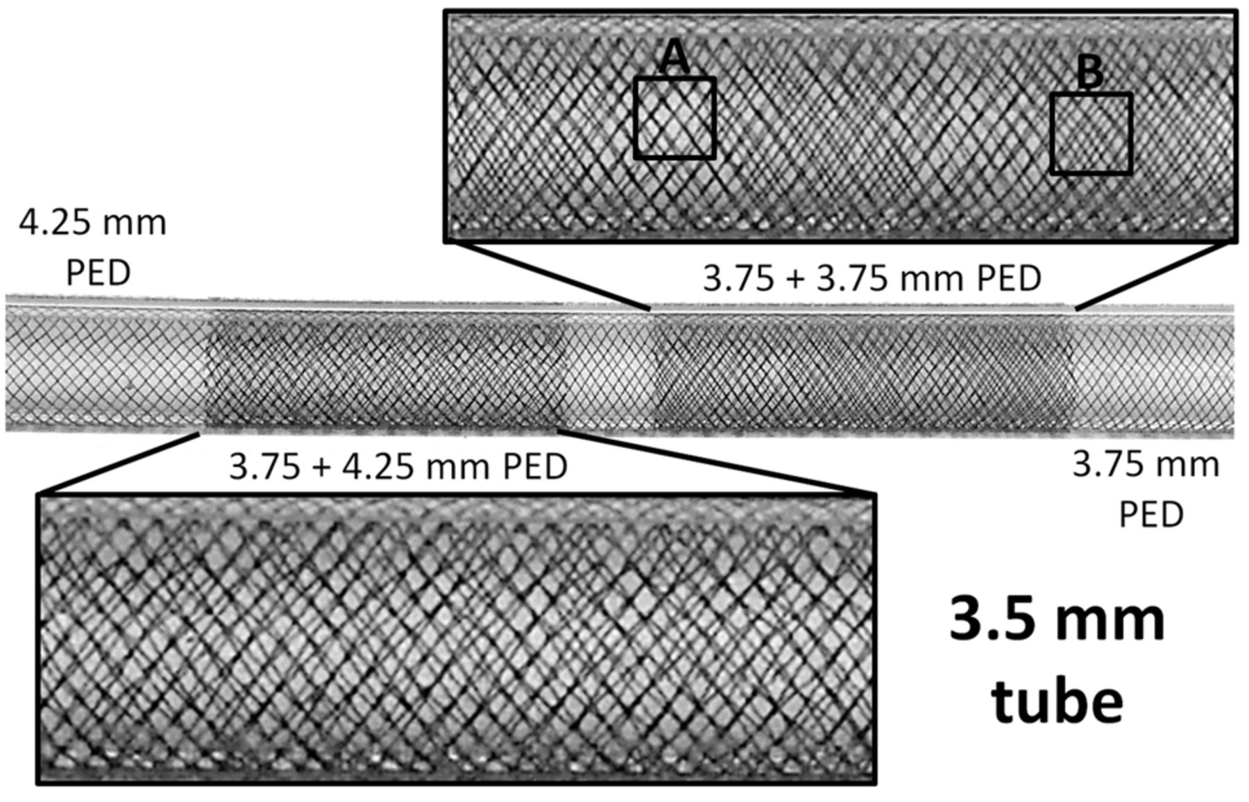
    Photographs of overlapping 3.75- and 4.25-mm-diameter devices deployed in a 3.5-mm tube. Visual appreciation of nonuniform coverage for two 3.75-mm devices: Area A shows near-perfect overlap of both device braids, with no practical increase in coverage for this segment, whereas the braid phase shift in area B produces significantly higher coverage. In contrast, overlap of 3.75- and 4.25-mm devices yields a more consistent pattern, with less potential variation in coverage values. The absolute value of such coverage is between the minimal and maximal potential values expected from overlapping two 3.75-mm devices.

  • Fig 6.
    • Download figure
    • Open in new tab
    • Download powerpoint
    Fig 6.

    Example of triple coverage with 3 different-diameter stents. Note that despite overall relatively high coverage, areas of lower coverage and significantly larger pore size remain (white rectangle).

Tables

  • Figures
    • View popup
    Table 1:

    Correlation between manual and estimated coverage values for the construct pictured in Fig 5a

    3.5-mm-Vessel Manual SegmentationManualEstimated
    3.75 PED22%
    3.75 + 4.25 PED36%–37%36%–37%
    4.25 PED18%–19%
    • ↵a Manual segmentation was done by tracing curved lines over each braid in the double-coverage area and comparing the result with values estimated by overlapping segmented images from separate 3.75- and 4.25-mm devices individually deployed within a 3.5-mm artificial vessel. The 2 methods are in excellent agreement.

    • View popup
    Table 2:

    The range of coverage values expected from overlap of various size devices in tubes of 3, 3.5, 4, and 4.5-mm diametersa

    3-mm Vessel
    3.253.754.254.75
    3.2530%–47%30%–40%31%–37%34%–37%
    3.7520%–37%30%–37%30%–35%
    4.2521%–35%31%–34%
    4.7520%–36%
    3.5-mm Vessel
    3.754.254.75
    3.7524%–41%36%–37%33%–34%
    4.2522%–36%31%–32%
    4.7518%–31%
    4-mm Vessel
    3.754.254.75
    3.7538%–46%37%–39%36%–37%
    4.2521%–35%29%–33%
    4.7522%–32%
    4.5-mm Vessel
    4.254.75
    4.2536%–57%40%–44%
    4.7525%–34%
    • ↵a Note that the broadest range of coverage for each device/artery combination is always observed during overlap of identical-diameter devices. Use of devices with progressively different diameters produces correspondingly narrower ranges of coverage (due to the more consistent misregistration of strands arising from the differing pitches imposed on devices of different diameters constrained within a given diameter vessel), with absolute coverage values falling in-between the potential minima and maxima expected from overlap of identical devices.

PreviousNext
Back to top

In this issue

American Journal of Neuroradiology: 35 (8)
American Journal of Neuroradiology
Vol. 35, Issue 8
1 Aug 2014
  • Table of Contents
  • Index by author
  • Complete Issue (PDF)
Advertisement
Print
Download PDF
Email Article

Thank you for your interest in spreading the word on American Journal of Neuroradiology.

NOTE: We only request your email address so that the person you are recommending the page to knows that you wanted them to see it, and that it is not junk mail. We do not capture any email address.

Enter multiple addresses on separate lines or separate them with commas.
Building Multidevice Pipeline Constructs of Favorable Metal Coverage: A Practical Guide
(Your Name) has sent you a message from American Journal of Neuroradiology
(Your Name) thought you would like to see the American Journal of Neuroradiology web site.
CAPTCHA
This question is for testing whether or not you are a human visitor and to prevent automated spam submissions.
Cite this article
M. Shapiro, E. Raz, T. Becske, P.K. Nelson
Building Multidevice Pipeline Constructs of Favorable Metal Coverage: A Practical Guide
American Journal of Neuroradiology Aug 2014, 35 (8) 1556-1561; DOI: 10.3174/ajnr.A3902

Citation Manager Formats

  • BibTeX
  • Bookends
  • EasyBib
  • EndNote (tagged)
  • EndNote 8 (xml)
  • Medlars
  • Mendeley
  • Papers
  • RefWorks Tagged
  • Ref Manager
  • RIS
  • Zotero
0 Responses
Respond to this article
Share
Bookmark this article
Building Multidevice Pipeline Constructs of Favorable Metal Coverage: A Practical Guide
M. Shapiro, E. Raz, T. Becske, P.K. Nelson
American Journal of Neuroradiology Aug 2014, 35 (8) 1556-1561; DOI: 10.3174/ajnr.A3902
del.icio.us logo Twitter logo Facebook logo Mendeley logo
  • Tweet Widget
  • Facebook Like
  • Google Plus One
Purchase

Jump to section

  • Article
    • Abstract
    • ABBREVIATIONS:
    • Materials and Methods
    • Results
    • Discussion
    • Conclusions
    • Footnotes
    • REFERENCES
  • Figures & Data
  • Info & Metrics
  • Responses
  • References
  • PDF

Related Articles

  • No related articles found.
  • PubMed
  • Google Scholar

Cited By...

  • Early experience with the Drivewire 24: a newly FDA-approved steerable microwire
  • Comprehensive Analysis of Post-Pipeline Endothelialization and Remodeling
  • SuperDyna: Unlocking the Potential of Post-Treatment Device Evaluation
  • Pipeline Embolization Device for intracranial aneurysms presenting with mass effect: a large Chinese cohort
  • SuperDyna: Unlocking the Potential of Post-Treatment Device Evaluation
  • Pipeline Embolization Device for intracranial aneurysms presenting with mass effect: a large Chinese cohort
  • Placement of a Stent within a Flow Diverter Improves Aneurysm Occlusion Rates
  • Aneurysm Remnants after Flow Diversion: Clinical and Angiographic Outcomes
  • Comparison of Pipeline Embolization Device Sizing Based on Conventional 2D Measurements and Virtual Simulation Using the Sim&Size Software: An Agreement Study
  • Toward Better Understanding of Flow Diversion in Bifurcation Aneurysms
  • A comparison between the new Low-profile Visualized Intraluminal Support (LVIS Blue) stent and the Flow Redirection Endoluminal Device (FRED) in bench-top and cadaver studies
  • Basilar artery perforator aneurysms (BAPAs): review of the literature and classification
  • p64 Flow Modulation Device in the treatment of intracranial aneurysms: initial experience and technical aspects
  • New Pipeline Flex device: initial experience and technical nuances
  • Endoluminal Reconstruction for Nonsaccular Aneurysms of the Proximal Posterior Cerebral Artery with the Pipeline Embolization Device
  • Anterior Choroidal Artery Patency and Clinical Follow-Up after Coverage with the Pipeline Embolization Device
  • Crossref
  • Google Scholar

This article has not yet been cited by articles in journals that are participating in Crossref Cited-by Linking.

More in this TOC Section

  • Connectivity for remote neurovascular procedures
  • Cavernous dural AVF treated by transfacial route
  • A Retrospective Study in Tentorial DAVFs
Show more NEUROINTERVENTION

Similar Articles

Advertisement

Indexed Content

  • Current Issue
  • Accepted Manuscripts
  • Article Preview
  • Past Issues
  • Editorials
  • Editor's Choice
  • Fellows' Journal Club
  • Letters to the Editor
  • Video Articles

Cases

  • Case Collection
  • Archive - Case of the Week
  • Archive - Case of the Month
  • Archive - Classic Case

More from AJNR

  • Trainee Corner
  • Imaging Protocols
  • MRI Safety Corner
  • Book Reviews

Multimedia

  • AJNR Podcasts
  • AJNR Scantastics

Resources

  • Turnaround Time
  • Submit a Manuscript
  • Submit a Video Article
  • Submit an eLetter to the Editor/Response
  • Manuscript Submission Guidelines
  • Statistical Tips
  • Fast Publishing of Accepted Manuscripts
  • Graphical Abstract Preparation
  • Imaging Protocol Submission
  • Evidence-Based Medicine Level Guide
  • Publishing Checklists
  • Author Policies
  • Become a Reviewer/Academy of Reviewers
  • News and Updates

About Us

  • About AJNR
  • Editorial Board
  • Editorial Board Alumni
  • Alerts
  • Permissions
  • Not an AJNR Subscriber? Join Now
  • Advertise with Us
  • Librarian Resources
  • Feedback
  • Terms and Conditions
  • AJNR Editorial Board Alumni

American Society of Neuroradiology

  • Not an ASNR Member? Join Now

© 2025 by the American Society of Neuroradiology All rights, including for text and data mining, AI training, and similar technologies, are reserved.
Print ISSN: 0195-6108 Online ISSN: 1936-959X

Powered by HighWire