
Article Figures & Data
Figures
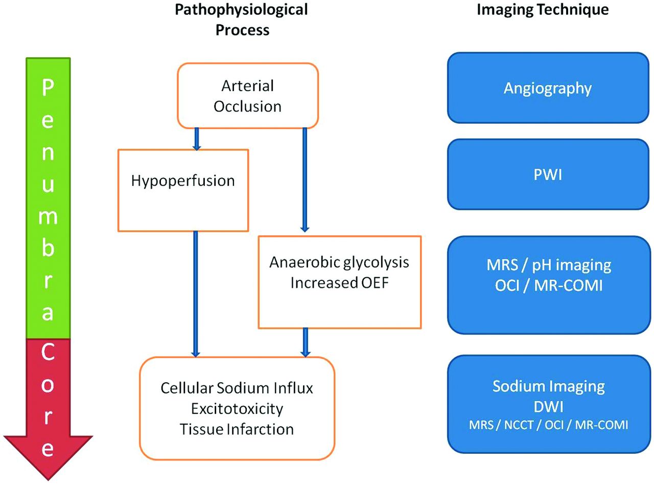
- Fig 1.
Approach to metabolic imaging of acute stroke. On the left, the tissue compartments (penumbra/core) are delineated. In the middle, the pathophysiologic process is outlined. On the right, and aligned in the horizontal meridian, imaging techniques that may image the respective pathophysiologic process are documented.
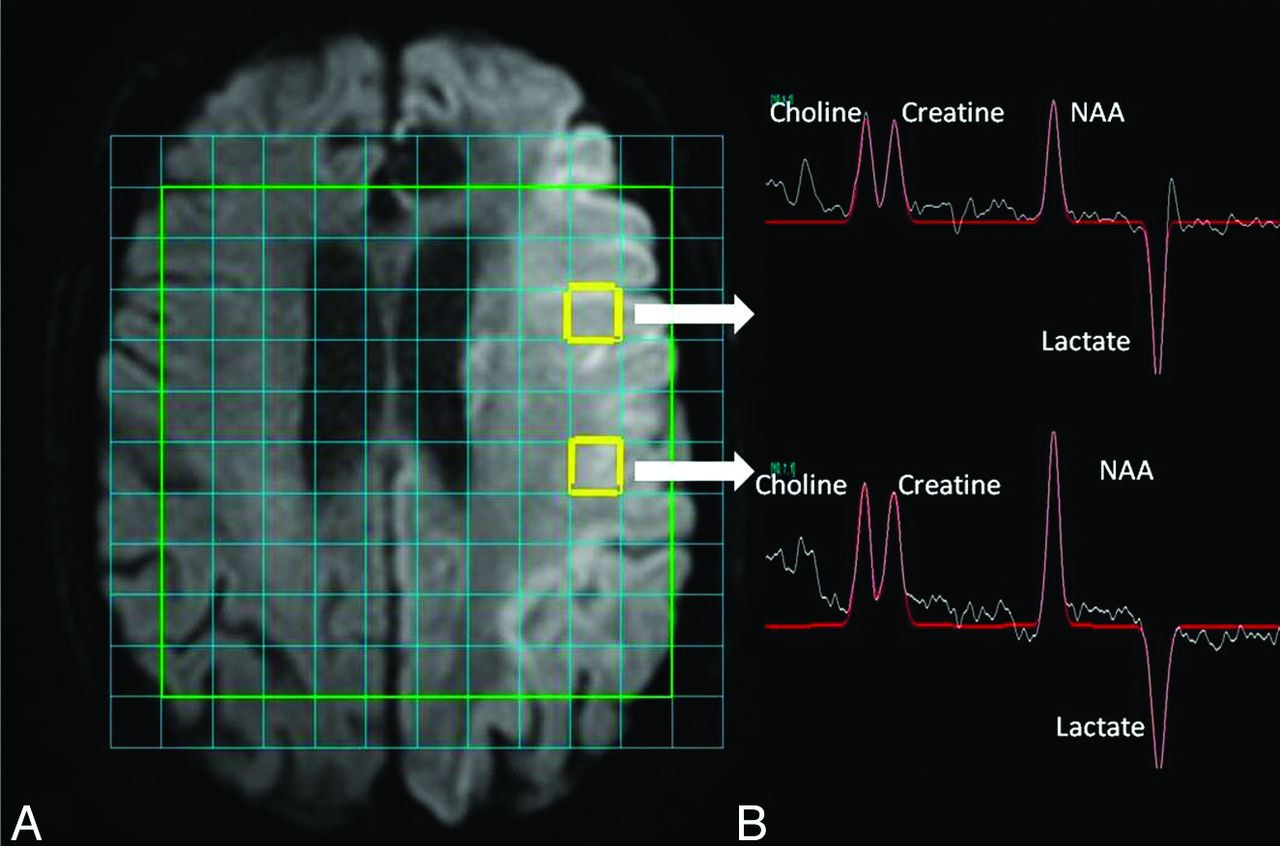
- Fig 2.
Data from MRS. A, Acute stroke lesion of the left hemisphere on DWI. There is a multi-voxel spectroscopy grid overlaid onto the time. Users can select data from any voxel. The 2 voxels highlighted in yellow show the regions from which spectroscopy data were derived and are illustrated in on the right-hand side. B, Spectroscopy data for the 4 major metabolites that are annotated. NAA appears low, and lactate concentration is elevated. Area under the peaks indicates relative metabolite concentration. Although the lesion on DWI appears relatively uniform throughout, the NAA concentration is less in the anteriorly placed voxel compared with the posteriorly placed voxel.
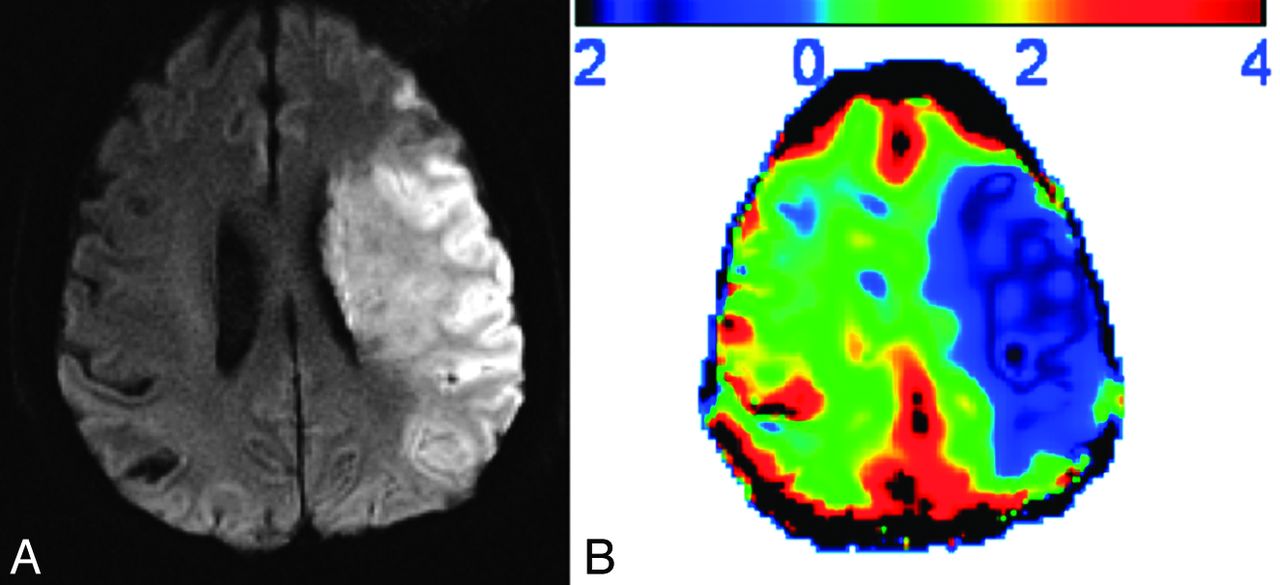
- Fig 3.
An example of oxygen challenge imaging. A, Stroke lesion of the left hemisphere on DWI from a subject last seen well approximately 24 hours before imaging. B, Results from oxygen challenge imaging. The percentage change in T2*WI signal is indicated in the scale bar at the top (positive results indicate an increase in signal; negative results a decrease in signal). There is a diminished response to oxygen challenge in regions corresponding to the established lesion on DWI, suggesting a core pattern.
- Fig 4.
Current and potential markers of core. A, MR images of DWI (right) and FLAIR image (left) showing extensive established infarct core; B, list of potential imaging profiles that may refine definitions of core. OCI indicates oxygen challenge imaging; MR-COMI, MR cerebral oxygen metabolic index; CMRO2, cerebral metabolic rate for oxygen.
- Fig 5.
Current and potential markers of penumbra. A, MR images of DWI (right) and PWI (left) showing a region of PWI-DWI mismatch; B, list of potential imaging profiles that may refine definitions of penumbra. OCI indicates oxygen challenge imaging; MR-COMI, MR cerebral oxygen metabolic index; CMRO2, cerebral metabolic rate for oxygen.


